. 2010 Oct;30(19):4732-43.
doi: 10.1128/MCB.00413-10. Epub 2010 Aug 2.

BRCT domain interactions with phospho-histone H2A target Crb2 to chromatin at double-strand breaks and maintain the DNA damage checkpoint

Affiliations

BRCT domain interactions with phospho-histone H2A target Crb2 to chromatin at double-strand breaks and maintain the DNA damage checkpoint

Sevil Sofueva et al. Mol Cell Biol. 2010 Oct.

Abstract

Relocalization of checkpoint proteins to chromatin flanking DNA double-strand breaks (DSBs) is critical for cellular responses to DNA damage. Schizosaccharomyces pombe Crb2, which mediates Chk1 activation by Rad3(ATR), forms ionizing radiation-induced nuclear foci (IRIF). Crb2 C-terminal BRCT domains (BRCT(2)) bind histone H2A phosphorylated at a C-terminal SQ motif by Tel1(ATM) and Rad3(ATR), although the functional significance of this interaction is controversial. Here, we show that polar interactions of Crb2 serine-548 and lysine-619 with the phosphate group of phospho-H2A (γ-H2A) are critical for Crb2 IRIF formation and checkpoint function. Mutations of these BRCT(2) domain residues have additive effects when combined in a single allele. Combining either mutation with an allele that eliminates the threonine-215 cyclin-dependent kinase phosphorylation site completely abrogates Crb2 IRIF and function. We propose that cooperative phosphate interactions in the BRCT(2) γ-H2A-binding pocket of Crb2, coupled with tudor domain interactions with lysine-20 dimethylation of histone H4, facilitate stable recruitment of Crb2 to chromatin surrounding DSBs, which in turn mediates efficient phosphorylation of Chk1 that is required for a sustained checkpoint response. This mechanism of cooperative interactions with the γ-H2A/X phosphate is likely conserved in S. pombe Brc1 and human Mdc1 genome maintenance proteins.


Figures
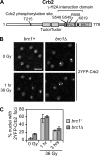
FIG. 1.
FIG. 1.
Crb2 forms IRIF independently of Brc1. (A) Domain organization of Crb2. Tandem tudor domains, which are conserved in mammalian 53BP1 DNA damage response protein, bind histone H4-K20me2 and are required for Crb2 IRIF formation (1, 32). The phosphate group of γ-H2A.1 pSer129 binds in a small pocket of Crb2-BRCT2 and makes polar contacts with the main and side chains of Ser548 and Lys619 (16). These contacts have direct functional counterparts in mammalian Mdc1 (35). The crystal structure of Crb2-BRCT2 in complex with the γ-H2A.1 pSer129 peptide also identified contacts between the phosphate and the peptide nitrogen of Gly549 and a water-bridged interaction with Arg558 (16). Residue Thr215 is a Cdc2 (CDK) phosphorylation site important for proper checkpoint response but not for IRIF formation (8, 24). (B) Crb2 IRIF in asynchronous brc1+ and brc1Δ cells irradiated with 36 Gy IR. 2YFP-Crb2 localization was observed 1 h (shown) and 3 h after irradiation. Bar, 5 μm. (C) Quantification of the percentage of nuclei with 2YFP-Crb2 foci. A total of 150 nuclei were scored for each time point. Error bars represent results of three independent experiments. The strains used are LLD3260 (brc1+) and YJW4896 (brc1Δ).
FIG. 2.
FIG. 2.
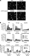
Crb2 focus induction by IR is largely dependent on the interaction with γ-H2A. (A) Representative images of 2YFP-Crb2 localization in IR-treated cells 1 h post-irradiation with 36 Gy. Bar, 5 μm. WT, wild type. (B) Quantitative representation of the numbers of foci in the different mutants. Exponentially growing asynchronous cultures were subjected to 36 Gy IR and nuclei with 1, 2, or more foci were counted as a percentage of the total. Each experiment was done at least twice. About 200 nuclei were scored for each time point. (C) Quantification of IR-iduced focus formation (36 Gy) by Crb2 point mutants in brc1Δ cells. A total of 150 nuclei were scored for each time point. Error bars represent three independent experiments. The strains used for the experiments shown in this figure are LLD3260 (crb2+), LLD4897 (crb2-T215A), LLD4898 (hta-AQ), LLD4901 (crb2-S548A), LLD4902 (crb2-K619M), SAS4908 (crb2-S548AK619M), SAS4922 (crb2-T215A brc1Δ), SAS4923 (crb2-S548A brc1Δ), and SAS4924 (crb2-K619M brc1Δ).
FIG. 3.
FIG. 3.
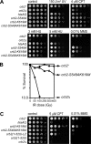
The Crb2 S548A and K619M mutations are additive for genotoxin sensitivity. (A) Fivefold serial dilutions of wild-type and mutant strains on YES plates were treated as indicated. (B) IR survival curves showing the sensitivity of crb2-S548AK619M compared to the wild type and crb2Δ. Log-phase cultures were irradiated and plated in triplicate to determine cell viability. (C) Replacement of Crb2-BRCT2 with a leucine zipper (LZ) dimerization domain leads to genotoxin sensitivity similar to that of crb2-S548AK619M. Control plates, UV treatment plates, 3 mM HU, and 4 μM CPT plates were photographed after 2 to 3 days at 30°C. The 5 μM CPT, 5 mM HU, and 0.01% MMS plates were photographed after 3 to 4 days. The strains used for the experiments shown in this figure are LLD3260 (crb2+), LLD3259 (crb2Δ), LLD4898 (hta-AQ), LLD4901 (crb2-S548A), LLD4902 (crb2-K619M), SAS4908 (crb2-S548AK619M), LLD3495 [crb2(1-520)], and LLD3496 [crb2(1-520)-LZ].
FIG. 4.
FIG. 4.
Mutating Ser548 or Lys619 in the crb2-T215A background leads to synergistic sensitivity to genotoxic stress. (A) Abolishing both the histone modification-dependent and -independent pathways of recruiting Crb2 to DNA damage in the crb2-T215AS548A and crb2-T215AK619M mutants results in a phenotype similar to crb2Δ. All plates were photographed after 2 to 3 days at 30°C. (B) IR survival curves confirm strong synergistic interactions of T215A with S548A or K619M. (C) No Crb2 IRIF are detectable in crb2-T215AK619M cells. Exponentially growing cultures were irradiated with 36 Gy and photographed 1 h after irradiation. Very similar images were obtained with crb2-T215AS548A cells (Sofueva and Russell, unpublished). The strains used for experiments shown in this figure are LLD3260 (crb2+), LLD3259 (crb2Δ), LLD4901 (crb2-S548A), LLD4902 (crb2-K619M), LLD4897 (crb2-T215A), SAS4903 (crb2-T215AS548A), SAS4916 (crb2-T215AK619M), and LLD4899 (crb2-T215A hta-AQ). WT, wild type. Bar, 5 μm.
FIG. 5.
FIG. 5.
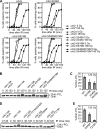
Crb2-BRCT2 mutants have a defective IR checkpoint response. (A) The checkpoint arrest triggered by IR is abbreviated in crb2-S548A, crb2-K619M, and crb2-S548AK619M cells. The indicated mutants in a cdc25-22 background were synchronized at late G2 phase by incubation at 35.5°C for 2.5 h. Following 180 Gy IR irradiation, cultures were returned to the permissive temperature of 25°C. Cell division (septation) was assessed by staining cells with Calcofluor. The data shown are representative of multiple experiments. (B) Chk1 phosphorylation is impaired in crb2-K619M and crb2-S548AK619M mutants. Exponentially growing asynchronous cultures were irradiated with 0, 30, and 120 Gy and harvested immediately following irradiation. (C) ImageJ quantification of the data shown in panel B. (D) Chk1 phosphorylation is ablated in crb2-T215AS548A cells. (E) ImageJ quantification of the data shown in panel D. Error bars represent the standard deviations of results from three independent experiments. The strains used for the experiments shown in this figure are SAS4904 (crb2+), LLD3628 (crb2Δ), SAS4905 (crb2-S548A), SAS4906 (crb2-K619M), SAS4917 (crb2-S548AK619M), SAS4909 (chk1-HA crb2Δ), SAS4914 (chk1-HA crb2+), SAS4910 (chk1-HA crb2-S548A), SAS4911 (chk1-HA crb2-K619M), SAS4912 (chk1-HA crb2-T215AS548A), SAS4913 (chk1-HA crb2-S548AK619M), and SAS4915 (chk1-HA crb2-T215A). WT, wild type.
FIG. 6.
FIG. 6.
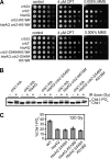
The Crb2-BRCT2 mutations are epistatic with htaAQ. (A) The htaAQ crb2-K619M and htaAQ crb2-S548AK619M mutants are as sensitive to genotoxins as htaAQ. All plates were photographed after 2 to 3 days at 30°C. (B) Chk1 phosphorylation in htaAQ is impaired similarly to the Crb2-BRCT2 mutants. Abolishing H2A phosphorylation in the Crb2-BRCT2 mutant backgrounds does not lead to further decrease in Chk1 phosphorylation levels. −, no IR; +, 120 Gy IR. (C) ImageJ quantification of the data shown in panel B. Error bars represent the standard deviations of results from three independent experiments. The strains used for the experiments shown in this figure are LLD3259 (crb2Δ), LLD3260 (crb2+), LLD4898 (htaAQ), LLD4902 (crb2-K619M), SAS4921 (htaAQ crb2-K619M), SAS4908 (crb2-S548AK619M), SAS4920 (htaAQ crb2-S548AK619M), SAS4914 (chk1-HA crb2+), OL4925 (chk1-HA htaAQ), OL4926 (chk1-HA htaAQ crb2-S548A), OL4927 (chk1-HA htaAQ crb2-K619M), and OL4928 (chk1-HA htaAQ crb2-S548AK619M). WT, wild type.
FIG. 7.
FIG. 7.
The Crb2-BRCT2 mutants form foci at DSBs created by the HO endonuclease. Cells carrying the HO cleavage site were grown in the absence of thiamine at 25°C for 27 h, permitting expression of HO. The occurrence of the break was confirmed by visualizing Rad22-2CFP foci (5, 6). (A) As expected, wild-type (WT) or mutant 2YFP-Crb2 impaired in the histone modification-dependent pathway localized to the break site. (B) No Crb2 foci were detected in the crb2-T215AS548A mutant, consistent with abrogation of both the histone modification-dependent and -independent pathways of Crb2 recruitment. (C) Quantification of the percentage of Rad22-2CFP foci colocalizing with a 2YFP-Crb2 focus. Nearly all 2YFP-Crb2 foci colocalized with a Rad22-2CFP focus. The strains used are LLD3650 (crb2+), LLD3652 (hta-AQ), LLD4900 (crb2-T215A), SAS4918 (crb2-S548A), OL4930 (crb2-K619M), OL4931 (crb2-S548AK619M), OL4929 (htaAQ crb2-K619M), OL4932 (htaAQ crb2-S548AK619M), and SAS4919 (crb2-T215AS548A).
FIG. 8.
FIG. 8.
Genetic interactions of Crb2 tudor and BRCT mutations. (A) Quantification of the percent nuclei with 2YFP-Crb2 foci in crb2-F400A and crb2-F400AS548A cells. Exponentially growing asynchronous cultures were subjected to 36 Gy IR, and nuclei with 1, 2, or more foci were counted as a percentage of the total. About 200 nuclei were scored for each time point. (B) Epistasis analysis of strains carrying mutations within the tudor and BRCT domains. Control plates, UV treatment plates, 5 μM CPT, and 0.0075% MMS plates were photographed after 2 to 3 days at 30°C. The 5 mM HU plate was photographed after 3 to 4 days. (C and D) Chk1 phosphorylation is further impaired in strains with simultaneously mutated tudor and BRCT domains. (E and F) ImageJ quantification of the data shown in panels C and D. Error bars represent the standard deviations of results from three independent experiments. The strains used for the experiments presented in this figure are LLD3643 (crb2-F400A), SAS4942 (crb2-F400AS548A), SAS4933 (crb2+), LLD3259 (crb2Δ), SAS4935 (crb2-F400A), SAS4934 (crb2-K619M), SAS4936 (crb2-F400AK619M), SAS4907 (crb2-S548AK619M), SAS4937 (crb2-F400AS548AK619M), SAS4914 (chk1-HA crb2+), SAS4910 (chk1-HA crb2-S548A), SAS4938 (chk1-HA crb2-F400A), SAS4939 (chk1-HA crb2-F400AS548A), SAS4911 (chk1-HA crb2-K619M), SAS4940 (chk1-HA crb2-F400AK619M), SAS4913 (chk1-HA crb2-S548AK619M), and SAS4941 (chk1-HA crb2-F400AS548AK619M). WT, wild type.

Similar articles

Cited by

References

    1. Botuyan, M. V., J. Lee, I. M. Ward, J. E. Kim, J. R. Thompson, J. Chen, and G. Mer. 2006. Structural basis for the methylation state-specific recognition of histone H4-K20 by 53BP1 and Crb2 in DNA repair. Cell 127:1361-1373. - PMC - PubMed
    1. Downs, J. A., S. Allard, O. Jobin-Robitaille, A. Javaheri, A. Auger, N. Bouchard, S. J. Kron, S. P. Jackson, and J. Cote. 2004. Binding of chromatin-modifying activities to phosphorylated histone H2A at DNA damage sites. Mol. Cell 16:979-990. - PubMed
    1. Downs, J. A., N. F. Lowndes, and S. P. Jackson. 2000. A role for Saccharomyces cerevisiae histone H2A in DNA repair. Nature 408:1001-1004. - PubMed
    1. Du, L. L., B. A. Moser, and P. Russell. 2004. Homo-oligomerization is the essential function of the tandem BRCT domains in the checkpoint protein Crb2. J. Biol. Chem. 279:38409-38414. - PubMed
    1. Du, L. L., T. M. Nakamura, B. A. Moser, and P. Russell. 2003. Retention but not recruitment of Crb2 at double-strand breaks requires Rad1 and Rad3 complexes. Mol. Cell. Biol. 23:6150-6158. - PMC - PubMed

Publication types

MeSH terms