Human anti-HIV-neutralizing antibodies frequently target a conserved epitope essential for viral fitness
- PMID: 20679402
- PMCID: PMC2931156
- DOI: 10.1084/jem.20101176
Human anti-HIV-neutralizing antibodies frequently target a conserved epitope essential for viral fitness
Abstract
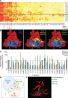
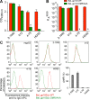
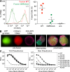
The identification and characterization of conserved epitopes on the HIV-1 viral spike that are immunogenic in humans and targeted by neutralizing antibodies is an important step in vaccine design. Antibody cloning experiments revealed that 32% of all HIV-neutralizing antibodies expressed by the memory B cells in patients with high titers of broadly neutralizing antibodies recognize one or more "core" epitopes that were not defined. Here, we show that anti-core antibodies recognize a single conserved epitope on the gp120 subunit. Amino acids D474, M475, R476, which are essential for anti-core antibody binding, form an immunodominant triad at the outer domain/inner domain junction of gp120. The mutation of these residues to alanine impairs viral fusion and fitness. Thus, the core epitope, a frequent target of anti-HIV-neutralizing antibodies, including the broadly neutralizing antibody HJ16, is conserved and indispensible for viral infectivity. We conclude that the core epitope should be considered as a target for vaccine design.
Figures


Similar articles
-
Increased Epitope Complexity Correlated with Antibody Affinity Maturation and a Novel Binding Mode Revealed by Structures of Rabbit Antibodies against the Third Variable Loop (V3) of HIV-1 gp120.J Virol. 2018 Mar 14;92(7):e01894-17. doi: 10.1128/JVI.01894-17. Print 2018 Apr 1. J Virol. 2018. PMID: 29343576 Free PMC article.
-
Bacterially expressed HIV-1 gp120 outer-domain fragment immunogens with improved stability and affinity for CD4-binding site neutralizing antibodies.J Biol Chem. 2018 Sep 28;293(39):15002-15020. doi: 10.1074/jbc.RA118.005006. Epub 2018 Aug 9. J Biol Chem. 2018. PMID: 30093409 Free PMC article.
-
Conserved Role of an N-Linked Glycan on the Surface Antigen of Human Immunodeficiency Virus Type 1 Modulating Virus Sensitivity to Broadly Neutralizing Antibodies against the Receptor and Coreceptor Binding Sites.J Virol. 2015 Oct 28;90(2):829-41. doi: 10.1128/JVI.02321-15. Print 2016 Jan 15. J Virol. 2015. PMID: 26512079 Free PMC article.
-
GP120: target for neutralizing HIV-1 antibodies.Annu Rev Immunol. 2006;24:739-69. doi: 10.1146/annurev.immunol.24.021605.090557. Annu Rev Immunol. 2006. PMID: 16551265 Review.
-
[Role of the HIV-1 gp120 V1/V2 domains in the induction of neutralizing antibodies].Enferm Infecc Microbiol Clin. 2009 Nov;27(9):523-30. doi: 10.1016/j.eimc.2008.02.010. Epub 2009 May 1. Enferm Infecc Microbiol Clin. 2009. PMID: 19409660 Review. Spanish.
Cited by
-
Somatic mutations of the immunoglobulin framework are generally required for broad and potent HIV-1 neutralization.Cell. 2013 Mar 28;153(1):126-38. doi: 10.1016/j.cell.2013.03.018. Cell. 2013. PMID: 23540694 Free PMC article.
-
Restriction of HIV-1 Escape by a Highly Broad and Potent Neutralizing Antibody.Cell. 2020 Feb 6;180(3):471-489.e22. doi: 10.1016/j.cell.2020.01.010. Epub 2020 Jan 30. Cell. 2020. PMID: 32004464 Free PMC article.
-
Viral escape from HIV-1 neutralizing antibodies drives increased plasma neutralization breadth through sequential recognition of multiple epitopes and immunotypes.PLoS Pathog. 2013 Oct;9(10):e1003738. doi: 10.1371/journal.ppat.1003738. Epub 2013 Oct 31. PLoS Pathog. 2013. PMID: 24204277 Free PMC article. Clinical Trial.
-
Candidate antibody-based therapeutics against HIV-1.BioDrugs. 2012 Jun 1;26(3):143-62. doi: 10.2165/11631400-000000000-00000. BioDrugs. 2012. PMID: 22462520 Free PMC article. Review.
-
B cell responses to HIV-1 infection and vaccination: pathways to preventing infection.Trends Mol Med. 2011 Feb;17(2):108-16. doi: 10.1016/j.molmed.2010.10.008. Epub 2010 Nov 26. Trends Mol Med. 2011. PMID: 21112250 Free PMC article.
References
-
- Barbas C.F., III, Björling E., Chiodi F., Dunlop N., Cababa D., Jones T.M., Zebedee S.L., Persson M.A., Nara P.L., Norrby E., et al. . 1992. Recombinant human Fab fragments neutralize human type 1 immunodeficiency virus in vitro. Proc. Natl. Acad. Sci. USA. 89:9339–9343. 10.1073/pnas.89.19.9339 - DOI - PMC - PubMed
-
- Burton D.R., Barbas C.F., III, Persson M.A., Koenig S., Chanock R.M., Lerner R.A.. 1991. A large array of human monoclonal antibodies to type 1 human immunodeficiency virus from combinatorial libraries of asymptomatic seropositive individuals. Proc. Natl. Acad. Sci. USA. 88:10134–10137. 10.1073/pnas.88.22.10134 - DOI - PMC - PubMed
Publication types
MeSH terms
Substances
Grants and funding
LinkOut - more resources
Full Text Sources
Other Literature Sources

