

The aryl hydrocarbon receptor interacts with c-Maf to promote the differentiation of type 1 regulatory T cells induced by IL-27
- PMID: 20676095
- PMCID: PMC2940320
- DOI: 10.1038/ni.1912
The aryl hydrocarbon receptor interacts with c-Maf to promote the differentiation of type 1 regulatory T cells induced by IL-27
Abstract
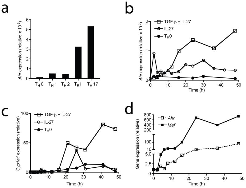
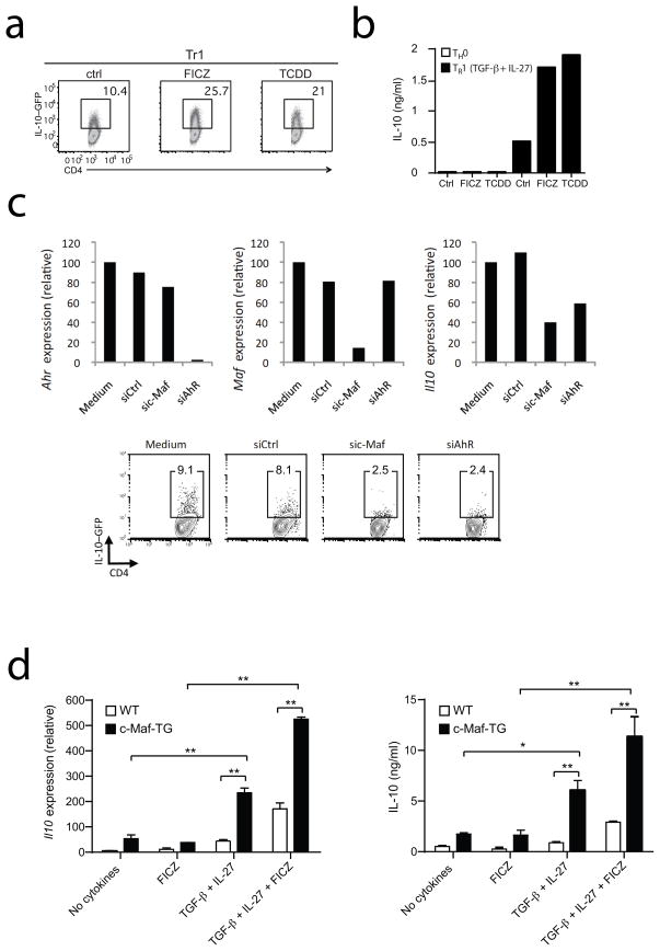
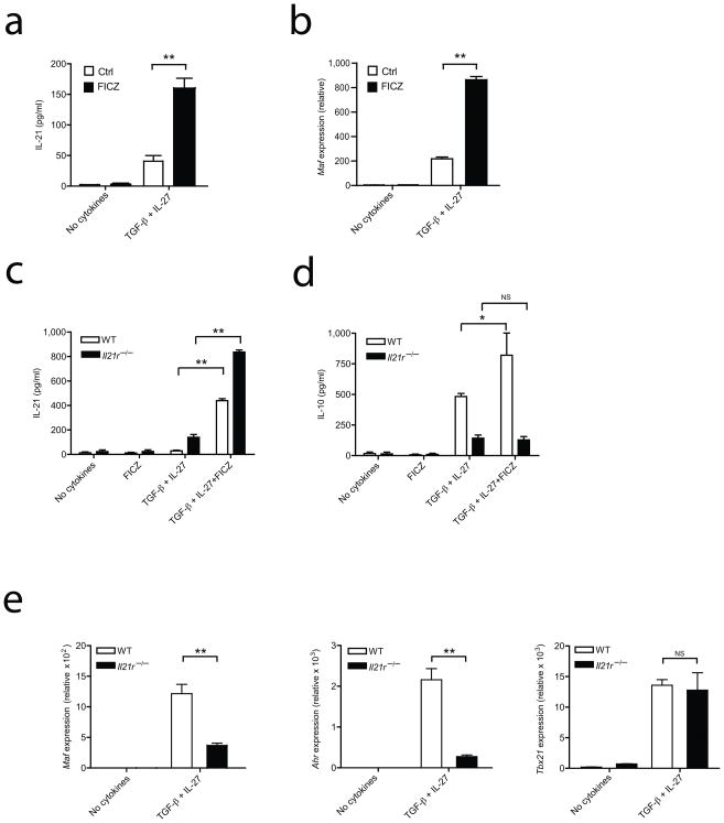
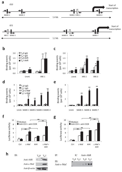
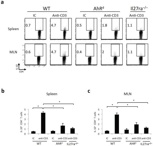
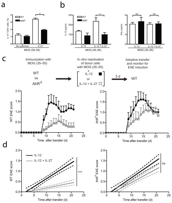
Type 1 regulatory T cells (Tr1 cells ) that produce interleukin 10 (IL-10) are instrumental in the prevention of tissue inflammation, autoimmunity and graft-versus-host disease. The transcription factor c-Maf is essential for the induction of IL-10 by Tr1 cells, but the molecular mechanisms that lead to the development of these cells remain unclear. Here we show that the ligand-activated transcription factor aryl hydrocarbon receptor (AhR), which was induced by IL-27, acted in synergy with c-Maf to promote the development of Tr1 cells. After T cell activation under Tr1-skewing conditions, the AhR bound to c-Maf and promoted transactivation of the Il10 and Il21 promoters, which resulted in the generation of Tr1 cells and the amelioration of experimental autoimmune encephalomyelitis. Manipulating AhR signaling could therefore be beneficial in the resolution of excessive inflammatory responses.
Figures
Comment in
-
A toxin-sensitive receptor able to reduce immunopathology.Nat Immunol. 2010 Sep;11(9):779-81. doi: 10.1038/ni0910-779. Nat Immunol. 2010. PMID: 20720583 No abstract available.
-
Collaborative control of induced regulators.Nat Rev Immunol. 2010 Sep;10(9):620. doi: 10.1038/nri2839. Nat Rev Immunol. 2010. PMID: 21080598 No abstract available.
Similar articles
-
Tr1 Cells, but Not Foxp3+ Regulatory T Cells, Suppress NLRP3 Inflammasome Activation via an IL-10-Dependent Mechanism.J Immunol. 2015 Jul 15;195(2):488-97. doi: 10.4049/jimmunol.1403225. Epub 2015 Jun 8. J Immunol. 2015. PMID: 26056255
-
Type 1 regulatory T cells (Tr1) in autoimmunity.Semin Immunol. 2011 Jun;23(3):202-8. doi: 10.1016/j.smim.2011.07.005. Epub 2011 Aug 12. Semin Immunol. 2011. PMID: 21840222 Free PMC article. Review.
-
Prostaglandin E2 inhibits Tr1 cell differentiation through suppression of c-Maf.PLoS One. 2017 Jun 12;12(6):e0179184. doi: 10.1371/journal.pone.0179184. eCollection 2017. PLoS One. 2017. PMID: 28604806 Free PMC article.
-
AhR activation increases IL-2 production by alloreactive CD4+ T cells initiating the differentiation of mucosal-homing Tim3+ Lag3+ Tr1 cells.Eur J Immunol. 2017 Nov;47(11):1989-2001. doi: 10.1002/eji.201747121. Epub 2017 Sep 15. Eur J Immunol. 2017. PMID: 28833046 Free PMC article.
-
Molecular pathways in the induction of interleukin-27-driven regulatory type 1 cells.J Interferon Cytokine Res. 2010 Jun;30(6):381-8. doi: 10.1089/jir.2010.0047. J Interferon Cytokine Res. 2010. PMID: 20540648 Free PMC article. Review.
Cited by
-
The Aryl Hydrocarbon Receptor Controls IFNγ-Induced Immune Checkpoints PD-L1 and IDO via the JAK/STAT Pathway in Lung Adenocarcinoma.bioRxiv [Preprint]. 2024 Aug 13:2024.08.12.607602. doi: 10.1101/2024.08.12.607602. bioRxiv. 2024. PMID: 39185148 Free PMC article. Preprint.
-
Biology of IL-27 and its role in the host immunity against Mycobacterium tuberculosis.Int J Biol Sci. 2015 Jan 5;11(2):168-75. doi: 10.7150/ijbs.10464. eCollection 2015. Int J Biol Sci. 2015. PMID: 25561899 Free PMC article. Review.
-
Type I interferons and microbial metabolites of tryptophan modulate astrocyte activity and central nervous system inflammation via the aryl hydrocarbon receptor.Nat Med. 2016 Jun;22(6):586-97. doi: 10.1038/nm.4106. Epub 2016 May 9. Nat Med. 2016. PMID: 27158906 Free PMC article.
-
An Aryl Hydrocarbon Receptor-Mediated Amplification Loop That Enforces Cell Migration in ER-/PR-/Her2- Human Breast Cancer Cells.Mol Pharmacol. 2016 Nov;90(5):674-688. doi: 10.1124/mol.116.105361. Epub 2016 Aug 29. Mol Pharmacol. 2016. PMID: 27573671 Free PMC article.
-
T cell cytokine signatures: Biomarkers in pediatric multiple sclerosis.J Neuroimmunol. 2016 Aug 15;297:1-8. doi: 10.1016/j.jneuroim.2016.04.015. Epub 2016 Apr 30. J Neuroimmunol. 2016. PMID: 27397070 Free PMC article.
References
-
- Roncarolo MG, et al. Interleukin-10-secreting type 1 regulatory T cells in rodents and humans. Immunol Rev. 2006;212:28–50. - PubMed
-
- Pflanz S, et al. IL-27, a heterodimeric cytokine composed of EBI3 and p28 protein, induces proliferation of naive CD4(+) T cells. Immunity. 2002;16:779–790. - PubMed
-
- Villarino A, et al. The IL-27R (WSX-1) is required to suppress T cell hyperactivity during infection. Immunity. 2003;19:645–655. - PubMed
-
- Batten M, et al. Interleukin 27 limits autoimmune encephalomyelitis by suppressing the development of interleukin 17-producing T cells. Nat Immunol. 2006;7:929–936. - PubMed
-
- Awasthi A, et al. A dominant function for interleukin 27 in generating interleukin 10-producing anti-inflammatory T cells. Nat Immunol. 2007;8:1380–1389. - PubMed
Publication types
MeSH terms
Substances
Grants and funding
- P01AI056299/AI/NIAID NIH HHS/United States
- K99 AI075285/AI/NIAID NIH HHS/United States
- P01 AI039671-140009/AI/NIAID NIH HHS/United States
- R01 NS030843-19/NS/NINDS NIH HHS/United States
- R00 AI075285/AI/NIAID NIH HHS/United States
- P01 NS038037-10/NS/NINDS NIH HHS/United States
- R37NS030843/NS/NINDS NIH HHS/United States
- P01 AI039671-130009/AI/NIAID NIH HHS/United States
- R01 NS030843/NS/NINDS NIH HHS/United States
- P01-ES11624/ES/NIEHS NIH HHS/United States
- K99 AI075285-01A2/AI/NIAID NIH HHS/United States
- R37 NS030843-16/NS/NINDS NIH HHS/United States
- P01 AI056299/AI/NIAID NIH HHS/United States
- P01 NS038037-08/NS/NINDS NIH HHS/United States
- P01 ES011624/ES/NIEHS NIH HHS/United States
- P01 NS038037-06A20004/NS/NINDS NIH HHS/United States
- NS38037/NS/NINDS NIH HHS/United States
- R37 NS030843-17/NS/NINDS NIH HHS/United States
- AI435801/AI/NIAID NIH HHS/United States
- P01 NS038037-080004/NS/NINDS NIH HHS/United States
- P01 NS038037-09/NS/NINDS NIH HHS/United States
- R56 AI093903/AI/NIAID NIH HHS/United States
- P01NS038037/NS/NINDS NIH HHS/United States
- P01 AI039671/AI/NIAID NIH HHS/United States
- R37 NS030843/NS/NINDS NIH HHS/United States
- P01 NS038037-090004/NS/NINDS NIH HHS/United States
- R01 AI093903/AI/NIAID NIH HHS/United States
- R01 NS030843-08/NS/NINDS NIH HHS/United States
- RG4111A1/RG/CSR NIH HHS/United States
- P01 NS038037-070004/NS/NINDS NIH HHS/United States
- R01 NS030843-18A2/NS/NINDS NIH HHS/United States
- R37 NS030843-15/NS/NINDS NIH HHS/United States
- 1K99AI075285/AI/NIAID NIH HHS/United States
- P01AI039671/AI/NIAID NIH HHS/United States
- P01 NS038037/NS/NINDS NIH HHS/United States
LinkOut - more resources
Full Text Sources
Other Literature Sources
Molecular Biology Databases
