. 2010 Oct 1;285(40):30523-30.
doi: 10.1074/jbc.M110.150755. Epub 2010 Jul 30.

Hrs controls sorting of the epithelial Na+ channel between endosomal degradation and recycling pathways

Affiliations

Hrs controls sorting of the epithelial Na+ channel between endosomal degradation and recycling pathways

Ruifeng Zhou et al. J Biol Chem. .

Abstract

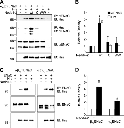
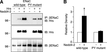
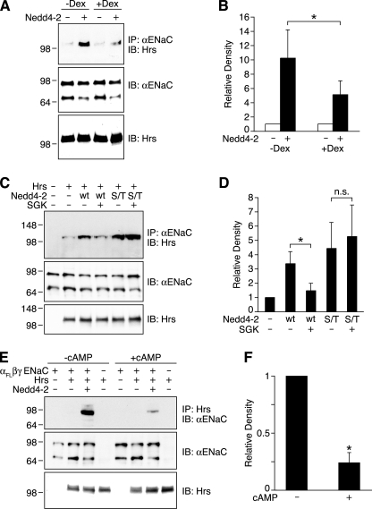
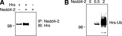
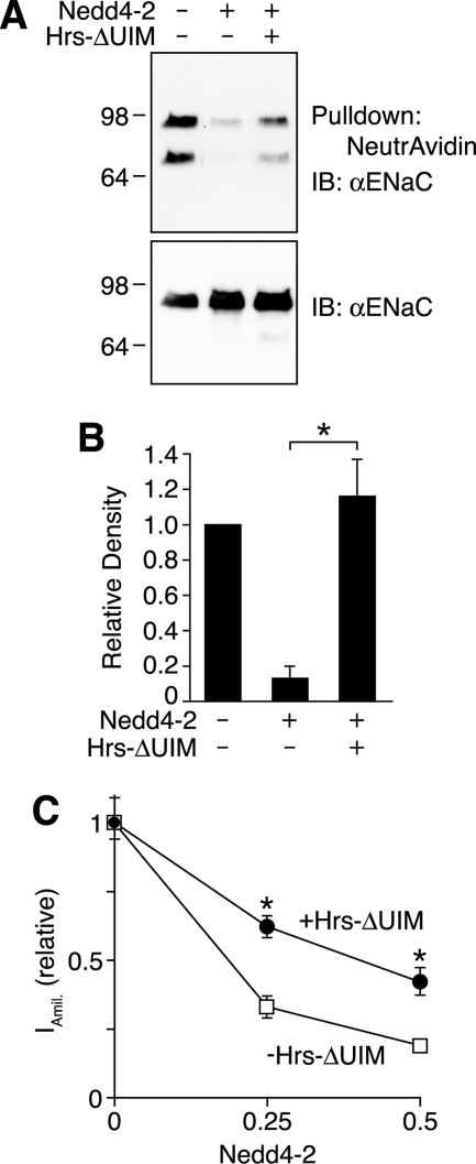
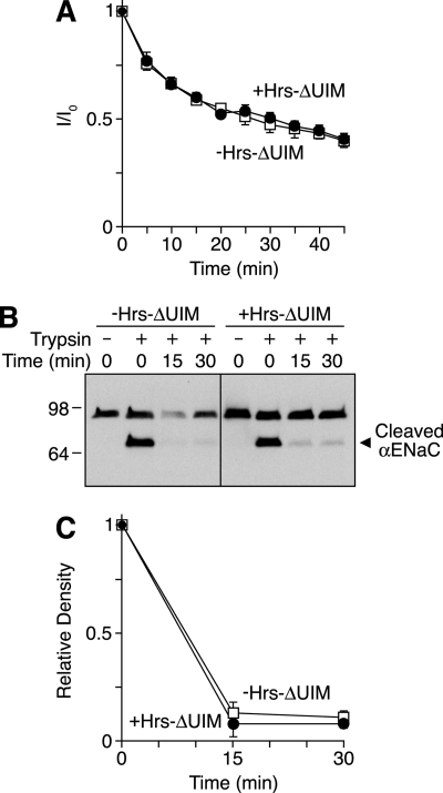
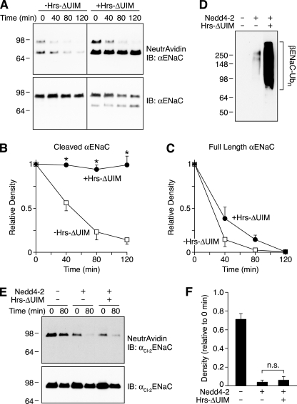
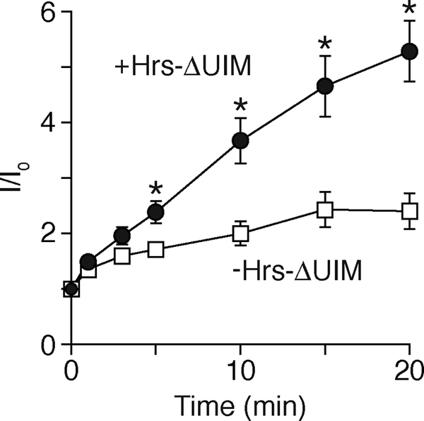
Epithelial Na(+) absorption is regulated by Nedd4-2, an E3 ubiquitin ligase that reduces expression of the epithelial Na(+) channel (ENaC) at the cell surface. Defects in this regulation cause Liddle syndrome, an inherited form of hypertension. Previous work found that Nedd4-2 functions through two distinct effects on trafficking, enhancing both ENaC endocytosis and ENaC degradation in lysosomes. To investigate the mechanism by which Nedd4-2 targets ENaC to lysosomes, we tested the role of hepatocyte growth factor-regulated tyrosine kinase substrate (Hrs), a component of the endosomal sorting complexes required for transport (ESCRT)-0 complex. We found that α-, β-, and γENaC each interact with Hrs. These interactions were enhanced by Nedd4-2 and were dependent on the catalytic function of Nedd4-2 as well as its WW domains. Mutation of ENaC PY motifs, responsible for inherited hypertension (Liddle syndrome), decreased Hrs binding to ENaC. Moreover, binding of ENaC to Hrs was reduced by dexamethasone/serum- and glucocorticoid-inducible kinase and cAMP, which are signaling pathways that inhibit Nedd4-2. Nedd4-2 bound to Hrs and catalyzed Hrs ubiquitination but did not alter Hrs protein levels. Expression of a dominant negative Hrs lacking its ubiquitin-interacting motif (Hrs-ΔUIM) increased ENaC surface expression and current. This occurred through reduced degradation of the cell surface pool of proteolytically activated ENaC, which enhanced its recycling to the cell surface. In contrast, Hrs-ΔUIM had no effect on degradation of uncleaved inactive channels. The data support a model in which Nedd4-2 induces binding of ENaC to Hrs, which mediates the sorting decision between ENaC degradation and recycling.


Figures

FIGURE 1.
FIGURE 1.
ENaC interacts with Hrs. A and B, coimmunoprecipitation of αENaC and Hrs in HEK 293T cells transfected with α-FLAG, β-, and γENaC (1.5 μg each), with or without Hrs-HA (0.5 μg) and Nedd4-2 (wt, wild type; C, C821A; WW, WW domain mutant) (0.5 μg). Total cDNA was held constant using GFP cDNA. A, representative immunoblots. In the top two panels, cell lysates were immunoprecipitated (IP) and immunoblotted (IB) as indicated with antibodies against αENaC (anti-FLAG) or Hrs (anti-HA). The bottom two panels show immunoblots of cell lysates. B, quantitation of αENaC that coprecipitated with Hrs (black bars) and of Hrs that coprecipitated with ENaC (white bars), relative to the group expressing ENaC and Hrs without Nedd4-2. Data are mean ± S.E. (n = 3–8, *, p < 0.03). C and D, coimmunoprecipitation of β-FLAG or γ-FLAG (coexpressed with the other two untagged ENaC subunits) and Hrs in HEK 293T cells using the methods described in panel A. C shows representative immunoblots, and D shows quantitation of Hrs coprecipitated with ENaC. Data are mean ± S.E. (n = 4–5, *, p < 0.003).
FIGURE 2.
FIGURE 2.
ENaC PY motifs are required for Nedd4-2 to enhance ENaC-Hrs interaction. A and B, coimmunoprecipitation of βENaC and Hrs in HEK 293T cells transfected with α-, β-FLAG, and γENaC (wild-type or αY644AβY620AγY627A, 1.5 μg each), with or without Hrs-HA (0.5 μg) and Nedd4-2 (0.5 μg). A, representative immunoblots. In the top panel, βENaC was immunoprecipitated with anti-FLAG antibody followed by immunoblot for Hrs with anti-HA antibody. The bottom two panels show immunoblots of cell lysates to detect Hrs (anti-HA) or βENaC (anti-FLAG), as indicated. B, quantitation of Hrs that coprecipitated with βENaC (relative to the group expressing ENaC and Hrs without Nedd4-2). Data are mean ± S.E. (n = 4, *, p < 0.04).
FIGURE 3.
FIGURE 3.
Phosphorylation regulates ENaC binding to Hrs. A–F, coimmunoprecipitation of αENaC and Hrs in HEK 293T cells transfected with α-FLAG, β-, and γENaC (1.5 μg each), with or without Hrs-HA (0.5 μg) and Nedd4-2 (wt, wild type; S/T, phosphorylation mutant) (0.5 μg). Total cDNA was held constant using GFP cDNA. Immunoprecipitation (IP) and immunoblot (IB) utilized anti-FLAG (αENaC) and anti-HA (Hrs) antibodies, as indicated. A, cells were incubated in serum-free medium with or without dexamethasone (Dex, 100 nm) for 4 h prior to harvest. Data for coprecipitation are quantitated in B (mean ± S.E., n = 3, *, p < 0.03). C, cells were cotransfected with or without SGK (1 μg), as indicated. For groups expressing ENaC and Hrs, data for coprecipitation are quantitated in D (mean ± S.E., n = 4, *, p < 0.04, n.s. indicates that the p value was not statistically significant). E, cells were treated with or without cAMP agonists (200 μm 8-(4-chlorophenylthio)-cAMP sodium, 100 μm 3-isobutyl-1-methylxanthine, 10 μm forskolin) for 2 h, as indicated. For groups expressing ENaC with Hrs and Nedd4-2, coimmunoprecipitation data are quantitated in F (mean ± S.E., n = 4, *, p < 0.002).
FIGURE 4.
FIGURE 4.
Nedd4-2 binds to and ubiquitinates Hrs. A, coimmunoprecipitation of Hrs and Nedd4-2 in COS 7 cells transfected with or without Hrs-HA (2 μg) and Nedd4-2 (2 μg). Nedd4-2 was immunoprecipitated (IP) (anti-WW1 antibody) followed by immunoblot (IB) for Hrs (anti-HA antibody). B, ubiquitinated Hrs in HEK 293T cells transfected with Hrs-HA (2 μg), ubiquitin-His (1 μg), and the indicated amount of Nedd4-2 (0–2 μg). Ubiquitinated Hrs (Hrs-Ub) was pulled down with Ni-NTA beads and detected by immunoblot (anti-HA antibody). The immunoblots are representative of three experiments.
FIGURE 5.
FIGURE 5.
Effect of Hrs on ENaC surface expression. A, immunoblots (IB) (anti-FLAG) of biotinylated (top panel) and total (bottom panel) αENaC in HEK 293T cells transfected with α-FLAG, β-, and γENaC (1 μg each), with or without Nedd4-2 (0.06 μg) and Hrs-ΔUIM (4 μg). Total cDNA was held constant using GFP cDNA. Irrelevant lanes were removed from the images. αENaC surface expression is quantitated in B, relative to the group without Nedd4-2 and Hrs-ΔUIM (mean ± S.E., n = 5, *, p < 0.003). C, amiloride-sensitive short circuit current (relative to the group without Nedd4-2) in FRT cells transfected with α-, β-, and γENaC, with or without Nedd4-2 and Hrs-ΔUIM (cDNA ratio 1:0–0.5:4; total cDNA was held constant using GFP cDNA). Data are mean ± S.E. (n = 10–11, *, p < 0.001).
FIGURE 6.
FIGURE 6.
Effect of Hrs on ENaC endocytosis. A, trypsin-activated amiloride-sensitive short circuit current versus time after trypsin removal in FRT cells transfected with αCl-1, β-, and γClENaC (0.25 μg each) and Hrs-ΔUIM (black circles) or empty vector (white squares) (0.25 μg). Data are mean ± S.E. (n = 8). B, immunoblot (anti-FLAG) of biotinylated αENaC in HEK 293T cells transfected with αCl-2-FLAG, β-, and γENaC (1 μg each), Nedd4-2 (0.06 μg), and with or without Hrs-ΔUIM (4 μg). The cells were incubated with or without trypsin (5 μg/ml for 5 min), incubated at 37 °C for 0–30 min, and then biotinylated. The cleaved αENaC bands are quantitated in C, relative to 0 min (mean ± S.E., n = 3).
FIGURE 7.
FIGURE 7.
Effect of Hrs on degradation of cell surface ENaC. A, immunoblots (IB) (anti-FLAG) of biotinylated (top panel) and total (bottom panel) αENaC in HEK 293T cells transfected with α-FLAG, β-, and γENaC (1 μg each) with Nedd4-2 (0.06 μg) and with or without Hrs-ΔUIM (4 μg). Cell surface proteins were pulse-labeled with biotin, and then the cells were incubated at 37 °C for 0–120 min. Biotinylated αENaC at each time point was quantitated (relative to 0 min) in B (cleaved αENaC) and C (full-length αENaC). Data are mean ± S.E. (n = 3–4, *, p < 0.02). D, immunoblot of ubiquitinated βENaC (βENaC-Ub) in HEK 293T cells transfected with α-, β-FLAG, and γENaC (1 μg each), ubiquitin-His (1 μg), with or without Nedd4-2 (0.06 μg) and Hrs-ΔUIM (3 μg). The cells were incubated with 10 μm N-acetyl-Leu-Leu-norleucinal for 2 h prior to lysis. Ubiquitinated proteins were isolated with Ni-NTA beads followed by immunoblot for βENaC with anti-FLAG. Irrelevant lanes were removed from the image. E, immunoblots (anti-FLAG) of biotinylated (top panel) and total (bottom panel) αENaC in HEK 293T cells transfected with αCl-2-FLAG, β-, and γENaC (1 μg each) with or without Nedd4-2 (0.06 μg) and Hrs-ΔUIM (4 μg). Cell surface proteins were pulse-labeled with biotin, and then the cells were incubated at 37 °C for 0 or 80 min. Biotinylated αENaC at 80 min was quantitated (relative to 0 min) in F. Data are mean ± S.E. (n = 3, n.s. indicates that the p value was not statistically significant).
FIGURE 8.
FIGURE 8.
Effect of Hrs on ENaC recycling. Plot of recovery of benzamil-sensitive short circuit current versus time after MTSET removal in FRT cells transfected with αCl-1, β-, and γCl-G536C ENaC (0.25 μg each) and Hrs-ΔUIM or empty vector (0.25 μg). The channels were activated by trypsin (10 μg/ml for 30 min), and then active channels remaining at the cell surface were irreversibly blocked with MTSET (1 mm). Data are mean ± S.E. (n = 11–12, *, p < 0.02).

Similar articles

Cited by

References

    1. Schild L. (2004) Rev. Physiol. Biochem. Pharmacol. 151, 93–107 - PubMed
    1. Snyder P. M. (2005) Endocrinology 146, 5079–5085 - PubMed
    1. Lifton R. P. (1996) Science 272, 676–680 - PubMed
    1. Boucher R. C., Stutts M. J., Knowles M. R., Cantley L., Gatzy J. T. (1986) J. Clin. Invest. 78, 1245–1252 - PMC - PubMed
    1. Snyder P. M. (2009) Sci. Signal. 2, pe41. - PubMed

Publication types

MeSH terms

Substances

LinkOut - more resources