

Schistosomiasis-induced experimental pulmonary hypertension: role of interleukin-13 signaling
- PMID: 20671265
- PMCID: PMC2928984
- DOI: 10.2353/ajpath.2010.100063
Schistosomiasis-induced experimental pulmonary hypertension: role of interleukin-13 signaling
Abstract
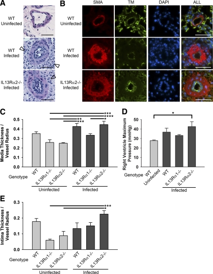
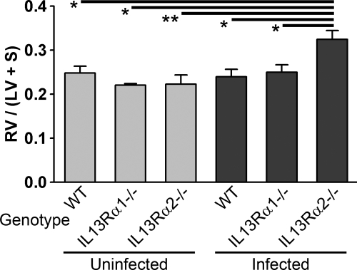
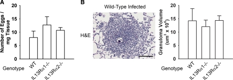
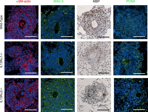
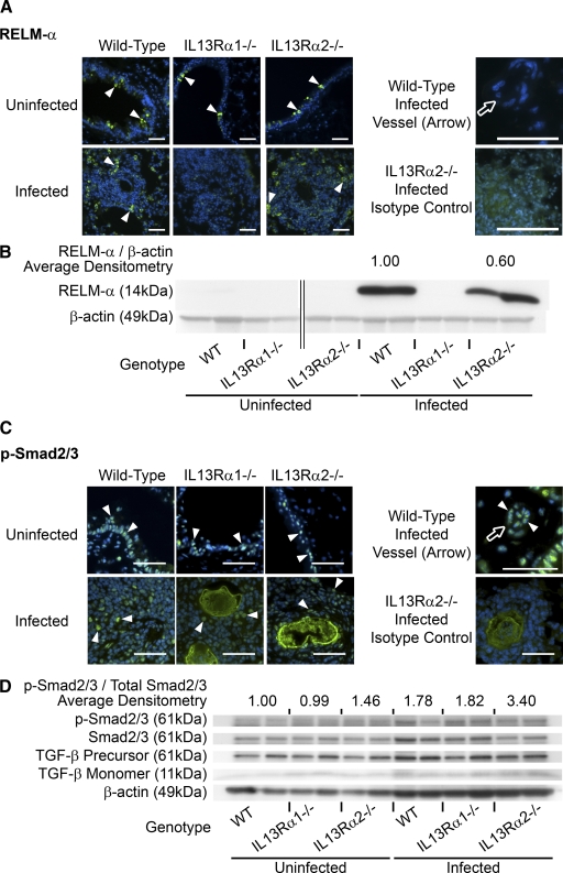
The mechanisms underlying schistosomiasis-induced pulmonary hypertension (PH), one of the most common causes of PH worldwide, remain unclear. We sought to determine whether Schistosoma mansoni causes experimental PH associated with pulmonary vascular remodeling in an interleukin (IL)-13-dependent manner. IL-13Ralpha1 is the canonical IL-13 signaling receptor, whereas IL-13Ralpha2 is a competitive nonsignaling decoy receptor. Wild-type, IL-13Ralpha1(-/-), and IL-13Ralpha2(-/-) C57BL/6J mice were percutaneously infected with S. mansoni cercariae, followed by i.v. injection of eggs. We assessed PH with right ventricular catheterization, histological evaluation of pulmonary vascular remodeling, and detection of IL-13 and transforming growth factor-beta signaling. Infected mice developed pulmonary peri-egg granulomas and arterial remodeling involving predominantly the vascular media. In addition, gain-of-function IL-13Ralpha2(-/-) mice had exacerbated vascular remodeling and PH. Mice with loss of IL-13Ralpha1 function did not develop PH and had reduced pulmonary vascular remodeling. Moreover, the expression of resistin-like molecule-alpha, a target of IL-13 signaling, was increased in infected wild-type and IL-13Ralpha2(-/-) but not IL-13Ralpha1(-/-) mice. Phosphorylated Smad2/3, a target of transforming growth factor-beta signaling, was increased in both infected mice and humans with the disease. Our data indicate that experimental schistosomiasis causes PH and potentially relies on up-regulated IL-13 signaling.
Figures

Similar articles
-
The Causal Role of IL-4 and IL-13 in Schistosoma mansoni Pulmonary Hypertension.Am J Respir Crit Care Med. 2015 Oct 15;192(8):998-1008. doi: 10.1164/rccm.201410-1820OC. Am J Respir Crit Care Med. 2015. PMID: 26192556 Free PMC article.
-
Protective role of IL-6 in vascular remodeling in Schistosoma pulmonary hypertension.Am J Respir Cell Mol Biol. 2013 Dec;49(6):951-9. doi: 10.1165/rcmb.2012-0532OC. Am J Respir Cell Mol Biol. 2013. PMID: 23815102 Free PMC article.
-
Transforming growth factor-β signaling promotes pulmonary hypertension caused by Schistosoma mansoni.Circulation. 2013 Sep 17;128(12):1354-64. doi: 10.1161/CIRCULATIONAHA.113.003072. Epub 2013 Aug 19. Circulation. 2013. PMID: 23958565 Free PMC article.
-
The Role of Type 2 Inflammation in Schistosoma-Induced Pulmonary Hypertension.Front Immunol. 2019 Jan 24;10:27. doi: 10.3389/fimmu.2019.00027. eCollection 2019. Front Immunol. 2019. PMID: 30733718 Free PMC article. Review.
-
Cellular and chemokine-mediated regulation in schistosome-induced hepatic pathology.Trends Parasitol. 2014 Mar;30(3):141-50. doi: 10.1016/j.pt.2013.12.009. Epub 2014 Jan 13. Trends Parasitol. 2014. PMID: 24433721 Review.
Cited by
-
Systems-level regulation of microRNA networks by miR-130/301 promotes pulmonary hypertension.J Clin Invest. 2014 Aug;124(8):3514-28. doi: 10.1172/JCI74773. Epub 2014 Jun 24. J Clin Invest. 2014. PMID: 24960162 Free PMC article.
-
Right ventricular systolic pressure measurements in combination with harvest of lung and immune tissue samples in mice.J Vis Exp. 2013 Jan 16;(71):e50023. doi: 10.3791/50023. J Vis Exp. 2013. PMID: 23354416 Free PMC article.
-
Sex Dimorphism in Pulmonary Hypertension: The Role of the Sex Chromosomes.Antioxidants (Basel). 2021 May 14;10(5):779. doi: 10.3390/antiox10050779. Antioxidants (Basel). 2021. PMID: 34068984 Free PMC article. Review.
-
TGF beta and IL13 in Schistosomiasis mansoni associated pulmonary arterial hypertension; a descriptive study with comparative groups.BMC Infect Dis. 2014 May 21;14:282. doi: 10.1186/1471-2334-14-282. BMC Infect Dis. 2014. PMID: 24886277 Free PMC article.
-
Type 2 cytokines: mechanisms and therapeutic strategies.Nat Rev Immunol. 2015 May;15(5):271-82. doi: 10.1038/nri3831. Epub 2015 Apr 17. Nat Rev Immunol. 2015. PMID: 25882242 Review.
References
-
- Cool CD, Rai MD, Yeager ME, Hernandez-Saavedra D, Serls AE, Bull TM, Geraci MW, Brown KK, Routes JM, Tuder RM, Voelkel NF. Expression of human herpesvirus 8 in primary pulmonary hypertension. N Engl J Med. 2003;349:1113–1122. - PubMed
-
- Richter A, Yeager ME, Zaiman A, Cool CD, Voelkel NF, Tuder RM. Impaired transforming growth factor-β signaling in idiopathic pulmonary arterial hypertension. Am J Respir Crit Care Med. 2004 Dec 15;170:1340–1348. - PubMed
Publication types
MeSH terms
Substances
Grants and funding
LinkOut - more resources
Full Text Sources
Medical
Molecular Biology Databases
