. 2010 Jul 30;39(2):259-68.
doi: 10.1016/j.molcel.2010.07.005.

The FANCM/FAAP24 complex is required for the DNA interstrand crosslink-induced checkpoint response

Affiliations

The FANCM/FAAP24 complex is required for the DNA interstrand crosslink-induced checkpoint response

Min Huang et al. Mol Cell. .

Abstract

Cells from Fanconi anemia (FA) patients are extremely sensitive to DNA interstrand crosslinking (ICL) agents, but the molecular basis of the hypersensitivity remains to be explored. FANCM (FA complementation group M), and its binding partner, FAAP24, anchor the multisubunit FA core complex to chromatin after DNA damage and may contribute to ICL-specific cellular response. Here we show that the FANCM/FAAP24 complex is specifically required for the recruitment of replication protein A (RPA) to ICL-stalled replication forks. ICL-induced RPA foci formation requires the DNA-binding activity of FAAP24 but not the DNA translocase activity of FANCM. Furthermore, FANCM/FAAP24-dependent RPA foci formation is required for efficient ATR-mediated checkpoint activation in response to ICL. Therefore, we propose that FANCM/FAAP24 plays a role in ICL-induced checkpoint activation through regulating RPA recruiment at ICL-stalled replication forks.


Figures

Figure 1
Figure 1. ICL fails to generate ssDNA at stalled replication forks
(A) A schematic model comparing stalled DNA replication forks caused by ICL and HU/UV. (B) Representative images showing less ssDNA generated by MMC treatment compared to HU or UV treatments. HeLa cells were labeled with 20 μM BrdU for 36 hr, then were treated with MMC (1 μM, 6 hr), HU (2 mM, 6 hr) or UV (30 J/m2, 6 hr post irradiation), respectively. ssDNA was detected using anti-BrdU antibody without denaturation. The BrdU intensity under the denaturing condition (2 M HCl) shows total BrdU incorporation. (C) Quantification of ssDNA generation shown in (B). The percentage of BrdU-positive cells was determined by counting at least 500 cells from each sample. Data are represented as mean ± SD from three independent experiments. (D) Immunoblot showing similar activation of DNA damage response in the conditions described in (B).
Figure 2
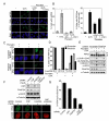
Figure 2. FANCM and FAAP24 are selectively required for MMC-induced RPA foci formation and RPA2 phosphorylation
(A) Representative images showing the requirement of FANCM and FAAP24 for MMC-induced RPA foci assembly but not other replication stress. HeLa cells were treated with MMC (1 μM, 6 hr), HU (2 mM, 6 hr), CPT (500 nM, 6 hr) or UV (30 J/m2, 6 hr post irradiation) respectively at 48 hr after transfection with either FANCM or FAAP24 siRNAs. RPA foci were detected using anti-RPA2 antibody. (B) Quantification for RPA foci shown in (A). The percentage of cells containing RPA foci was determined by counting at least 200 cells from each sample. Data are represented as mean ± SD from three independent experiments. (C) Immunoblot showing that depletion of either FANCM or FAAP24 more severely reduced RPA2 phosphorylation following MMC treatment. HeLa cells were treated as described in (A). Different mobility of RPA2 caused by phosphorylation is indicated as “B” (baseline) and “H” (hyperphosphorylated), respectively. (D) Time-course of RPA2 phosphorylation in FAAP24- or FANCM-depleted cells. HeLa cells transfected with indicated siRNAs were harvested at 0, 6, or 12 hr post treatment with MMC (1 μM) or HU (2 mM). (E) Dose-response of RPA2 phosphorylation in FAAP24- or FANCM-depleted cells. HeLa cells transfected with siRNAs were treated with MMC or HU at indicated concentrations for 8 hr.
Figure 3
Figure 3. FANCM and FAAP24 are required for RPA foci formation and RPA2 phosphorylation following PUVA-induced ICL formation
(A) Representative images of ssDNA staining and RPA foci showing that PUVA treatment induced RPA foci but not ssDNA. ssDNA and RPA foci were detected using anti-BrdU antibody or anti-RPA2 antibody 4 hr after PUVA treatment in HeLa cells. The immunostaining intensities are compared to those of UVC treatment. (B) Quantification of ssDNA staining and RPA foci shown in (A). The percentages of cells containing ssDNA (left panel) or cells containing RPA foci (right panel) were determined by counting at least 200 cells from each sample. Data are represented as mean ± SD from three independent experiments. (C) Representative images showing FAAP24-dependent RPA foci formation after PUVA treatment. RPA foci were detected in HeLa cells with scramble shRNA or FAAP24 shRNA 4 hr after PUVA treatment. (D) Quantification of RPA foci shown in (C). The percentage of cells containing RPA foci was determined by counting at least 200 cells from each sample. Data are represented as mean ± SD from three independent experiments. (E) Immunoblot showing reduced RPA2 phosphorylation in FAAP24-depleted cells. Cells were treated as described in (C). Different mobility of RPA2 caused by phosphorylation is indicated as “B” (baseline) and “H” (hyperphosphorylated), respectively. (F) Representative images showing that CtIP depletion resulted in a mild reduction in RPA foci compared to the depletion of FAAP24. HeLa cells were treated with PUVA at 48 hr after transfection with indicated siRNAs. Immunoblot showing the depletion efficiency of indicated proteins (upper panel). After co-staining with RPA and γ-H2AX, RPA foci formation was compared in CtIP-, FAAP24-, or CtIP- and FAAP24-depleted cells (lower panel). (G) Quantification of RPA foci formation shown in (F). The percentage of cells containing RPA foci among γ-H2AX-positive cells was determined by counting at least 200 γ-H2AX-positive cells from each sample. Data are represented as mean ± SD from three independent experiments.
Figure 4
Figure 4. The DNA translocase activity of FANCM is dispensable for ICL-induced RPA foci assembly
(A) Representative images showing that K117R FANCM rescued the defective RPA foci formation in FANCM-depleted cells. Non-targeting control siRNAmir, FANCM siRNAmir, and wild-type FANCM or K117R FANCM-corrected FANCM siRNAmir HEK293 Flip-in cells were treated with MMC (10 μM) for 12 hr. RPA foci were detected using anti-RPA2 antibody. (B) Quantification of RPA foci induced by 2 and 10 μM MMC treatment as described in (A). The percentage of cells containing RPA foci was determined by counting at least 200 cells from each sample. Data are represented as mean ± SD from three independent experiments. (C) Immunoblot comparing the defects in RPA2 phosphorylation in the condition as described in (A). Different mobility of RPA2 caused by phosphorylation is indicated as “B” (baseline) and “H” (hyperphosphorylated), respectively. (D) Immunoblot (left panel) and quantification (right panel) for the levels of CDC25A showing that K117R FANCM-corrected FANCM siRNAmir cells had a moderate defect in checkpoint activation. Cells were treated with MMC and harvested at indicated time points. The level of CDC25A was quantified and normalized with that of α-Tubulin. Data are represented as mean ± SD from three independent experiments.
Figure 5
Figure 5. DNA binding activity of FAAP24 is required for ICL-induced RPA foci formation
(A) A scheme showing full-length and C-terminal truncated FAAP24. (B) Immunoblot showing wild-type FAAP24 (WT) but not C-terminal truncated FAAP24 mutant (N150) rescued defective RPA2 phosphorylation in FAAP24-depleted HeLa cells. FAAP24 shRNA cells, transfected with empty-vector (EV), Myc-tagged WT or N150 FAAP24, were treated with 1 μM of MMC for 10 hr. Whole cell extracts (WCE) and chromatin enriched extracts (Chromatin) were analyzed by immunoblotting using indicated antibodies. (C) Quantification showing recovered RPA foci by WT but not N150 FAAP24. Cells as described in (B) were treated with MMC 1 μM for 6 hr. RPA foci were detected using anti-RPA2 antibody. The percentage of cells containing RPA foci was determined by counting at least 200 cells from each sample. Data are represented as mean ± SD from three independent experiments.
Figure 6
Figure 6. RPA binding to ICL DNA is dependent on FAAP24
(A) Migration of ICL DNA on a denaturing gel. (B) Immunoblot showing that FAAP24 wild-type (WT) but not the C-terminal truncated FAAP24 mutant (N150) binds to ICL DNA. Biotinylated control DNA or ICL-DNA (100 ng) were attached to streptavidin-coated beads and incubated with purified His-tagged WT FAAP24 or N150 FAAP24. (C) Immunoblot showing that RPA loading to ICL DNA was decreased in FAAP24-depleted nuclear extracts. Nuclear extracts (100 μg) derived from HeLa scramble or FAAP24 shRNA cells were incubated with biotinylated control DNA or ICL-DNA (100 ng) respectively. The DNA-bound FAAP24, RPA2 and KU70 were detected. (D) Proposed working model for the role of FANCM/FAAP24 in the ICL-induced checkpoint response.

Similar articles

Cited by

References

    1. Anantha RW, Vassin VM, Borowiec JA. Sequential and synergistic modification of human RPA stimulates chromosomal DNA repair. J. Biol. Chem. 2007;282:35910–35923. - PubMed
    1. Andreassen PR, D’Andrea AD, Taniguchi T. ATR couples FANCD2 monoubiquitination to the DNA-damage response. Genes Dev. 2004;18:1958–1963. - PMC - PubMed
    1. Auerbach AD. Fanconi anemia and its diagnosis. Mutat. Res. 2009;668:4–10. - PMC - PubMed
    1. Banerjee S, Smith S, Oum JH, Liaw HJ, Hwang JY, Sikdar N, Motegi A, Lee SE, Myung K. Mph1p promotes gross chromosomal rearrangement through partial inhibition of homologous recombination. J. Cell Biol. 2008;181:1083–1093. - PMC - PubMed
    1. Bartek J, Lukas C, Lukas J. Checking on DNA damage in S phase. Nat. Rev. Mol. Cell. Biol. 2004;5:792–804. - PubMed

Publication types

MeSH terms

LinkOut - more resources