. 2010 Sep 15;21(18):3171-81.
doi: 10.1091/mbc.E10-01-0073. Epub 2010 Jul 21.

A phosphatidic acid binding/nuclear localization motif determines lipin1 function in lipid metabolism and adipogenesis

Affiliations

A phosphatidic acid binding/nuclear localization motif determines lipin1 function in lipid metabolism and adipogenesis

Hongmei Ren et al. Mol Biol Cell. .

Abstract

Lipins are phosphatidic acid phosphatases with a pivotal role in regulation of triglyceride and glycerophospholipid metabolism. Lipin1 is also an amplifier of PGC-1α, a nuclear coactivator of PPAR-α responsive gene transcription. Lipins do not contain recognized membrane-association domains, but interaction of these enzymes with cellular membranes is necessary for access to their phospholipid substrate. We identified a role for a conserved polybasic amino acid motif in an N-terminal domain previously implicated as a determinant of nuclear localization in selective binding of lipin1β to phosphatidic acid, using blot overlay assays and model bilayer membranes. Studies using lipin1β polybasic motif variants establish that this region is also critical for nuclear import and raise the possibility that nuclear/cytoplasmic shuttling of lipin1β is regulated by PA. We used pharmacological agents and lipin1β polybasic motif mutants to explore the role of PA-mediated membrane association and nuclear localization on lipin1β function in phospholipid metabolism and adipogenic differentiation. We identify a role for the lipin1 polybasic motif as both a lipid binding motif and a primary nuclear localization sequence. These two functions are necessary for full expression of the biological activity of the protein in intracellular lipid metabolism and transcriptional control of adipogenesis.


Figures
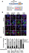
Figure 1.
Figure 1.
The Lipin1β polybasic motif is required for nuclear localization. (A) Sequence alignment showing the conserved polybasic motif in mammalian lipins, and the polybasic motif mutants “9A” “4A” in lipin1β. In the 4A and 9A mutants, the polybasic motif was disrupted by substitution of conserved basic residues with four or nine alanines, respectively. In the “D712E” mutant the catalytic motif aspartic acid residue was substituted with glutamic acid. NLIP is the conserved lipin N-terminal homology domain, and HAD is the lipin catalytic domain with homology to halo acid dehalogenases. (B) HEK293 cells were transfected with vectors for expression of the indicated lipin1β variants. 24 h after transfection, cells were treated with vehicle or 40 nM leptomycin B for 4 h. Subcellular localization of HA-tagged lipin1β (green) was examined by confocal microscopy to compare the localization with the nuclear marker DAPI (blue) and the endoplasmic reticulum (ER) marker calnexin (red). (C) Percentage of the number of transfected cells examined in which lipin1β was localized in cytoplasm, nucleus, or both cytoplasm and nucleus (mean ± SD of at least 100 cells counted in 10 distinct fields).
Figure 2.
Figure 2.
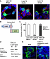
Deletion of the lipin1β C terminus promotes nuclear localization. (A) Subcellular localization of truncated lipin1β (lipin1βΔC) with wt 4A and 9A polybasic motif sequences (green) compared with the nuclear marker DAPI (blue) and endoplasmic reticulum marker calnexin (red). (B) Structural organization of lipin1β C-terminal truncated mutants and lipin polybasic motif GFP fusion. (C) Percentage of the number of transfected cells examined in which lipin1β was localized in cytoplasm, nucleus, or both cytoplasm and nucleus (mean ± SD of at least 100 cells counted in 10 distinct fields). (D) Association of lipin1βΔC with a wt or “9A” polybasic motif sequence with importin-α was determined as described in the Materials and Methods. (E) Subcellular localization of GFP or a lipin 1β polybasic motif GFP fusion protein (green) and the nuclear marker DAPI (blue).
Figure 3.
Figure 3.
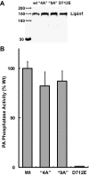
Lipin 1 polybasic motif mutants are catalytically active. (A) Western blot of purified lipin1β wt and the indicated mutants expressed in insect cells using baculovirus vectors and detected by using an anti-HA antibody. (B) Specific PA phosphatase activity of wt lipin1β and mutants were determined by using [32P]-labeled PA substrate, and activity was monitored by measuring the release of water soluble [32P]-PO42−. Data are means ± SD of triplicate determinations normalized to activity of wt lipin1β.
Figure 4.
Figure 4.
Selective binding of lipin 1 to PA in bilayer liposomes is attenuated by mutation of the polybasic motif. (A, B). The indicated lipids were immobilized on a nylon membrane (Triacylglycerol [TG]; Diacylglycerol [DG]; Phosphatidic Acid [PA]; Phosphatidylserine [PS]; Phosphatidylethanolamine [PE]; Phosphatidylglycerol [PG]; Cardiolipin [CL]; Phosphatidylinositol [PI']; Phosphatidylinositol 4-phosphate [PI4P]; Phosphatidylinositol (4,5)-bisphosphate: [PI(4,5)P2]; Phosphatidylinositol (3,4,5)-trisphosphate [PI(3,4,5P3]; Cholesterol ester [CE]; Sphingomyelin [SM]; 3-sulfogalactosylceramide [3SGC]. Binding of recombinant purified lipin1β was monitored using an HA-tag selective primary antibody and fluorescently conjugated secondary antibody with quantitation using the Li-Cor Odyssey imaging system. (C) Binding of lipin1β to sucrose loaded phosphatidylcholine (PC) vesicles containing 10 mol % of the indicated phospholipids was determined by measuring PA phosphatase activity remaining in the supernatant after sedimentation of the vesicles by ultracentrifugation. (D) Comparison of the binding of wild-type lipin1β and the 4A and 9A polybasic motif mutants to sucrose loaded PC vesicles containing 10 mol % PA.
Figure 5.
Figure 5.
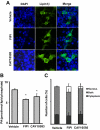
Nuclear localization of lipin1 is promoted by pharmacological inhibition of phospholipase D. (A) HEK 293 cells expressing wild-type lipin1β and the 4A and 9A polybasic motif mutants were treated with 1.5 μM of the dual PLD1/PLD2 inhibitor FIPI or 50 μM of the PLD2 selective inhibitor CAY10593 for 4h. Subcellular localization of lipin1β (green) was visualized by indirect immunofluorescence and compared with the nuclear marker DAPI (blue). (B) Total PA levels in cells incubated with vehicle or the indicated PLD inhibitors at the concentrations used for the experiment shown in panel A and total PA levels (the sum of 16 abundant PA species) quantitated by HPLC ESI MS/MS. (C) Percent of total number of transfected cells, which lipin1β was localized in cytoplasm, nucleus, or both cytoplasm and nucleus (mean ± SD of at least 100 cells counted in 10 distinct fields).
Figure 6.
Figure 6.
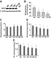
Effects of overexpression of lipin1β wt and mutants on diacylglycerol (DG), phosphatidylcholine (PC), and phosphatidic acid (PA) levels. (A) HA-tagged wt lipin1β and the indicated mutants were overexpressed in HepG2 cells and detected by Western blotting. (B) HepG2 cells expressing wt lipin1β and the indicated mutants were radiolabeled with [3H]palmitic acid for 24 h. DG levels were determined after separation by TLC, and quantitation by scintillation counting. (C–E) Levels of 16 abundant molecular species of PA, DG, and PC were determined in HepG2 cells overexpressing lipin1β wt, and mutants incubated with 1 μM palmitic acid were measured by HPLC ESI MS/MS. Data are means ± SD of at least three independent determinations.
Figure 7.
Figure 7.
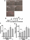
The polybasic motif plays an important role in complementation of the adipogenesis defect of fld/fld mouse embryo fibroblasts by lipin1β. (A) Oil red-O staining (red) of differentiated wt and fld/fld mouse embryo fibroblasts expressing wild-type lipin1β, the indicated lipin1β mutants, or a GFP control using lentivirus vectors at day 6 postinduction of differentiation. (B) Expression of lipin 1 variants was quantitated by Western blotting. (C) Adipogenic differentiation was quantitated by counting the number of oil red-O–stained colonies per field. (D) Adipogenic differentiation was quantitated by measuring total TG levels (the sum of 36 abundant TG species) using HPLC ESI MS/MS. Data shown are means ± SD of three or more independent determinations. Statistically significant differences between cells expressing wt lipin 1 and the indicated lipin 1 mutants are indicated by asterisks. Note that TG accumulation or the number of differentiated colonies was not significantly different (N.S.) between cells expressing GFP or Lipin1 D712E.
Figure 8.
Figure 8.
The polybasic motif is required for complementation of the adipogenic gene transcription defect of fld/fld mouse embryo fibroblasts by lipin1β. RT-PCR was used to quantify PPAR-γ (A), PPAR-α (B), adiponectin (C), and aP2 mRNA expression (D) in MEFs isolated from wt mice, and fld/fld mice infected with lipin1β wt, lipin 1β D712E, and lipin1β “9A” lentivirus. Samples for RNA analysis were prepared from cells at 6 d postinduction of differentiation. Values are normalized (= 1.0) to RNA levels in control virus–infected fld/fld MEFs.
Figure 9.
Figure 9.
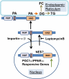
Regulation of nucleo-cytoplasmic lipin1β traffic by phosphatidic acid. Our results suggest a model in which lipin1β actively shuttles between the nucleus and cytoplasm. This process is controlled by a nuclear import sequence identified here as the polybasic motif and putative nuclear export signal localized elsewhere in the C terminus of the protein. Interactions between lipin1β and PA mediated by the polybasic motif antagonize the nuclear import process and are important for proper expression of lipin1β catalytic activity required for synthesis of glycerophospholipids. NLIP is the conserved lipin N-terminal homology domain, HAD is the lipin catalytic domain with homology to halo acid dehalogenases and PMB is the polybasic motif. NES designates a putative nuclear export sequence.

Similar articles

Cited by

References

    1. Bou Khalil M., Sundaram M., Zhang H. Y., Links P. H., Raven J. F., Manmontri B., Sariahmetoglu M., Tran K., Reue K., Brindley D. N., Yao Z. The level and compartmentalization of phosphatidate phosphatase-1 (lipin-1) control the assembly and secretion of hepatic VLDL. J. Lipid Res. 2009;50:47–58. - PubMed
    1. Buser C. A., McLaughlin S. Ultracentrifugation technique for measuring the binding of peptides and proteins to sucrose-loaded phospholipid vesicles. Methods Mol. Biol. 1998;84:267–281. - PubMed
    1. Carman G. M., Han G. S. Phosphatidic acid phosphatase, a key enzyme in the regulation of lipid synthesis. J. Biol. Chem. 2009;284:2593–2597. - PMC - PubMed
    1. Carman G. M., Henry S. A. Phosphatidic acid plays a central role in the transcriptional regulation of glycerophospholipid synthesis in Saccharomyces cerevisiae. J. Biol. Chem. 2007;282:37293–37297. - PMC - PubMed
    1. Donkor J., Sariahmetoglu M., Dewald J., Brindley D. N., Reue K. Three mammalian lipins act as phosphatidate phosphatases with distinct tissue expression patterns. J. Biol. Chem. 2007;282:3450–3457. - PubMed

Publication types

MeSH terms

LinkOut - more resources