. 2010 Oct;6(7):929-35.
doi: 10.4161/auto.6.7.12785. Epub 2010 Oct 24.

Quantitation of "autophagic flux" in mature skeletal muscle

Affiliations

Quantitation of "autophagic flux" in mature skeletal muscle

Jeong-Sun Ju et al. Autophagy. 2010 Oct.

Abstract

Reliable and quantitative assays to measure in vivo autophagy are essential. Currently, there are varied methods for monitoring autophagy; however, it is a challenge to measure "autophagic flux" in an in vivo model system. Conversion and subsequent degradation of the microtubule-associated protein 1 light chain 3 (MAP1-LC3/LC3) to the autophagosome associated LC3-II isoform can be evaluated by immunoblot. However, static levels of endogenous LC3-II protein may render possible misinterpretations since LC3-II levels can increase, decrease or remain unchanged in the setting of autophagic induction. Therefore, it is necessary to measure LC3-II protein levels in the presence and absence of lysomotropic agents that block the degradation of LC3-II, a technique aptly named the "autophagometer." In order to measure autophagic flux in mouse skeletal muscle, we treated animals with the microtubule depolarizing agent colchicine. Two days of 0.4 mg/kg/day intraperitoneal colchicine blocked autophagosome maturation to autolysosomes and increased LC3-II protein levels in mouse skeletal muscle by >100%. The addition of an autophagic stimulus such as dietary restriction or rapamycin led to an additional increase in LC3-II above that seen with colchicine alone. Moreover, this increase was not apparent in the absence of a "colchicine block." Using this assay, we evaluated the autophagic response in skeletal muscle upon denervation induced atrophy. Our studies highlight the feasibility of performing an "in vivo autophagometer" study using colchicine in skeletal muscle.


Figures

Figure 1
Figure 1
(A) 3-month-old male mice were treated with vehicle, 50 mg/kg/day chloroquine, 0.4 mg/kg/day colchicine or 2 mg/kg/day vinblastine for two days and tibialis anterior (TA) muscle lysates were evaluated by LC3 immunoblot. Actin is shown as a loading control. Blot is representative of 3 independent experiments. (B) TA from similarly treated mice as in (A) was subjected to immunofluorescence using an LC3 antibody. Colchicine treated mice had an increase in LC3 positive puncta. Images were taken at the same camera setting and exposure time. Scale is 40 µM. (C) The TA muscle from 3-month-old male mice was electroporated with a tandemly tagged mCherry-GFP-LC3 reporter construct. Mice were either treated for two days with vehicle or 0.4 mg/kg/day colchicine. Vehicle treated mice had diffuse LC3 fluorescence while colchicine treated mice generated GFP and mCherry positive co-localizing puncta consistent with colchicine impairing autophagosome maturation to an acidic organelle. Scale is 25 µM.
Figure 2
Figure 2
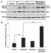
(A) Similar mice were treated with vehicle, 0.4 mg/kg/day colchicine, nutrient deprivation (starvation) or nutrient deprivation plus 0.4 mg/kg/day colchicine for two days. TA muscle lysates were evaluated by LC3 and p62 immunoblot. Note that colchicine treatment significantly increases LC3-II levels. These levels are augmented by nutrient deprivation. Blot is representative of three independent experiments. (B) LC3-II/actin ratios were quantitated via densitometry from six mice per treatment condition. Error bars represent standard error and **denote p values of <0.005 as obtained by student paired t-test.
Figure 3
Figure 3
(A) Differentiated C2C12 myotubes were treated with vehicle, 200 nM BafilomycinA1, 10 µg/mL rapamycin or 10 µg/mL rapamycin plus 200 nM BafilomycinA1 for 6 hours. Lysates were subjected to LC3 and p62 immunoblots. Rapamycin did not increase LC3-II levels further than that seen with BafilomycinA1 alone. The same lysates were subjected to immunoblot with antibodies to phospho-S6 (Ser235/236) and S6 kinase demonstrating that rapamycin is capable of mTOR inhibition in these cells. Blot is representative of two independent experiments. (B) 3-month-old male mice were treated with vehicle, 0.4 mg/kg/day colchicine for two days, 10 mg/kg/day rapamycin for 7 days or 10 mg/kg/day rapamycin for 7 days and 0.4 mg/kg/day colchicine for two days beginning on day 5 and TA lysates were subjected to immunoblotting with LC3, p62, phospho-S6, total S6 kinase or actin. Blot is representative of four independent experiments. (C) LC3-II/actin ratios were quantitated via densitometry from eight mice per treatment condition. Error bars represent standard error and **denote p values of <0.005 as obtained by student paired t-test. (D) A similar experiment to (B) was performed using 20 mg/kg/day temsirolimus/CCI-779 instead of rapamycin. Note that the addition of temsirolimus/CCI-779 to colchicine further increased LC3-II levels consistent with an enhancement in autophagic flux.
Figure 4
Figure 4
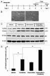
Denervation-induced atrophy enhances autophagic flux. (A) Schematic of experimental design to assess autophagic flux post denervation. Briefly, 3-month-old male mice had a sham or complete sciatic nerve transaction (denervation) for seven days prior to muscle harvesting. Mice were also treated with vehicle or 0.4 mg/kg/day colchicine for two days prior to harvesting. (B) Hematoxylin and eosin staining of TA muscle from 7 day sham or denervated mice. Note an increase in atrophic angular fibers in denervated animal (arrows). Scale is 40 µM. (C) TA muscle lysates from each group were subjected to LC3, p62 or actin immunoblots. Blots are representative of two independent experiments. There was an augmentation of LC3-II levels above that seen with colchicine alone after seven days of denervation. (D) LC3-II/actin ratios were quantitated via densitometry from muscle lysates from two independent experiments with two mice per treatment condition. Error bars represent standard error and *denote p values of <0.05 as obtained by student paired t-test.

Similar articles

Cited by

  • IL-6 deletion decreased REV-ERBα protein and influenced autophagy and mitochondrial markers in the skeletal muscle after acute exercise.
    Pinto AP, Muñoz VR, da Rocha AL, Rovina RL, Ferrari GD, Alberici LC, Simabuco FM, Teixeira GR, Pauli JR, de Moura LP, Cintra DE, Ropelle ER, Freitas EC, Rivas DA, da Silva ASR. Pinto AP, et al. Front Immunol. 2022 Oct 13;13:953272. doi: 10.3389/fimmu.2022.953272. eCollection 2022. Front Immunol. 2022. PMID: 36311768 Free PMC article.
  • Measurement of autophagy flux in benign prostatic hyperplasia in vitro.
    Oh SH, Lee DW, Choi YB, Lee YH, Ju JS. Oh SH, et al. Prostate Int. 2020 Jun;8(2):70-77. doi: 10.1016/j.prnil.2019.11.004. Epub 2020 Feb 26. Prostate Int. 2020. PMID: 32647643 Free PMC article.
  • Why should autophagic flux be assessed?
    Zhang XJ, Chen S, Huang KX, Le WD. Zhang XJ, et al. Acta Pharmacol Sin. 2013 May;34(5):595-9. doi: 10.1038/aps.2012.184. Epub 2013 Mar 11. Acta Pharmacol Sin. 2013. PMID: 23474710 Free PMC article. Review.
  • Inhibition of autophagy inhibits the conversion of cardiac fibroblasts to cardiac myofibroblasts.
    Gupta SS, Zeglinski MR, Rattan SG, Landry NM, Ghavami S, Wigle JT, Klonisch T, Halayko AJ, Dixon IM. Gupta SS, et al. Oncotarget. 2016 Nov 29;7(48):78516-78531. doi: 10.18632/oncotarget.12392. Oncotarget. 2016. PMID: 27705938 Free PMC article.
  • Guidelines for the use and interpretation of assays for monitoring autophagy (3rd edition).
    Klionsky DJ, Abdelmohsen K, Abe A, Abedin MJ, Abeliovich H, Acevedo Arozena A, Adachi H, Adams CM, Adams PD, Adeli K, Adhihetty PJ, Adler SG, Agam G, Agarwal R, Aghi MK, Agnello M, Agostinis P, Aguilar PV, Aguirre-Ghiso J, Airoldi EM, Ait-Si-Ali S, Akematsu T, Akporiaye ET, Al-Rubeai M, Albaiceta GM, Albanese C, Albani D, Albert ML, Aldudo J, Algül H, Alirezaei M, Alloza I, Almasan A, Almonte-Beceril M, Alnemri ES, Alonso C, Altan-Bonnet N, Altieri DC, Alvarez S, Alvarez-Erviti L, Alves S, Amadoro G, Amano A, Amantini C, Ambrosio S, Amelio I, Amer AO, Amessou M, Amon A, An Z, Anania FA, Andersen SU, Andley UP, Andreadi CK, Andrieu-Abadie N, Anel A, Ann DK, Anoopkumar-Dukie S, Antonioli M, Aoki H, Apostolova N, Aquila S, Aquilano K, Araki K, Arama E, Aranda A, Araya J, Arcaro A, Arias E, Arimoto H, Ariosa AR, Armstrong JL, Arnould T, Arsov I, Asanuma K, Askanas V, Asselin E, Atarashi R, Atherton SS, Atkin JD, Attardi LD, Auberger P, Auburger G, Aurelian L, Autelli R, Avagliano L, Avantaggiati ML, Avrahami L, Awale S, Azad N, Bachetti T, Backer JM, Bae DH, Bae JS, Bae ON, Bae SH, Baehrecke EH, Baek SH, Baghdiguian S, Bagniewska-Zadworna A, Bai H, Bai J, Bai XY, Bailly Y, Balaji KN, … See abstract for full author list ➔ Klionsky DJ, et al. Autophagy. 2016;12(1):1-222. doi: 10.1080/15548627.2015.1100356. Autophagy. 2016. PMID: 26799652 Free PMC article. No abstract available.

References

    1. Klionsky DJ. Autophagy: from phenomenology to molecular understanding in less than a decade. Nat Rev Mol Cell Biol. 2007;8:931–937. - PubMed
    1. Mizushima N, Yoshimori T, Levine B. Methods in mammalian autophagy research. Cell. 140:313–326. - PMC - PubMed
    1. Klionsky DJ, Abeliovich H, Agostinis P, Agrawal DK, Aliev G, Askew DS, et al. Guidelines for the use and interpretation of assays for monitoring autophagy in higher eukaryotes. Autophagy. 2008;4:151–175. - PMC - PubMed
    1. Rubinsztein DC, Cuervo AM, Ravikumar B, Sarkar S, Korolchuk V, Kaushik S, et al. In search of an “autophagomometer.”. Autophagy. 2009;5:585–589. - PubMed
    1. Kuncl RW, Bilak MM, Craig SW, Adams R. Exocytotic “constipation” is a mechanism of tubulin/lysosomal interaction in colchicine myopathy. Exp Cell Res. 2003;285:196–207. - PubMed

Publication types

MeSH terms