. 2010 Sep 30;405(2):579-91.
doi: 10.1016/j.virol.2010.06.043. Epub 2010 Jul 23.

Three-dimensional structure of Rubella virus factories

Affiliations

Three-dimensional structure of Rubella virus factories

Juan Fontana et al. Virology. .

Abstract

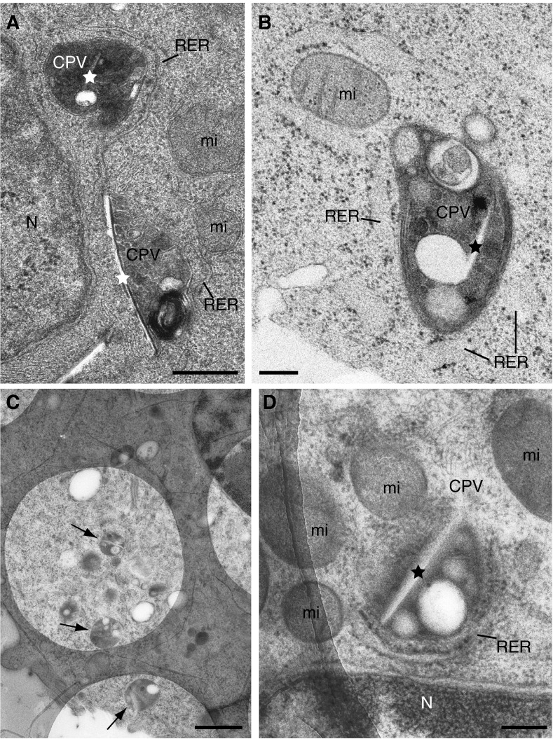
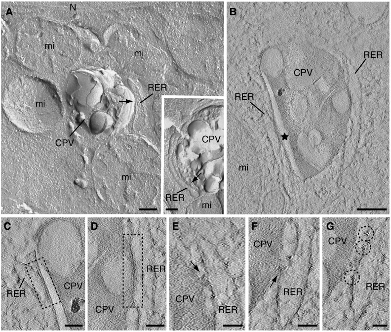
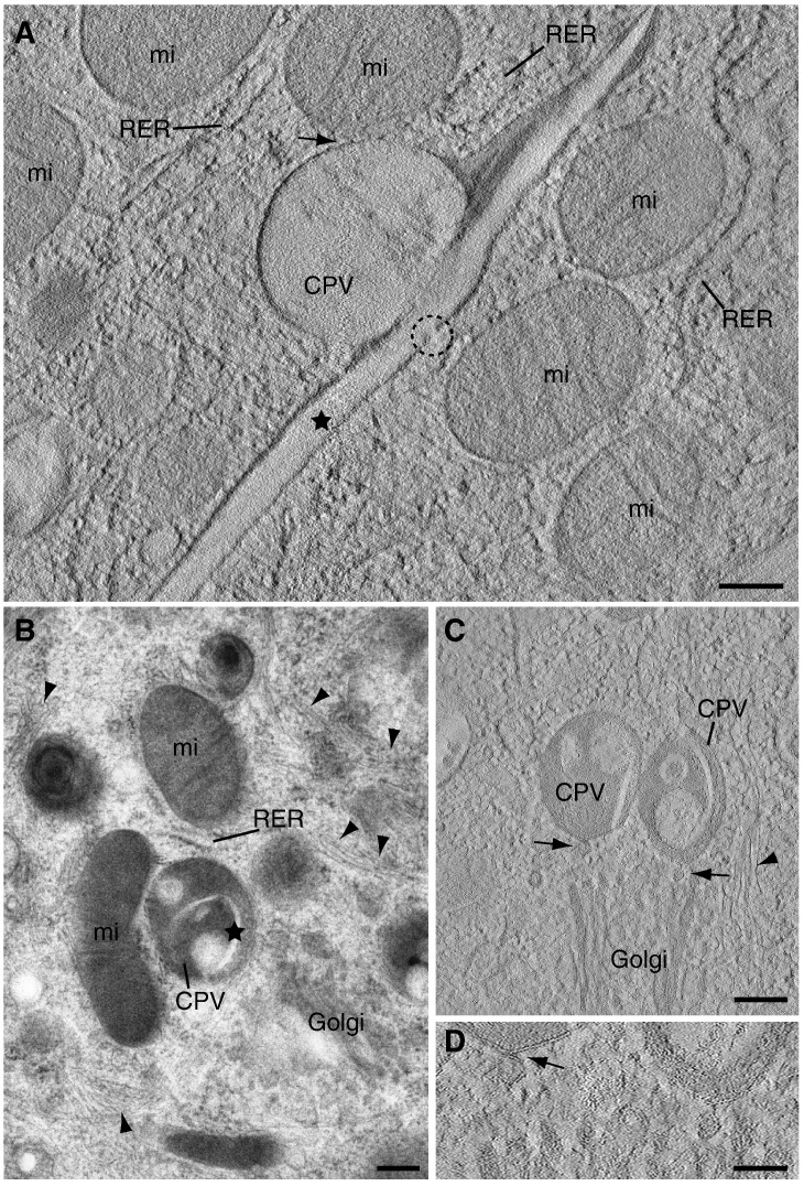
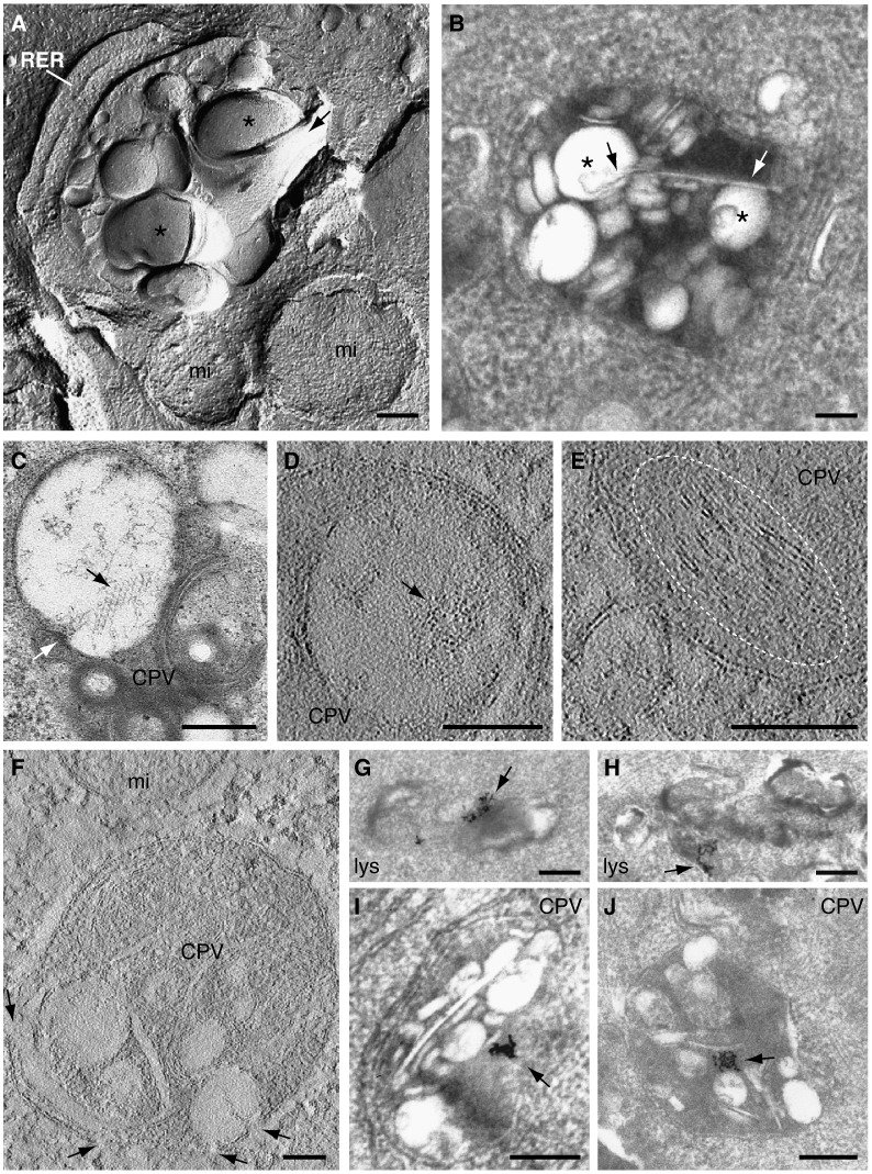
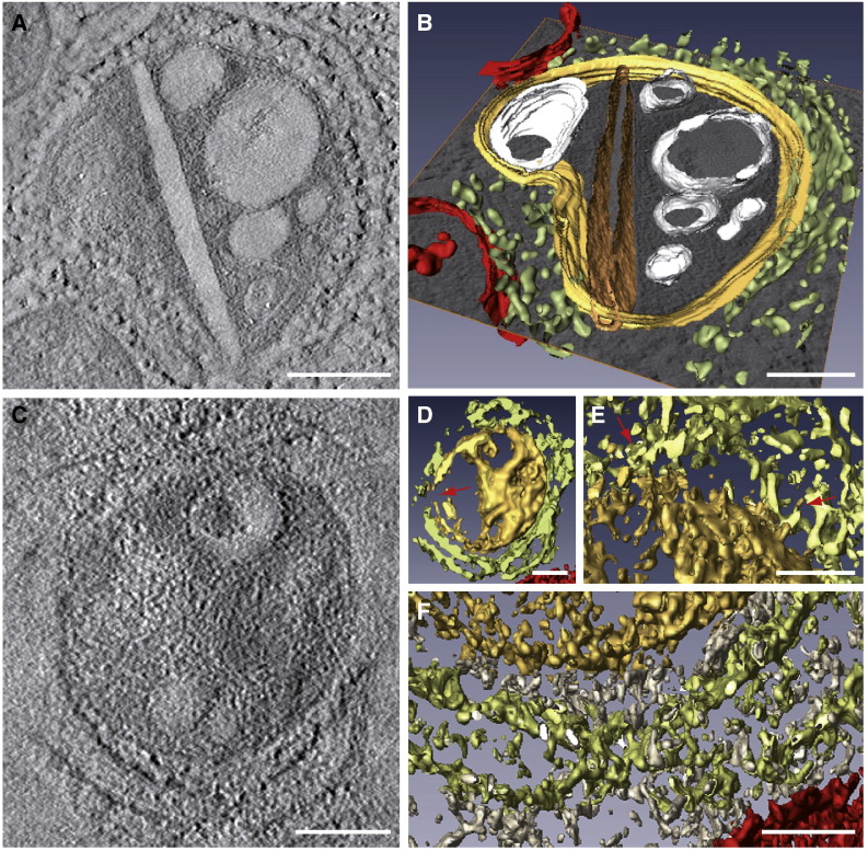
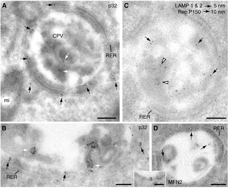
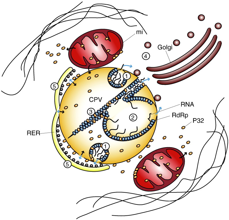
Viral factories are complex structures in the infected cell where viruses compartmentalize their life cycle. Rubella virus (RUBV) assembles factories by recruitment of rough endoplasmic reticulum (RER), mitochondria and Golgi around modified lysosomes known as cytopathic vacuoles or CPVs. These organelles contain active replication complexes that transfer replicated RNA to assembly sites in Golgi membranes. We have studied the structure of RUBV factory in three dimensions by electron tomography and freeze-fracture. CPVs contain stacked membranes, rigid sheets, small vesicles and large vacuoles. These membranes are interconnected and in communication with the endocytic pathway since they incorporate endocytosed BSA-gold. RER and CPVs are coupled through protein bridges and closely apposed membranes. Golgi vesicles attach to the CPVs but no tight contacts with mitochondria were detected. Immunogold labelling confirmed that the mitochondrial protein p32 is an abundant component around and inside CPVs where it could play important roles in factory activities.


Figures

Fig. 1
Fig. 1
Two-dimensional ultrastructure of RUBV factories: high-pressure freezing, freeze-substitution and sectioning. (A) Conventional EM shows two CPVs (frontal view for the one on the top and side view for the CPV on the bottom) in the perinuclear area of a replicon-transfected cell. Both CPVs exhibit internal vesicles and straight elements (stars) and locally recruited RER and mitochondria. (B to D) Suspensions of BHK-21 cells stably transfected with a RUBV replicon were cryofixed by HPF without cryoprotectants. Cell in (B) was freeze-substituted in 1% OsO4, 0.25% glutaraldehyde in acetone and embedded in EML-812 while cells in (C) and (D) were freeze-substituted in 0.5% UA in acetone and embedded in Lowicryl HM23. (B) Detail of a CPV surrounded by RER cisternae and one mitochondrion. The CPV exhibits a very heterogeneous content with numerous vesicles of variable size and density, vacuoles and a rigid membrane (star). (C) Low magnification view showing large areas of the cell and several CPVs (arrows) over the holes of the carbon film. (D) Image at 0º of a CPV surrounded by RER and mitochondria ready for ET. Ultra-thin (~ 50 nm) post-stained sections are shown in (A) and (B), while (C) and (D) show semi-thick (~ 200 nm) sections collected on Quantifoil® holey carbon grids and visualized without post-staining. RER, rough endoplasmic reticulum; mi, mitochondria; N, nucleus. Bars, 200 nm in A, B, and D; 1 μm in C.
Fig. 2
Fig. 2
Three-dimensional views of RUBV factories as visualized by freeze-fracture and electron tomography. (A) Metal replica of BHK-21 cells stably transfected with the RUBrep/GFP/neo replicon and processed by freeze-fracture followed by etching. Mitochondria and RER cisternae surround the CPVs. Although these organelles are very close to the CPVs no clear contacts are detected (arrows in mainfield and inset). (B to G) A computational tomographic slice (~ 1.2 nm) corresponding to the tomogram of a semi-thick (~ 250 nm) section obtained from suspensions of trypsinized cells processed by HPF, FS and embedding in Lowicryl HM23 and visualized without post-staining. (B) CPV with a characteristic rigid membrane (star) surrounded by RER. (C to G) Expanded areas from the CPV shown in (B). (C and D) RER–CPV contacts seen as closely apposed membranes (dashed rectangles). (E and F) Protein bridges between RER and CPV are also observed (arrows). (G) Close to these complexes several ribosomes facing the CPV are found (dashed circles). RER, rough endoplasmic reticulum; mi, mitochondria. Bars, 200 nm in A and B; 50 nm in C to G.
Fig. 3
Fig. 3
CPV contacts with recruited mitochondria and Golgi stacks. (A, C and D) Computational tomographic slices (~ 1.2 nm) and (B) semi-thick (~ 200 nm) section visualized by TEM. (A) Side view of a CPV completely surrounded by mitochondria. Holes or channels in the CPV rigid membrane (dashed circle) apparently open the CPV to the cytoplasm. (B) Characteristic RUBV factory with a CPV containing rigid membranes (the star labels one of them) surrounded by RER, mitochondria and Golgi. The Golgi stack approaches laterally to the CPV but no clear contacts are detected. (C and D) Computational tomographic slice of a similar factory showing contacts between Golgi vesicles and the CPVs (arrows). Arrowheads in B and C point to filaments around the factories. RER, rough endoplasmic reticulum; mi, mitochondria. Bars, 200 nm in A to C; 100 nm in D.
Fig. 4
Fig. 4
Complex internal organization of CPVs and connection with the endo-lysosomal pathway. (A) Metal replica of a RUBV factory processed by freeze-etching showing the interior of a CPV with a rigid membrane (black arrow) wrapping two large vacuoles (asterisks). (B and C) Ultra-thin sections of CPVs as visualized by TEM after staining. (B) Rigid membrane (arrows) contacting two vacuoles (asterisks). (C) A section along the rigid membrane showing features compatible with a close packing of particles (black arrow). The white arrow points to the spot where the rigid membrane interacts with the membrane of the large vacuole. (D to F) Computational slices (~ 0.6 nm for D and E, ~ 1.2 nm for F) of two tomograms of CPVs filtered with three rounds of a median filter. (D) Groups of particles associated with a large vacuole (arrow) and (E) stacked membranes (white dashed ellipse) are shown. (F) Openings to the cytosol (arrows) in the peripheral membrane of a CPV (frontal view). Thin-sections of lysosomes from control BHK-21 cells (G and H) and CPVs from stably transfected cells (I and J) after treatment with BSA-gold in culture and posterior fixation and embedding. Both lysosomes (lys) from control cells and CPVs contain BSA-gold particles (arrows). RER, rough endoplasmic reticulum; mi, mitochondria; Bars, 200 nm.
Fig. 5
Fig. 5
Analysis of ET volumes. Computational tomographic slices (A and C) and 3D models after segmentation and visualization with AMIRA (B and D to F). Color code is as follows: CPV, yellow; straight sheet, brown; RER, light green; mitochondria, red; vesicles and vacuoles, white; cytoplasm, grey. (A) A computational tomographic slice and the corresponding 3D model (B) of a CPV surrounded by the RER and containing a number of vacuoles, vesicles and a rigid straight sheet that is connected with the periphery of the CPV. The 3D model was built using a combination of masking, isosurface, and manual tracing using Amira segmentation tools. (C) A computational tomographic slice and two 3D models (D, E-F) of a CPV almost completely surrounded by the RER and filled with abundant material. The 3D model in (D) was built with a combination of masking and isosurface after strong filtering of the volume with nonlinear diffusion. The internal material of the CPV was solid rendered, keeping the vacuoles hollow, thus revealing the dense content of the vacuole at the top. This representation also shows that the CPV is opened to the cytosol (red arrow). The other 3D model in (E–F) shows contacts RER-CPV (red arrows in E, this area corresponds to the upper part of the model in (D)) and the “cytosolic macromolecules” (grey) filling the gap between the RER and the CPV (F, corresponding to the lower part of the model in (D)). This model was prepared as that in (D), but without nonlinear diffusion, to show the complex material surrounding the CPV. Bars, 200 nm in A–D; 100 nm in E and F.
Fig. 6
Fig. 6
Immunogold labeling of CPVs. (A and B) correspond to Tokuyasu cryosections while (C and D) are Lowicryl resin sections. (A) Labeling with an antibody specific for the mitochondrial protein p32 showing signals in the CPVs' internal membranes (white arrows) and in the gap between the CPV and the RER (black arrows). (B) Higher magnification view of a CPV labeled with anti-p32 antibodies showing intense signals in CPV's internal membranes (white arrows) and around the CPV (black arrows). (C) Double labeling with a mixture of two mouse monoclonal antibodies specific for lysosomal markers (LAMP1 and LAMP2, white arrowheads) and a rabbit polyclonal antibody specific for P150 protein, one of the two components of the RUBV replicase complex (black arrows). Labeling concentrates in the interior of CPVs. (D) Immunogold labelling with an antibody specific for Mitofusin 2 (MFN2) (mainfield and inset). A weak signal is observed on the internal periphery of CPVs (arrows). Bars, 200 nm in A, C and D; 100 nm in B and inset in H.
Fig. 7
Fig. 7
A model for RUVB factory organization. CPVs and recruited organelles build functional factories that would work as follows: Viral RNA would replicate in vesicles (1), vacuoles (2) and rigid membranes (3). Newly synthesized RNA molecules would be transported to the Golgi as RNPs (ribonucleoproteins) with the help of capsid protein molecules for the assembly of new viruses (4) and to the RER for the synthesis of new viral proteins (5). Recruited mitochondria will provide energy and co-factors such as p32 while cytoskeletal elements would build a cage-like structure around the viral factory. Blue arrows: movements of RNA/RNPs. Black arrows: movements of proteins. CPV, cytopathic vacuole; RdRp, RNA dependent- RNA polymerase; RER, rough endoplasmic reticulum; mi, mitochondria. (For interpretation of the references to colour in this figure legend, the reader is referred to the web version of this article.)

Similar articles

Cited by

References

    1. Barajas D., Jiang J., Nagy P.D. A unique role for the host ESCRT proteins in replication of Tomato bushy stunt virus. PLoS Pathog. 2009;5(12):e1000705. - PMC - PubMed
    1. Beatch M.D., Everitt J.C., Law L.J., Hobman T.C. Interactions between Rubella virus capsid and host protein p32 are important for virus replication. J. Virol. 2005;79:10807–10820. - PMC - PubMed
    1. Cabezas P., Risco C. Studying cellular architecture in three dimensions with improved resolution: Ta replicas revisited. Cell Biol. Int. 2006;30:747–754. - PubMed
    1. Cardone G., Grünewald K., Steven A.C. A resolution criterion for electron tomography based on cross-validation. J. Struct. Biol. 2005;151:117–129. - PubMed
    1. Carlson L.-A., Briggs J.A.G., Glass B., Riches J.D., Simon M.N., Johnson M.C., Müller B., Grünewald K., Kräusslich H.-G. Three-dimensional analysis of budding sites and released virus suggests a revised model for HIV-1 morphogenesis. Cell Host Microbe. 2008;4:592–599. - PMC - PubMed

Publication types

MeSH terms