. 2010 Sep 24;285(39):30115-25.
doi: 10.1074/jbc.M110.141549. Epub 2010 Jul 22.

Arresting a transient receptor potential (TRP) channel: beta-arrestin 1 mediates ubiquitination and functional down-regulation of TRPV4

Affiliations

Arresting a transient receptor potential (TRP) channel: beta-arrestin 1 mediates ubiquitination and functional down-regulation of TRPV4

Arun K Shukla et al. J Biol Chem. .

Abstract

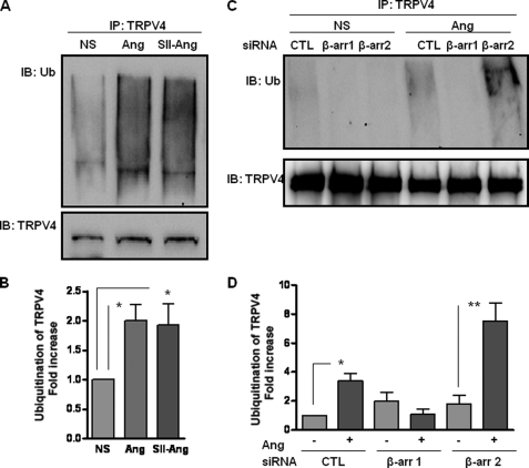
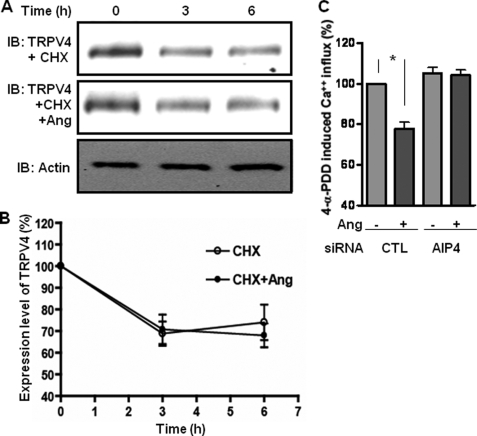
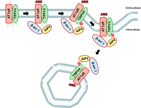
β-Arrestins, originally discovered to desensitize activated G protein-coupled receptors, (aka seven-transmembrane receptors, 7TMRs) also mediate 7TMR internalization and G protein-independent signaling via these receptors. More recently, several regulatory roles of β-arrestins for atypical 7TMRs and non-7TM receptors have emerged. Here, we uncover an entirely novel regulatory role of β-arrestins in cross-talk between the angiotensin receptor (AT1aR) and a member of the transient receptor potential (TRP) ion channel family, TRPV4. AT1aR and TRPV4 form a constitutive complex in the plasma membrane, and angiotensin stimulation leads to recruitment of β-arrestin 1 to this complex. Surprisingly, angiotensin stimulation results in ubiquitination of TRPV4, a process that requires β-arrestin 1, and subsequently to internalization and functional down-regulation of TRPV4. β-Arrestin 1 interacts with, and acts as an adaptor for AIP4, an E3 ubiquitin ligase responsible for TRPV4 ubiquitination. Thus, our data provide the first evidence of a functional link between β-arrestins and TRPV4 and uncovers an entirely novel mechanism to maintain appropriate intracellular Ca(2+) concentration to avoid excessive Ca(2+) signaling.


Figures

FIGURE 1.
FIGURE 1.
Angiotensin stimulation leads to formation of a multiprotein complex involving β-arrestin, TRPV4, and AT1aR. A–D, interaction of β-arrestins (β-arr) and TRPV4 upon angiotensin (Ang) stimulation. A, rVSMCs were stimulated with angiotensin (1 μm) for the indicated time points and coimmunoprecipitated using a goat polyclonal TRPV4 antibody. β-Arrestins were detected in the immunoprecipitate using rabbit polyclonal A1CT antibody. Blots were reprobed with rabbit anti-TRPV4 polyclonal antibody. B, quantification of coimmunoprecipitated β-arrestin (shown in panel A) by densitometry. Data are expressed as -fold over basal interaction detected between β-arrestin and TRPV4. C, rVSMCs were stimulated with angiotensin for the indicated time points and coimmunoprecipitated using rabbit polyclonal A1CT antibody. TRPV4 was detected in the immunoprecipitate using goat polyclonal anti-TRPV4 antibody. Blots were reprobed with goat anti-TRPV4 polyclonal antibody. D, quantification of coimmunoprecipitated TRPV4 (shown in panel C) by densitometry. Data are expressed as -fold over basal interaction detected between β-arrestin and TRPV4. Data presented in panels B and D show mean ± S.E. from at least 3 independent experiments. E, colocalization analysis of AT1aR and TRPV4 by confocal microscopy. rVSMCs were transfected with HA-AT1aR, fixed, permeabilized, and immunostained using goat polyclonal anti-TRPV4 antibody (green) and a polyclonal rabbit anti-HA antibody (blue). F and G, interaction of AT1aR and TRPV4 in rVSMCs. F, rVSMCs were stimulated with angiotensin (1 μm) for the indicated time points and subsequently coimmunoprecipitated using a goat polyclonal anti-TRPV4 antibody. AT1aR was detected in the immunoprecipitate using a rabbit polyclonal anti-HA antibody. Blots were reprobed with rabbit anti-TRPV4 polyclonal antibody. G, rVSMCs were stimulated with angiotensin for the indicated time points and coimmunoprecipitated using a mouse monoclonal anti-HA antibody, and TRPV4 was detected in the immunoprecipitate using a goat polyclonal anti-TRPV4 antibody. Representative blots and confocal images are shown from at least 3 independent experiments.
FIGURE 2.
FIGURE 2.
β-Arrestin 1 is required for angiotensin-induced internalization and functional down-regulation of TRPV4. A–C, angiotensin-induced internalization of TRPV4. A, rVSMCs were stimulated with angiotensin (Ang) (1 μm) for the indicated time period, fixed, permeabilized, and stained using goat polyclonal anti-TRPV4 antibody followed by Alexa Fluor 488-conjugated secondary antibody (green: TRPV4). B, HEK-293 cells expressing HA-AT1aR and FLAG-TRPV4 were stimulated with angiotensin for the indicated time points, and surface TRPV4 was labeled with NHS-S-S-biotin. The cells were lysed, immunoprecipitated using NeutrAvidin beads, and immunoblotted with a mouse monoclonal anti-FLAG antibody. C, densitometry analysis of angiotensin-induced TRPV4 internalization as measured by surface biotinylation (shown in panel B). Data are presented as the percentage of initial levels of TRPV4 at the cell surface and show mean ± S.E. from at 3 independent experiments. D–F, involvement of β-arrestin 1 in TRPV4 internalization. D, 4-α-PDD-induced Ca2+ influx measured in HEK-293 cells. The cells were prestimulated with either angiotensin or SII-angiotensin (SII-Ang), and subsequently 4-α-PDD-induced Ca2+ influx was measured using the Fluo-4 NW calcium assay kit. The data are presented as the percentage of control cells, i.e. cells prestimulated with saline. NS, non-stimulated. E, 4-α-PDD-induced Ca2+ influx measured in HEK-293 cells after depletion of β-arrestin 1 (β-arr 1) or β-arrestin 2 (β-arr 2) using siRNA. CTL is control scrambled siRNA. F, angiotensin-induced internalization was measured in HEK-293 cells by surface biotinylation after depletion of β-arrestin 1 or 2 using siRNA using mouse monoclonal anti-FLAG antibody. Representative blots are shown from at least 3 independent experiments. G, densitometry analysis of angiotensin-induced internalization of TRPV4 (shown in panel F). Data are normalized with respect to the non-treated control siRNA sample (100%). The data presented in panels D–F show the mean ± S.E. from 3–4 independent experiments, and the statistical analysis was carried out using one-way ANOVA with Bonferroni post test.
FIGURE 3.
FIGURE 3.
β-Arrestin mediates angiotensin-induced ubiquitination of TRPV4. A, rVSMCs were preincubated with MG132 (10 μm) for 2 h and then stimulated with angiotensin (Ang) (0.1 μm) or SII-angiotensin (SII-Ang) (10 μm) for 1 h. Subsequently, the cells were lysed and immunoprecipitated with goat polyclonal anti-TRPV4 antibody followed by Western blot using mouse monoclonal anti-ubiquitin antibody. The levels of TRPV4 in the lysate were detected by goat polyclonal anti-TRPV4 antibody. NS, non-stimulated; Ub, ubiquitin. B, densitometry analysis of angiotensin-induced TRPV4 ubiquitination (shown in panel A). Data are presented as -fold increase over the basal ubiquitination of TRPV4. C, rVSMCs transfected with either control or siRNA against β-arrestin 1 (β-arr 1) or β-arrestin 2 (β-arr 2) were preincubated with MG132 (10 μm) for 2 h and then stimulated with angiotensin (0.1 μm) for 1 h. Subsequently, the cells were lysed and immunoprecipitated with TRPV4 antibody followed by Western blot using anti-ubiquitin antibody. The levels of TRPV4 in the lysate were detected by rabbit polyclonal anti-TRPV4 antibody. CTL, control siRNA. D, densitometry analysis of angiotensin-induced TRPV4 ubiquitination (shown in panel C). Data are presented as -fold increase over the basal ubiquitination of TRPV4. Representative blots are shown from 4 independent experiments. Data presented as bar graphs show mean ± S.E. from 4 independent experiments. Statistical analysis was carried out using one-way ANOVA with Bonferroni post test.
FIGURE 4.
FIGURE 4.
AIP4 is required for angiotensin-induced ubiquitination of TRPV4, and β-arrestin 1 acts as an adaptor for AIP4. A, HEK-293 cells expressing AT1aR and TRPV4 were transfected with either control or siRNA against AIP4. Cells were preincubated with MG132 (10 μm) for 2 h and then stimulated with angiotensin (0.1 μm) for 1 h. Subsequently, the cells were lysed and immunoprecipitated with TRPV4 antibody followed by Western blot using anti-ubiquitin antibody. The blots were reprobed with anti-TRPV4 antibody, and lysate was probed with anti-AIP4 antibody. CTL, control siRNA; Ub, ubiquitin. B, densitometry analysis of angiotensin-induced TRPV4 ubiquitination (shown in panel A). Data are presented as -fold increase over the basal ubiquitination of TRPV4. C, HEK-293 cells expressing AT1aR and TRPV4 were transfected with either control or siRNA against β-arrestin 1 (β-arr). Cells were stimulated with angiotensin (0.1 μm) for 2 min. Subsequently, the cells were lysed and immunoprecipitated with AIP4 antibody followed by Western blot using anti-TRPV4 antibody. The blots were reprobed with anti-AIP4 antibody, and lysate was probed with anti-TRPV4 and anti-AIP4 antibody. D, densitometry analysis of angiotensin-induced coimmunoprecipitation of TRPV4 and AIP4 (shown in panel C). Data are presented as -fold increase over the basal interaction between TRPV4 and AIP4. Representative blots are shown from at 3 independent experiments, and the data presented as bar graphs show mean ± S.E. from at least 3 independent experiments. Statistical analysis was carried out using one-way ANOVA with Bonferroni post test.
FIGURE 5.
FIGURE 5.
Angiotensin-induced ubiquitination of TRPV4 promotes internalization but not degradation. A, HEK-293 cells expressing AT1aR and TRPV4 were incubated with cycloheximide (CHX) (10 μm) in the absence or presence of angiotensin (Ang) (0.1 μm) for the indicated time points. Cells were lysed, and the expression levels of TRPV4 in the lysate were detected by Western blot using mouse monoclonal anti-FLAG antibody. An anti-actin blot is shown as the loading control. B, densitometry analysis of angiotensin-induced degradation of TRPV4 normalized with levels of actin. Data are presented as the percentage of decrease in TRPV4 level over the basal level of TRPV4. C, 4-α-PDD-induced Ca2+ influx measured in HEK-293 cells transfected with control (CTL) or AIP4 siRNA. Cells were prestimulated with angiotensin (0.1 μm) for 1 h, and subsequently 4-α-PDD-induced Ca2+ influx was measured using the Fluo-4 NW kit. The data are presented as the percentage of control cells, i.e. cells prestimulated with saline. The data presented in panels B and C show mean ± S.E. from 3 independent experiments. Statistical analysis was carried out using one-way ANOVA with Bonferroni post test.
FIGURE 6.
FIGURE 6.
Schematic representation of β-arrestin 1-dependent ubiquitination and functional down-regulation of TRPV4 upon activation of AT1aR. This illustrative scheme shown for TRPV4 can be potentially applicable to β-arrestin-mediated down-regulation of other TRP channels as well as other ion channels and transporters. ANG, angiotensin; U, ubiquitin; β-arr 1, β-arrestin 1.

Similar articles

Cited by

References

    1. Takeda S., Kadowaki S., Haga T., Takaesu H., Mitaku S. (2002) FEBS Lett. 520, 97–101 - PubMed
    1. Pierce K. L., Premont R. T., Lefkowitz R. J. (2002) Nat. Rev. Mol. Cell Biol. 3, 639–650 - PubMed
    1. Lefkowitz R. J. (1998) J. Biol. Chem. 273, 18677–18680 - PubMed
    1. Pitcher J. A., Freedman N. J., Lefkowitz R. J. (1998) Annu. Rev. Biochem. 67, 653–692 - PubMed
    1. Shenoy S. K., Lefkowitz R. J. (2003) Biochem. J. 375, 503–515 - PMC - PubMed

Publication types

MeSH terms