. 2010 Oct;30(19):4712-21.
doi: 10.1128/MCB.00657-10. Epub 2010 Jul 20.

Myeloid deletion of SIRT1 induces inflammatory signaling in response to environmental stress

Affiliations

Myeloid deletion of SIRT1 induces inflammatory signaling in response to environmental stress

Thaddeus T Schug et al. Mol Cell Biol. 2010 Oct.

Abstract

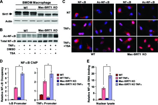
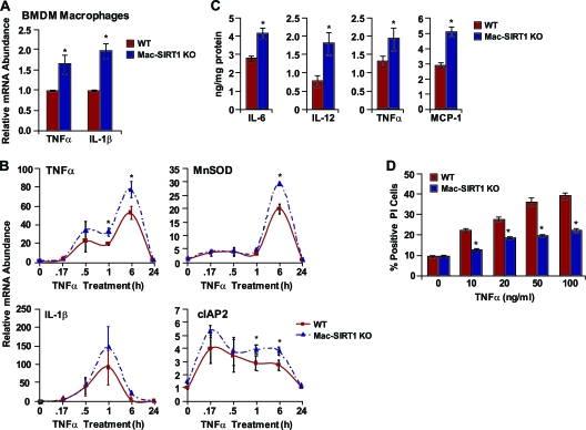
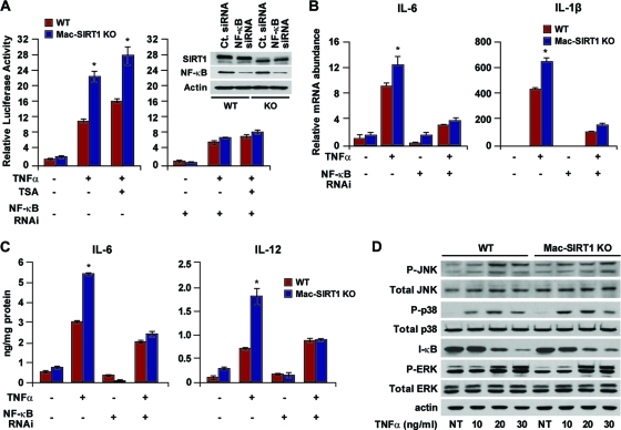
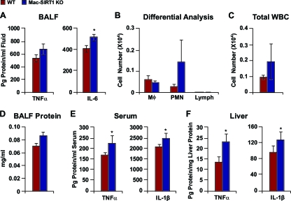
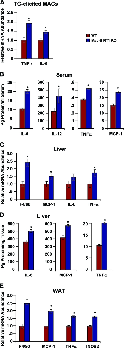
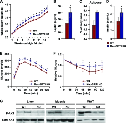
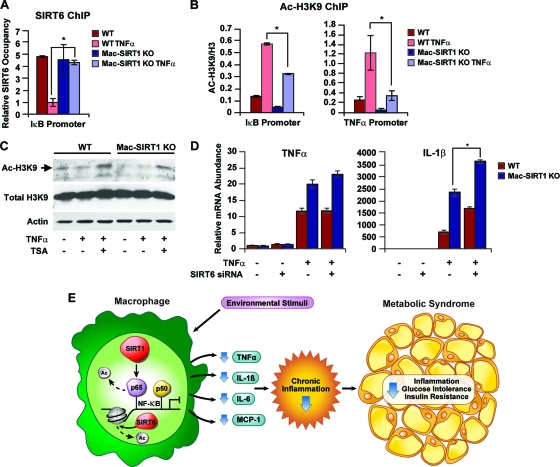
Macrophage activation and infiltration into resident tissues is known to mediate local inflammation and is a hallmark feature of metabolic syndrome. Members of the sirtuin family of proteins regulate numerous physiological processes, including those involved in nutrient regulation and the promotion of longevity. However, the important role that SIRT1, the leading sirtuin family member, plays in immune response remains unclear. In this study, we demonstrate that SIRT1 modulates the acetylation status of the RelA/p65 subunit of NF-κB and thus plays a pivotal role in regulating the inflammatory, immune, and apoptotic responses in mammals. Using a myeloid cell-specific SIRT1 knockout (Mac-SIRT1 KO) mouse model, we show that ablation of SIRT1 in macrophages renders NF-κB hyperacetylated, resulting in increased transcriptional activation of proinflammatory target genes. Consistent with increased proinflammatory gene expression, Mac-SIRT1 KO mice challenged with a high-fat diet display high levels of activated macrophages in liver and adipose tissue, predisposing the animals to development of systemic insulin resistance and metabolic derangement. In summary, we report that SIRT1, in macrophages, functions to inhibit NF-κB-mediated transcription, implying that myeloid cell-specific modulation of this sirtuin may be beneficial in the treatment of inflammation and its associated diseases.


Figures

FIG. 1.
FIG. 1.
Myeloid cell-specific deletion of SIRT1 leads to hyperactive NF-κB signaling. (A) Western blot (WB) analysis of SIRT1 protein in BMDMs from Mac-SIRT1 KO mice compared to those from control mice. WT, wild type. (B) Elevated levels of acetylated p65 in Mac-SIRT1 KO BMDMs after TNF-α treatment. BMDMs from control and Mac-SIRT1 KO mice were treated with TNF-α (10 ng/ml) in the presence or absence of TSA (100 nM). Levels of acetylated p65 were analyzed by Western blotting. (C) Ablation of SIRT1 results in higher levels of Ac-p65 in the nucleus following TNF-α and TSA treatment. Total p65/RelA and Ac-p65/RelA were stained (red) by indirect immunofluorescence, and the nuclei were stained (blue) with DAPI (4′,6-diamidino-2-phenylindole). NT, nontreated. (D) SIRT1 deficiency increases the association of p65/RelA with κB sites at the promoters of NF-κB target genes. BMDMs were treated with vehicle or TNF-α for 30 min and assayed using anti-p65/RelA antibodies. NF-κB occupancy (percentage of input) at κB sites of IκB and TNF-α promoters were analyzed by qPCR. (E) SIRT1 deficiency increases activated NF-κB. The data are represented as means ± SEM; n = 3; *, P < 0.05.
FIG. 2.
FIG. 2.
Loss of SIRT1 in macrophages promotes NF-κB-dependent cytokine release and inhibits NF-κB-mediated apoptosis. (A) Elevated proinflammatory cytokine message levels in Mac-SIRT1 KO BMDMs. (B) Increased mRNA expression of NF-κB target genes in Mac-SIRT1 KO cells following TNF-α (10 ng/ml) treatment at the indicated time points. (C) Increased release of proinflammatory cytokines from Mac-SIRT1 KO BMDMs measured by ELISA following 12-h TNF-α treatment. (D) SIRT1 deficiency protects BMDMs from TNF-α-induced apoptosis. BMDMS from control and Mac-SIRT1 KO mice were treated with TNF-α and stained with propidium iodide. The data are represented as means ± SEM; n = 3; *, P < 0.05.
FIG. 3.
FIG. 3.
Loss of SIRT1 induces elevated levels of proinflammatory cytokines through NF-κB. (A) Loss of SIRT1 induces NF-κB-dependent transactivation. (Left) BMDMs were cotransfected with 3× κB luciferase reporter, followed by treatment with TNF-α (10 ng/ml). (Right) NF-κB siRNA was cotransfected together with reporter plasmids. (Inset) Western blot demonstrating NF-κB knockdown in primary macrophages. RNAi, RNA interference. (B) Loss of SIRT results in elevated message levels of proinflammatory cytokines through NF-κB. (C) Cytokine protein levels are essentially equal in WT and Mac-SIRT1 KO cells following knockdown of NF-κB. (D) SIRT1 does not appear to regulate the MAP kinase signaling pathway. Shown is a representative Western blot of BMDMs from control and Mac-SIRT1 KO mice following TNF-α treatment, as indicated. The dsata are represented as means ± SEM; n = 3; *, P < 0.05.
FIG. 4.
FIG. 4.
SIRT1 deficiency augments proinflammatory cytokine levels in vivo. Mice were exposed to aerosolized LPS and sacrificed 2 h later. (A) BALF was collected and analyzed for TNF-α and IL-6 levels (n = 5 or 6). (B) Differential analysis of blood cells in BALF following aerosolized LPS exposure, as described in Materials and Methods (n = 6 per cohort). The data are represented as means ± SEM. (C) Total WBC counts from control and Mac-SIRT1 KO mice following aerosol LPS treatment, as described in Materials and Methods (n = 6 per cohort). The data are represented as means ± SEM. (D) BALF protein levels in control and Mac-SIRT1 KO mice following aerosol LPS treatment, as described in Materials and Methods (n = 6 per cohort). The data are represented as means ± SEM. (E) Elevated serum cytokine protein levels in Mac-SIRT1 KO mice (n = 9) given LPS injections (1 mg/kg). (F) Elevated liver tissue cytokine protein levels in Mac-SIRT1 KO mice (n = 9) given LPS injection (1 mg/kg). The data are represented as means ± SEM; *, P < 0.05.
FIG. 5.
FIG. 5.
Mac-SIRT1-deficient mice display enhanced proinflammatory cytokine levels on a high-fat diet. (A) Elevated message levels of proinflammatory cytokines in Mac-SIRT1 KO thioglycolate (TG)-elicited macrophages after high-fat feeding. (B) Elevated serum cytokine protein levels in Mac-SIRT1 KO mice after high-fat feeding. (C and D) Markers of macrophage infiltration and inflammation message levels (C) and protein levels (D) in liver. (E) Elevated proinflammatory cytokine mRNA levels and macrophage infiltration markers in white adipose tissue of Mac-SIRT1 KO mice. The mice were fed a high-fat diet for 14 weeks. The data are represented as means ± SEM; n = 5 or 6; *, P < 0.05.
FIG. 6.
FIG. 6.
Myeloid cell-specific deletion of SIRT1 impairs systemic metabolic homeostasis on a high-fat diet. (A) Mac-SIRT1 KO mice gain more weight on a Western-style high-fat diet (n = 14 per cohort). (B) Increased leptin levels in Mac-SIRT1 KO mice following high-fat diet feeding. (C) Increased white adipose tissue in Mac-SIRT1 KO mice upon high-fat diet feeding. (D) Increased resting insulin levels in Mac-SIRT1 KO mice following a high-fat diet. (E) Oral glucose tolerance tests (1 g/kg) were carried out after 14 weeks of high-fat diet in male Mac-SIRT1 KO and control mice (n = 14 per cohort). (F) Impairment in insulin action in Mac-SIRT1 KO mice measured by insulin tolerance tests. (G) Impaired insulin signaling in tissues of Mac-SIRT1 KO mice. Total cell lysates were immunoblotted for phospho-Akt (P-Akt) or total Akt in liver, quadriceps muscle, and white adipose tissue. The data are represented as means ± SEM; *, P < 0.05, unless noted.
FIG. 7.
FIG. 7.
Compensatory effects of SIRT6 on NF-κB activation in Mac-SIRT1 KO BMDMs. (A) Increased association of SIRT6 on the κB site of NF-κB target genes in Mac-SIRT1 KO BMDMs upon TNF-α treatment. Cell extracts from control and Mac-SIRT1 KO BMDMS were ChIPed using anti-SIRT6 antibodies. SIRT6 occupancy (percentage of input) at the κB site of IκB was analyzed by qPCR. (B) Decreased acetylation levels of H3K9 on the κB site of NF-κB target genes in BMDMs. ChIP assays were performed with anti-Ac-H3K9 antibodies following 1 h of TNF-α treatment. (C) Decreased total levels of Ac-H3K9 in SIRT1 KO BMDMS after TNF-α treatment (10 ng/ml). (D) Knockdown of SIRT6 enhances proinflammatory cytokine levels in SIRT1-deficient macrophages following TNF-α treatment. Control and Mac-SIRT1 KO BMDMS were transfected with control siRNA or siRNA specific to SIRT6, followed by TNF-α treatment. (E) Proposed model showing the role of SIRT1 in NF-κB-mediated signaling in the macrophage. Environmental stimuli activating NF-κB transcriptional activity trigger the immune response. SIRT1 suppresses NF-κB-mediated transcription by deacetylating the p65/RelA subunit of NF-κB, resulting in lower levels of inflammation and reduced likelihood of developing metabolic syndrome. The data are represented as means ± SEM; n = 3; *, P < 0.05.

Similar articles

Cited by

References

    1. Anastasiou, D., and W. Krek. 2006. SIRT1: linking adaptive cellular responses to aging-associated changes in organismal physiology. Physiology (Bethesda) 21:404-410. - PubMed
    1. Anderson, R. M., K. J. Bitterman, J. G. Wood, O. Medvedik, and D. A. Sinclair. 2003. Nicotinamide and PNC1 govern lifespan extension by calorie restriction in Saccharomyces cerevisiae. Nature 423:181-185. - PMC - PubMed
    1. Bäckesjä, C. M., Y. Li, U. Lindgren, and L. A. Haldosen. 2006. Activation of Sirt1 decreases adipocyte formation during osteoblast differentiation of mesenchymal stem cells. J. Bone Miner. Res. 21:993-1002. - PubMed
    1. Reference deleted.
    1. Caamaño, J., and C. A. Hunter. 2002. NF-kappaB family of transcription factors: central regulators of innate and adaptive immune functions. Clin. Microbiol. Rev. 15:414-429. - PMC - PubMed

Publication types

MeSH terms