. 2010 Nov 15;58(15):1806-15.
doi: 10.1002/glia.21050.

Regulation of system x(c)(-)activity and expression in astrocytes by interleukin-1β: implications for hypoxic neuronal injury

Affiliations

Regulation of system x(c)(-)activity and expression in astrocytes by interleukin-1β: implications for hypoxic neuronal injury

Nicole A Jackman et al. Glia. .

Abstract

We recently demonstrated that interleukin-1β (IL-1β) increases system x(c)(-) (cystine/glutamate antiporter) activity in mixed cortical cell cultures, resulting in an increase in hypoxic neuronal injury when glutamate clearance is impaired. Herein, we demonstrate that neurons, astrocytes, and microglia all express system x(c)(-) subunits (xCT, 4F2hc, RBAT) and are capable of cystine import. However, IL-1β stimulation increases mRNA for xCT--the light chain that confers substrate specificity--in astrocytes only; an effect blocked by the transcriptional inhibitor actinomycin D. Additionally, only astrocytes show an increase in cystine uptake following IL-1β exposure; an effect associated with a change in xCT protein. The increase in cystine uptake that follows IL-1β is lacking in astrocytes derived from mice harboring a mutation in Slc7a11 (sut gene), which encodes for xCT, and in wild-type astrocytes treated with the protein synthesis inhibitor cycloheximide. IL-1β does not regulate the light chain of the amino acid transporter, LAT2, or the expression and function of astrocytic excitatory amino acid transporters (EAATs), demonstrating some target selectivity. Finally, the enhanced neuronal vulnerability to hypoxia that followed IL-1β treatment in our mixed culture system was not observed in chimeric cultures consisting of wild-type neurons plated on top of sut astrocytes. Nor was it observed in wild-type cultures treated with a system x(c)(-) inhibitor or an NMDA receptor antagonist. Overall, our data demonstrate that IL-1β selectively regulates system x(c)(-) activity in astrocytes and that this change is specifically responsible for the deleterious, excitotoxic effects of IL-1β found under hypoxic conditions.


Figures

Figure 1
Figure 1. Cellular system xc expression
(A) Total RNA was isolated from unstimulated mixed cortical cell cultures (mix; lanes 1–2), pure astrocytes (lanes 3–5), pure neurons (lanes 6–8), and pure microglia (lanes 9–11), reverse transcribed, and PCR performed using specific primers for xCT (33 cycles), 4F2hc (33 cycles), RBAT (33 cycles) and β-actin (23 cycles) in separate reactions.
Figure 2
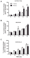
Figure 2. Astrocytes increase cystine uptake following IL-1β treatment
Astrocytes (A; n=4), neurons (B; n=4–8) and microglia (C; n=6–10) were treated with vehicle (white bars) or IL-1β (3 ng/ml; black bars) for 20–24 hr following which cells were washed and incubated with a buffer containing 14C-L-cystine (3 μM) and uptake was determined over time as indicated. Data are expressed as mean ± SEM 14C-L-cystine uptake in pmol/mg protein. An asterisk (*) indicates a significant between-group difference as determined by a two-way ANOVA followed by Bonferroni’s post hoc test. Significance was set at p < 0.05.
Figure 3
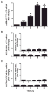
Figure 3. IL-1β selectively increases astrocytic xCT mRNA
(A) Pure astrocytes (n=4) were treated with IL-1β (3ng/ml) or its vehicle for the indicated durations and xCT mRNA assessed via qPCR. Data are expressed as mean ± SEM fold change in xCT mRNA compared to untreated cells (0 h). (B) Neurons (n=4) and microglia (inset; n=4) were treated with IL-1β (3ng/ml) or its vehicle for the indicated durations and xCT mRNA expression was assessed via qPCR. Data are expressed as mean ± SEM fold change in xCT mRNA compared to untreated cells (0 h). (C) Astrocytes (n=3) were treated with IL-1β (3ng/ml) or its vehicle for the indicated durations and 4F2hc and RBAT (inset) mRNA expression was assessed via qPCR. Data are expressed as mean ± SEM fold change in 4F2hc and RBAT mRNA compared to untreated cells (0 h). An asterisk (*) denotes values different from 0 hr as assessed by one-way ANOVA. Significance was set at p < 0.05.
Figure 4
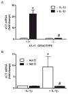
Figure 4. IL-1R1 signaling and transcription are required for the enhancement in astrocyte xCT mRNA expression
(A) Astrocytes (n=4–5 from single pup dissections) were treated with IL-1β (3 ng/ml) or its vehicle for 6 h. Thereafter, triplicate or quadruplicate culture wells were pooled, total RNA isolated, reverse transcribed and xCT and β-actin expression assessed using quantitative PCR. Data are expressed as mean ± SEM fold change in xCT mRNA compared to Il1r1 +/+ untreated cells (-IL-1β). (B) Astrocytes (n= 5) were treated with IL-1β (3 ng/ml) or its vehicle in the presence and absence of actinomycin D (Act D; 10 μg/ml) and xCT mRNA expression assessed via qPCR 6 hr later. Data are expressed as mean ± SEM fold change in xCT mRNA compared to untreated cells (-IL-1β, -Act D). An asterisk (*) denotes values different from control and a pound sign (#) indicates values that significantly differ from IL-1β-treated conditions as assessed by two-way ANOVA followed by Bonferroni’s post hoc test. Significance was set at p <0.05.
Figure 5
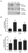
Figure 5. IL-1β increases xCT protein expression
(A) Pure astrocyte cultures were incubated with vehicle or 3 ng/ml IL-1β in the absence or presence of cycloheximide (CHX 1 μg/ml) for 20–24 hr. Cells were harvested, whole cell lysates prepared, and 100 μg protein was separated by SDS-PAGE (10% gel). Western blot analysis was performed using antibodies directed against xCT, 4F2hc, and β-actin (loading control). Protein from unstimulated C6 glioma cells was used as a positive control for xCT protein. Lane 1, Basal; Lane 2, IL-1β; Lane 3, IL-1β+CHX, Lane 4, C6 positive control. Representative of two blots. (B,C) Films were scanned and densitometry performed using Gelpro Analyzer software. (B) xCT and (C) 4F2hc protein levels were normalized to their corresponding β-actin protein levels and expressed as a fold increase (mean ± SEM; n = 2) over control (basal; set to 1).
Figure 6
Figure 6. Protein synthesis is required for the enhancement of astrocyte system xc activity that follows IL-1β treatment
Pure astrocyte cultures (n =11–12) were treated with actinomycin D (ACT D; 12.5 μg/ml) or cycloheximide (CHX; 1 μg/ml) in the absence (white bars) or presence (black bars) of IL-1β (3 ng/ml) for 20–24 hr following which 14C-L-cystine uptake (3 μM labeled + 27μM unlabeled; 25°C) was determined. Data are expressed as mean ± SEM 14C-L-cystine uptake in pmol/30 min/mg protein. An asterisk (*) denotes values different from control (-IL-1β) and a pound sign (#) indicates values different from IL-1β-treated conditions as assessed by two-way ANOVA followed by Bonferroni’s post hoc test. Significance was set at p < 0.05.
Figure 7
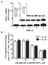
Figure 7. IL-1β does not regulate mRNA expression or activity of system XAG amino acid transporters
(A) Astrocytes (n= 3–4) were treated with IL-1β (3ng/ml) or its vehicle for the indicated durations and EAAT-1, EAAT-2, and LAT2 mRNA expression assessed via qPCR. Data are expressed as mean ± SEM fold change in mRNA compared to untreated cells (0 h). (B) Pure astrocyte cultures (n =10) were treated with vehicle (white bars) or IL-1β (3 ng/ml; black bars) for 20–24 hr following which 3H-D-aspartate (0.1 μCi/ml labeled + 1–100 μM unlabeled; 25°C) uptake was determined. Data are expressed as mean ± SEM 3H-D-aspartate uptake in cpm × 103/5 min/mg protein. No significant between-group differences were found via two-way ANOVA.
Figure 8
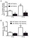
Figure 8. Cystine uptake and hypoxic neuronal cell death are reduced in cultures containing sut astrocytes
(A) Pure astrocyte cultures (n = 5–6) derived from either wild-type (white bars) or sut mice (black bars) [all cultured w/55 μM β-ME] were treated with vehicle or IL-1β (3 ng/ml) for 20–24 hr after which 14C-L-cystine uptake was determined. Data are expressed as mean ± SEM 14C-L -cystine uptake in pmol/30 min/mg protein. (B) Chimeric mixed cortical cell cultures were obtained by plating wild-type neurons on astrocytes derived from sut mice (black bars). These and control cultures (WT neurons on WT astrocytes; white bars) were treated with 1 ng/ml IL-1β or vehicle for 20–24 hr, washed, and then deprived of oxygen for 5 hr. The percentage of total neuronal cell death was determined 20–24 hr later (n = 4 cultures pooled from two independent experiments). An asterisk (*) indicates a significant within-group difference, while a pound (#) sign indicates a significant between-group difference as determined by a two-way ANOVA followed by Bonferroni’s post hoc test. Significance was set at p < 0.05.
Figure 9
Figure 9. Ionotropic glutamate receptor and system xc antagonism prevent IL-1β-mediated hypoxic neuronal injury
Mixed cortical cell cultures were treated with 3 ng/ml IL-1β for 20–24 hr, washed, and then deprived of oxygen for 4 hr. The ionotropic glutamate receptor antagonist MK-801 (10 μM) and the system xc antagonist LY367385 (50 μM) were added at the initiation of hypoxia. The percentage of total neuronal cell death was determined 20–24 hr later (n = 5–6 cultures pooled from 2 independent experiments). An asterisk (*) denotes values different from control untreated cultures (hypoxia) and a pound sign (#) indicates values different from IL-1β-treated conditions as assessed by one-way ANOVA followed by a Student-Newman-Keul’s post hoc test. Significance was set at p < 0.05.

Similar articles

Cited by

References

    1. Augustin H, Grosjean Y, Chen K, Featherstone DE. Nonvesicular release of glutamate by glial xCT transporters suppresses glutamate receptor clustering in vivo. J Neurosci. 2007;27(1):111–123. - PMC - PubMed
    1. Backus KH, Kettenmann H, Schachner M. Pharmacological characterization of the glutamate receptor in cultured astrocytes. J Neurosci Res. 1989;22(3):274–82. - PubMed
    1. Baker DA, Madayag A, Kristiansen LV, Meador-Woodruff JH, Haroutunian V, Raju I. Contribution of cystine-glutamate antiporters to the psychotomimetic effects of phencyclidine. Neuropsychopharmacology. 2008;33(7):1760–72. - PMC - PubMed
    1. Baker DA, Shen H, Kalivas PW. Cystine/glutamate exchange serves as the source for extracellular glutamate: modifications by repeated cocaine administration. Amino Acids. 2002a;23(1–3):161–2. - PubMed
    1. Baker DA, Xi Z, Shen H, Swanson CJ, Kalivas PW. The origin and neuronal function of in vivo nonsynaptic glutamate. J Neurosci. 2002b;22(20):9134–9141. - PMC - PubMed

Publication types

MeSH terms