Targeted nanoparticles enhanced flow electroporation of antisense oligonucleotides in leukemia cells
- PMID: 20630739
- PMCID: PMC3369826
- DOI: 10.1016/j.bios.2010.06.025
Targeted nanoparticles enhanced flow electroporation of antisense oligonucleotides in leukemia cells
Abstract
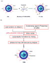
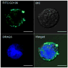
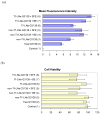
Liposome nanoparticles (LNs) with a targeting ligand were used in a semi-continuous flow electroporation (SFE) device to enhance in vitro delivery of exogenous oligonucleotides (ODN). Nanoparticles comprising transferrin-targeted lipoplex encapsulating ODN G3139 were mixed with K562 cells (a chronic myeloid leukemia cell line) and incubated for half an hour to accomplish nanoparticle binding. The mixture was then flowed through a SFE channel where electric pulses were given. Better ODN delivery efficiency was achieved with an increase of ∼24% to the case in combination of non-targeted LNs and SFE, and ∼60% to the case using targeted LNs alone, respectively. The MTS assay results confirmed cell viability greater than 75%.
Copyright © 2010 Elsevier B.V. All rights reserved.
Figures


Similar articles
-
Semicontinuous flow electroporation chip for high-throughput transfection on mammalian cells.Anal Chem. 2009 Jun 1;81(11):4414-21. doi: 10.1021/ac9002672. Anal Chem. 2009. PMID: 19419195 Free PMC article.
-
Transferrin receptor-targeted lipid nanoparticles for delivery of an antisense oligodeoxyribonucleotide against Bcl-2.Mol Pharm. 2009 Jan-Feb;6(1):221-30. doi: 10.1021/mp800149s. Mol Pharm. 2009. PMID: 19183107 Free PMC article.
-
Improving the intracellular delivery and molecular efficacy of antisense oligonucleotides in chronic myeloid leukemia cells: a comparison of streptolysin-O permeabilization, electroporation, and lipophilic conjugation.Blood. 1998 Jun 15;91(12):4738-46. Blood. 1998. PMID: 9616172
-
Gold nanoparticle-enhanced electroporation for leukemia cell transfection.Methods Mol Biol. 2014;1121:69-77. doi: 10.1007/978-1-4614-9632-8_6. Methods Mol Biol. 2014. PMID: 24510813
-
Microscale electroporation: challenges and perspectives for clinical applications.Integr Biol (Camb). 2009 Mar;1(3):242-51. doi: 10.1039/b819201d. Epub 2009 Jan 29. Integr Biol (Camb). 2009. PMID: 20023735 Free PMC article. Review.
Cited by
-
Bottom-Up Strategy to Forecast the Drug Location and Release Kinetics in Antitumoral Electrospun Drug Delivery Systems.Int J Mol Sci. 2023 Jan 12;24(2):1507. doi: 10.3390/ijms24021507. Int J Mol Sci. 2023. PMID: 36675021 Free PMC article.
-
Improvement of K562 Cell Line Transduction by FBS Mediated Attachment to the Cell Culture Plate.Biomed Res Int. 2019 Mar 27;2019:9540702. doi: 10.1155/2019/9540702. eCollection 2019. Biomed Res Int. 2019. PMID: 31032368 Free PMC article.
-
Individually addressable multi-chamber electroporation platform with dielectrophoresis and alternating-current-electro-osmosis assisted cell positioning.Biomicrofluidics. 2014 Apr 24;8(2):024117. doi: 10.1063/1.4873439. eCollection 2014 Mar. Biomicrofluidics. 2014. PMID: 24803966 Free PMC article.
-
Advances in microfluidic devices made from thermoplastics used in cell biology and analyses.Biomicrofluidics. 2017 Oct 24;11(5):051502. doi: 10.1063/1.4998604. eCollection 2017 Sep. Biomicrofluidics. 2017. PMID: 29152025 Free PMC article. Review.
-
Design strategies for physical-stimuli-responsive programmable nanotherapeutics.Drug Discov Today. 2018 May;23(5):992-1006. doi: 10.1016/j.drudis.2018.04.003. Epub 2018 Apr 10. Drug Discov Today. 2018. PMID: 29653291 Free PMC article. Review.
References
-
- Abdallah B, Hassan A, Benoist C, Goula D, Behr JP, Demeneix BA. Hum Gene Ther. 1996;7:1947–1954. - PubMed
-
- Allen TM, Sapra P, Moase E. Cell Mol Biol Lett. 2002;7:889–894. - PubMed
-
- Boussif O, Zanta MA, Behr JP. Gene Ther. 1996;3:1074–1080. - PubMed
-
- Chang DC, Chassy BM, Saunder JA. Guide to electroporation and electrofusion. Academic; San Diego: 1992.
-
- Chiu SJ, Liu SJ, Perrotti D, Marcucci G, Lee RJ. J Controlled Release. 2006;112:199–207. - PubMed
Publication types
MeSH terms
Substances
Grants and funding
LinkOut - more resources
Full Text Sources

