. 2010 Jul 9:11:37.
doi: 10.1186/1471-2172-11-37.

Differential and coordinated expression of defensins and cytokines by gingival epithelial cells and dendritic cells in response to oral bacteria

Affiliations

Differential and coordinated expression of defensins and cytokines by gingival epithelial cells and dendritic cells in response to oral bacteria

Lei Yin et al. BMC Immunol. .

Abstract

Background: Epithelial cells and dendritic cells (DCs) both initiate and contribute to innate immune responses to bacteria. However, much less is known about the coordinated regulation of innate immune responses between GECs and immune cells, particularly DCs in the oral cavity. The present study was conducted to investigate whether their responses are coordinated and are bacteria-specific in the oral cavity.

Results: The beta-defensin antimicrobial peptides hBD1, hBD2 and hBD3 were expressed by immature DCs as well as gingival epithelial cells (GECs). HBD1, hBD2 and hBD3 are upregulated in DCs while hBD2 and hBD3 are upregulated in GECs in response to bacterial stimulation. Responses of both cell types were bacteria-specific, as demonstrated by distinctive profiles of hBDs mRNA expression and secreted cytokines and chemokines in response to cell wall preparations of various bacteria of different pathogenicity: Fusobacterium nucleatum, Actinomyces naeslundii and Porphyromonas gingivalis. The regulation of expression of hBD2, IL-8, CXCL2/GRObeta and CCL-20/MIP3alpha by GECs was greatly enhanced by conditioned medium from bacterially activated DCs. This enhancement was primarily mediated via IL-1beta, since induction was largely attenuated by IL-1 receptor antagonist. In addition, the defensins influence DCs by eliciting differential cytokine and chemokine secretion. HBD2 significantly induced IL-6, while hBD3 induced MCP-1 to approximately the same extent as LPS, suggesting a unique role in immune responses.

Conclusions: The results suggest that cytokines, chemokines and beta-defensins are involved in interaction of these two cell types, and the responses are bacteria-specific. Differential and coordinated regulation between GECs and DCs may be important in regulation of innate immune homeostasis and response to pathogens in the oral cavity.


Figures

Figure 1
Figure 1
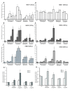
(A, B). Basal level of defensin gene expression in GECs and iDCs in the absence of bacteria. The absolute amount (picogram) of each defensin was determined using external plasmids standards in GECs (A) and iDCs (B). The expression of hBD1, hBD2 and hBD3 was normalized to housekeeping gene RPO. Results shown are the mean of three to five independent experiments. The error bars indicate SEM (standard error of mean).
Figure 2
Figure 2
Differential defensins expression in response to oral bacteria by DCs and GECs. DCs (A, B, C) and GECs (D, E, F) were stimulated with graded doses of oral bacteria cell wall extracts (A. naeslundii, AnCW; F. nucleatum, FnCW; and P. gingivalis, PgCW) for 24 h. The gene expression of hBD1, 2, and 3 was quantified by quantitative real-time PCR. LPS was used as a positive control. A, D. hBD1 expression; B, E. hBD2 expression; C. F. hBD3 expression. Results are shown as mean fold change ± SEM over unstimulated controls. The data are average of three independent experiments performed in duplicate. GECs were stimulated with bacterial preparation for 24 and 48 h. Proteins were harvested, and hBD2 and hBD3 proteins in GECs were measured by ELISA (G, H). The concentration of defensins range was pg/ml. Inductions of hBD2 and hBD3 proteins are as fold induction relative to unstimulated controls. The data are average of two independent experiments performed in duplicate. Error bars represent the mean ± SEM. Asterisks indicate statistically significant difference compared to unstimulated control (Ctl) (*p < 0.05).
Figure 3
Figure 3
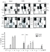
Gene expression of hBD2 and other innate immune makers in GECs in response to conditioned media from bacterially stimulated DCs. A. HBD2 mRNA upregulation in GECs in response to FnCW, and in the presence of conditioned medium (CM-DC, 1:2 dilution) from FnCW-stimulated DCs for 24 h. HBD2 mRNA was poorly expressed in GECs stimulated with PgCW (1 μg/ml), but expression was enhanced by the conditioned medium (CM-DC, 1:2 dilution) from PgCW-stimulated DCs. Controls include epithelial cells in GEC medium with or without added bacteria, and in DC medium with or without bacteria. Data are expressed as mean fold changes of experimental duplicates relative to unstimulated GECs after normalization to housekeeping gene RPO. Real-time PCR analysis of the additional markers for innate immunity (B, C, D) also show enhanced expression in GECs with conditioned medium from stimulated DCs (B: IL-8,C: CCL-20, D: CXCL2). Results are represented as mean ± SEM. *: p < 0.05 versus the relative control. Consistent results were obtained in two to three independent experiments for all markers.
Figure 4
Figure 4
Array analysis of the released cytokines and chemokines in response to F. nucleatum and P. gingivalis in DCs and GECs. Each cytokine or chemokine is represented by duplicate spots. Supernatants (1 ml) of unstimulated cells or cells stimulated for 24 h with FnCW or PgCW were applied to RayBio human cytokine protein array III as described in Materials and Methods. The rectangles in the upper left and lower right portions of the arrays indicate positive controls. The upregulated cytokines and chemokines are indicated by solid rectangles; down-regulated or absent products are indicated by dashed rectangles. The cytokine array image represents one of two independent experiments (A, B). ENA-78, epithelial neutrophil-activating protein 78; GRO, growth regulated oncogene; MCP, macrophage colony stimulating factor; MDC, macrophage-derived chemoattractant; SDF-1, stromal cell-derived factor; TARC, thymus and activation-regulated chemokine. Note the upregulation of IL-1β in DCs with both bacterial preparations (bold rectangle). C. IL-1β in the cultured media from DCs after FnCW or PgCW stimulation. The cells were stimulated with FnCW or PgCW at the indicated dose and time points. The culture media were collected and IL-1β was quantified by ELISA. Results are represented as mean ± SD for two determinations in a representative experiment. Similar results were obtained with conditioned media from three different donors. (**: p < 0.001; *: p < 0.05). Ctl: medium only.
Figure 5
Figure 5
Effects of IL-1ra on bacterially-induced hBD2 in cultured GECs. Cells were stimulated by conditioned medium from DCs (CM-DCs) treated with FnCW (A, B) at 1:2 (A) or 1:20 dilution (B); or PgCW at 1:2 (C) and at 1:20 dilution (D) with or without IL-1ra (100, 200, 400 ng/ml). Expression of mRNA was analyzed by real-time PCR. Data are expressed as mean fold change of experimental duplicates compared to the expression of unstimulated cells for each condition after normalization to housekeeping gene RPO. Results are represented as mean of fold change ± SD for two determinations in a representative experiment. Similar results were obtained with cells from three different donors. Values are significantly different compared to the respective control (*: p < 0.05).
Figure 6
Figure 6
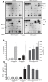
A. Cytokine profiling in DCs stimulated with hBD2, hBD3 and LPS B, C. Assay of IL-6 and MCP-1 in culture media from DCs stimulated by β-defensins by ELISA. A: The iDCs were treated with hBD2 (10 μg/ml), hBD3 (10 μg/ml), LPS (1 μg/ml) and untreated control for 24 h. Supernatants were analyzed by protein array as in Figure 4. The most differentially regulated cytokines are identified by arrow. B, C: The iDCs were stimulated with hBD2 (10 μg/ml) and hBD3 (10 μg/ml) at the indicated dose. The culture media were collected and IL-6 (B) and MCP-1 (C) were quantified by ELISA. Results are represented as mean ± SEM for three determinations from independent experiments. Note the difference in scale of B (right and left axis). HBD2 stimulated significantly greater IL-6 than hBD3, while hBD3 produced significantly greater MCP-1 than hBD2 (*: p < 0.05: **: p < 0.001).
Figure 7
Figure 7
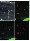
(A-D). HBD2 associates with DCs in an oral tissue model. HBD2 peptide was applied for 6 or 18 h to the surface of oral full thickness tissue model containing DCs. Immunohistochemical reaction of hBD2 polyclonal antibody (negative image) showing reaction in the upper layers of the epithelium and in a scattered distribution within the connective tissue. B-D. Double label immunofluroesence of an area similar to that indicated by the box in A, B. Double label; Anti-human HLA-DR monoclonal antibody with biotinylated anti-mouse IgG made in goat, streptavidin Alexafluor 594 (red) and anti-hBD2 polycloonal antibody made in rabbit with Alexafluor 488 labeled anti-rabbit IgG made in goat (green). C. HBD2 immunolocalization only (green). D. HLA-DR immunolocalization only (red). The solid green line visible in B and C is the supporting membrane for the tissue model. Controls without primary antibody were negative. Experimental control without added hBD2 showed no green fluorescence associated with HLA-DR positive cells. Original magnification: A: 20×, B, D: 60×.

Similar articles

Cited by

References

    1. Muller CA, Autenrieth IB, Peschel A. Innate defenses of the intestinal epithelial barrier. Cell Mol Life Sci. 2005;62:1297–307. doi: 10.1007/s00018-005-5034-2. - DOI - PubMed
    1. Ganz T. Defensins: antimicrobial peptides of innate immunity. Nat Rev Immunol. 2003;3:710–20. doi: 10.1038/nri1180. - DOI - PubMed
    1. Braff MH, Bardan A, Nizet V, Gallo RL. Cutaneous defense mechanisms by antimicrobial peptides. J Invest Dermatol. 2005;125:9–13. doi: 10.1111/j.0022-202X.2004.23587.x. - DOI - PubMed
    1. Chung WO, Dommisch H, Yin L, Dale BA. Expression of defensins in gingiva and their role in periodontal health and disease. Curr Pharm Des. 2007;13:3073–83. doi: 10.2174/138161207782110435. - DOI - PubMed
    1. Devine DA. Antimicrobial peptides in defence of the oral and respiratory tracts. Mol Immunol. 2003;40:431–43. doi: 10.1016/S0161-5890(03)00162-7. - DOI - PubMed

Publication types

MeSH terms

LinkOut - more resources