CD137 is required for M cell functional maturation but not lineage commitment
- PMID: 20616340
- PMCID: PMC2913358
- DOI: 10.2353/ajpath.2010.090811
CD137 is required for M cell functional maturation but not lineage commitment
Abstract
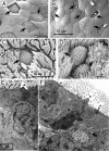
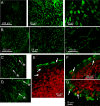
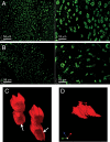
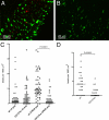
Mucosal immune surveillance depends on M cells that reside in the epithelium overlying Peyer's patch and nasopharyngeal associated lymphoid tissue to transport particles to underlying lymphocytes. M cell development is associated with B lymphocytes in a basolateral pocket, but the interactions between these cells are poorly understood. In a cell culture model of M cell differentiation, we found lymphotoxin/tumor necrosis factor alpha induction of CD137 (TNFRSF9) protein on intestinal epithelial cell lines, raising the possibility that CD137 on M cells in vivo might interact with CD137L expressed by B cells. Accordingly, while CD137-deficient mice produced UEA-1+ M cell progenitors in nasopharyngeal associated lymphoid tissue and Peyer's patch epithelium, they showed an abnormal morphology, including the absence of basolateral B cell pockets. More important, CD137-deficient nasopharyngeal associated lymphoid tissue M cells were defective in microparticle transcytosis. Bone marrow irradiation chimeras confirmed that while induction of UEA-1+ putative M cell precursors was not CD137-dependent, full M cell transcytosis function required expression of CD137 by radioresistant stromal cells as well as by bone marrow-derived cells. These results are consistent with a two-step model of M cell differentiation, with initial CD137-independent commitment to the M cell lineage followed by a CD137-CD137L interaction of M cells with CD137-activated B lymphocytes or dendritic cells for functional maturation.
Figures


Similar articles
-
Peyer's patch organogenesis is intact yet formation of B lymphocyte follicles is defective in peripheral lymphoid organs of mice deficient for tumor necrosis factor and its 55-kDa receptor.Proc Natl Acad Sci U S A. 1997 Jun 10;94(12):6319-23. doi: 10.1073/pnas.94.12.6319. Proc Natl Acad Sci U S A. 1997. PMID: 9177215 Free PMC article.
-
c-Rel is dispensable for the differentiation and functional maturation of M cells in the follicle-associated epithelium.Immunobiology. 2017 Feb;222(2):316-326. doi: 10.1016/j.imbio.2016.09.008. Epub 2016 Sep 18. Immunobiology. 2017. PMID: 27663963 Free PMC article.
-
Functional characteristics of Peyer's patch lymphoid cells. II. Lipopolysaccharide is thymus dependent.J Exp Med. 1974 Feb 1;139(2):407-13. doi: 10.1084/jem.139.2.407. J Exp Med. 1974. PMID: 4589992 Free PMC article.
-
The regulation of mucosal immune system.J Allergy Clin Immunol. 1982 Oct;70(4):225-30. doi: 10.1016/0091-6749(82)90057-4. J Allergy Clin Immunol. 1982. PMID: 6214574 Review. No abstract available.
-
Development of Peyer's patches, follicle-associated epithelium and M cell: lessons from immunodeficient and knockout mice.Semin Immunol. 1999 Jun;11(3):183-91. doi: 10.1006/smim.1999.0174. Semin Immunol. 1999. PMID: 10381864 Review.
Cited by
-
Microfold (M) cells: important immunosurveillance posts in the intestinal epithelium.Mucosal Immunol. 2013 Jul;6(4):666-77. doi: 10.1038/mi.2013.30. Epub 2013 May 22. Mucosal Immunol. 2013. PMID: 23695511 Free PMC article. Review.
-
Inducible Colonic M Cells Are Dependent on TNFR2 but Not Ltβr, Identifying Distinct Signalling Requirements for Constitutive Versus Inducible M Cells.J Crohns Colitis. 2017 Jun 1;11(6):751-760. doi: 10.1093/ecco-jcc/jjw212. J Crohns Colitis. 2017. PMID: 27932454 Free PMC article.
-
CD137 signaling enhances tight junction resistance in intestinal epithelial cells.Physiol Rep. 2014 Aug 5;2(8):e12090. doi: 10.14814/phy2.12090. Print 2014 Aug 1. Physiol Rep. 2014. PMID: 25096552 Free PMC article.
-
Prion disease and the innate immune system.Viruses. 2012 Dec;4(12):3389-419. doi: 10.3390/v4123389. Viruses. 2012. PMID: 23342365 Free PMC article. Review.
-
M Cells: Intelligent Engineering of Mucosal Immune Surveillance.Front Immunol. 2019 Jul 2;10:1499. doi: 10.3389/fimmu.2019.01499. eCollection 2019. Front Immunol. 2019. PMID: 31312204 Free PMC article. Review.
References
-
- Mebius RE. Organogenesis of lymphoid tissues. Nat Rev Immunol. 2003;3:292–303. - PubMed
-
- Cyster JG. Lymphoid organ development and cell migration. Immunol Rev. 2003;195:5–14. - PubMed
-
- Neutra MR, Mantis NJ, Kraehenbuhl JP. Collaboration of epithelial cells with organized mucosal lymphoid tissues. Nat Immunol. 2001;2:1004–1009. - PubMed
Publication types
MeSH terms
Substances
Grants and funding
LinkOut - more resources
Full Text Sources
Molecular Biology Databases
Research Materials

