. 2010 Jun 25;141(7):1208-19.
doi: 10.1016/j.cell.2010.05.015.

The conserved Bardet-Biedl syndrome proteins assemble a coat that traffics membrane proteins to cilia

Affiliations

The conserved Bardet-Biedl syndrome proteins assemble a coat that traffics membrane proteins to cilia

Hua Jin et al. Cell. .

Abstract

The BBSome is a complex of Bardet-Biedl Syndrome (BBS) proteins that shares common structural elements with COPI, COPII, and clathrin coats. Here, we show that the BBSome constitutes a coat complex that sorts membrane proteins to primary cilia. The BBSome is the major effector of the Arf-like GTPase Arl6/BBS3, and the BBSome and GTP-bound Arl6 colocalize at ciliary punctae in an interdependent manner. Strikingly, Arl6(GTP)-mediated recruitment of the BBSome to synthetic liposomes produces distinct patches of polymerized coat apposed onto the lipid bilayer. Finally, the ciliary targeting signal of somatostatin receptor 3 needs to be directly recognized by the BBSome in order to mediate targeting of membrane proteins to cilia. Thus, we propose that trafficking of BBSome cargoes to cilia entails the coupling of BBSome coat polymerization to the recognition of sorting signals by the BBSome.


Figures

Figure 1
Figure 1. The BBSome is the major effector of Arl6 in retinal extracts
(A) Arl6GTP specifically captures the BBSome from retinal extracts. Bovine retinal extracts were loaded onto GST-Arl6ΔN16[Q73L]GTP (“Arl6GTP”), GST-Arl6ΔN16GDP (“Arl6GDP”) or GST columns. Eluates were resolved by SDS-PAGE and silver stained. Eight bands unique to the Arl6GTP eluate (red dots) were excised and protein identification carried out by mass spectrometry revealed these eight proteins to be the eight subunits of the BBSome (listed on the right side of the gel). (B) The BBSome is one of only 2 protein entities recovered by Arl6GTP chromatography. Direct “in-solution” mass spectrometry analysis of the Arl6GTP eluate identified 186 proteins each represented by 1 to 136 peptides. 177 of these proteins were also identified by direct analysis of the Arl6GDP eluate and/ or of the GST eluate and therefore represent contaminants. The nine proteins identified only in the Arl6GTP eluate are listed in the table together with the total number of peptides and the number of unique peptides identified by tandem mass spectrometry for each protein. (C) One volume equivalent of retinal extract (Load), one volume equivalent of each flow-through (FT) and two volume equivalents of each eluates from the Arl6 affinity chromatographies were immunoblotted for the BBSome subunit BBS4. Over 75% of BBS4 is depleted by Arl6GTP column and close to 50% of BBS4 is recovered in the Arl6GTP eluate. The asterisk denotes a non specific band. (D) Arl6GTP specifically interacts with the β-propeller domain of BBS1. Extracts of HEK293 cells transfected with Myc-tagged BBSome subunits, or the β-propeller (a.a. 1–430) or C-terminal domains (a.a. 431–593) of BBS1 were applied to beads decorated with GST-Arl6ΔN16[Q73L]GTP. Total cell extracts (top panel) and captured materials (bottom panel) were immunoblotted for Myc. The BBSome subunit most efficiently recovered on Arl6GTP beads is BBS1 and within BBS1, only the N-terminal domain binds Arl6GTP.
Figure 2
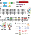
Figure 2. The BBSome and canonical coat complexes share a related structural organization
(A) Schematics of the predicted domain organization of each BBSome subunit. BBS4 and BBS8 respectively have 13 and 12.5 tetratricopeptide repeats (TPR). BBS1, BBS2, BBS7 and BBS9 each consist of a β-propeller followed by an amphipathic helical linker and a γ-adaptin ear domain (GAE). In BBS2, BBS7 and BBS9, the GAE is followed by an α/ β platform domain and α-helix domain. In BBS1, a four-helix bundle is inserted between the second and third blades. BBS5 contains two pleckstrin homology (PH) domains and a 3-helix bundle while BBIP10 is predicted to fold into two coiled-coils. (B) Structure-based alignment of the proposed GAE modules of BBS1, BBS2, BBS7 and BBS9. The structures used to generate the model of the GAE domain of BBS1 were those of GGA1, AP2©1, AP2®2, AP2α and γCOP. Only the N- and C-terminal six β-strands of the Ig-like folds (respectively labeled A–C and E–G) are shown, in a color gradient that matches the chain topology of the human BBS1 GAE domain model. While the primary sequence identity between solved or predicted structures remains quite low, hydrophobic positions (highlighted) are well conserved. See Supplemental Experimental Procedures for details on fold recognitions and secondary structure predictions. (C) The platform-like modules of BBS2, BBS7 and BBS9 are topological variants of the appendage domains. The C-terminal platform domains of γCOP, βCOP, AP2β2 and AP2α were structurally aligned to the proposed platform-like modules of BBS2, BBS7 and BBS9 as above. β-strands are shown as arrows (labeled I–M) and helices as cylinders (labeled 1–3), color matched to the AP2α structure and the modeled fold of the human BBS7 platform-like module. Intriguingly, β-strand H and helix 1 are missing from BBS2/ 7/ 9, but in turn these gain an additional, conserved C-terminal β-strand (labeled N) that is predicted to form an edge strand (grey) in the platform β-sheet. (D) Recurring membrane recruitment machinery and structural elements of the canonical coats and of the BBSome. The PIPs that participate in coat complexes binding to membranes in vitro are listed in orange. The Arf-like GTPases that recruit the coat complexes to membranes are listed in black. The β-propeller, α-solenoid and appendage domains are listed in blue, red and green respectively (McMahon and Mills, 2004; Owen et al., 2004).
Figure 3
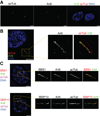
Figure 3. Arl6 is found in punctae together with the BBSome inside primary cilia
(A) Arl6 localizes to the primary cilium in RPE cells. Serum-starved RPE cells were immunostained for Arl6 and acetylated α-tubulin (acTub). Scale bars, 5µm. (B) Arl6 is found in punctae flanking the axoneme inside primary cilia. RPE cells immunostained as in (A) were imaged by structured illumination microscopy. Scale bar, 5µm. A 3D view of the same cilium is shown in movie S1. (C) Arl6 co-localizes with the BBSome inside cilia. Serum-starved RPE cells stably expressing GFP-tagged Arl6 were immunostained for BBS1 (top panel), BBIP10 (bottom panel), and acetylated α-tubulin (acTub). GFP-Arl6 was visualized in the green channel without antibody staining. Scale bars, 5 µm. A line scan through these cilia is shown in Figure S1B and C
Figure 4
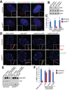
Figure 4. Arl6GTP targets the BBSome to primary cilia
(A) Arl6 is required for BBSome localization to cilia. RPE cells treated with control siRNA or siRNA targeting the coding sequence (ORF) of Arl6 were serum starved and immunostained for Arl6, BBS1 or BBIP10. Cilia were visualized by staining with acetylated α-tubulin (acTub) antibody. Insets show an enlargement of the Arl6, BBS1 or BBIP10 channel in the region around the cilium. (B) Protein extracts from cells treated with siRNAs targeting the Arl6 ORF, the Arl6 3‘ UTR or control siRNA were immunoblotted for Arl6, BBS4 and actin (loading control). Arl6 protein is depleted by both siRNAs targeting Arl6, while BBS4 protein levels remain unaffected. (C) RPE cells were prepared and stained as in (A). At least 150 cilia per experiment were counted and the percentages of Arl6-, BBS1- or BBIP10-positive cilia were plotted. Error bars represents standard deviations (SD) between three independent experiments. (D) GTP-binding but not hydrolysis by Arl6 is required for BBSome targeting to cilia. RPE, RPE-[Arl6-GFP], RPE-[Arl6(Q73L)-GFP], RPE-[Arl6(T31R)-GFP] clonal cell lines treated with control siRNA or siRNA against Arl6 3‘UTR were serum starved and immunostained for BBS1 and acetylated α-tubulin (acTub). Arl6-GFP was visualized in the green channel without antibody staining. (E) Protein extracts from cells prepared as in (D) were immunoblotted for Arl6. Endogenous Arl6, Arl6-GFP and a nonspecific band of 70 kDa (double asterisk) that serves as a loading control are shown. All three panels are extracted from the same exposure scan shown in Figure S3B. The asterisk denotes a weak non-specific band that runs slightly below Arl6-GFP. Quantitation of signal intensities shows that Arl6-GFP, Arl6[Q73L]-GFP, Arl6[T31R]-GFP are respectively 7.4-, 5.3- and 2.3-fold more abundant than endogenous Arl6. When cells are treated with Arl6UTR siRNA, endogenous Arl6 is depleted and Arl6-GFP proteins are slightly upregulated. (F) BBS1-positive cilia in the experiment shown in (D) were counted. At least 150 cilia were counted for each condition. Error bars represent SD between microscope fields.
Figure 5
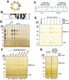
Figure 5. Arl6GTP recruits the BBSome to liposome made from pure lipids
(A) Helical-wheel representation of the N-terminal 13 amino acids of Arl6. Hydrophobic, non-polar residues are clustered on one side of the helix while charged or polar residues are on the opposite side. Non-polar residues are yellow, polar residues are purple, acidic residues are red, basic residues are blue and glycine is grey. The diameter of each circle is proportional to the bulk of each residue. (B) Sequence alignment of the N-terminus from select Arf/ Arl family members. Hydrophobic amino acids are highlighted in dark grey and basic residues are light green. (C) Arl6GTP binds to liposome through its N-terminus. 20 µg of liposomes made from brain lipids were incubated with 2 µM Arl6 or Arl6ΔN in the presence of 100 µM GMP-PNP or GDP in a 100 µl reaction at 30°C for 1 hr. The reactions were centrifuged at 385,000 × gave for 30 min at 24°C and equal portions of the resulting supernatants (S) and pellets (P) were resolved by SDS-PAGE and stained with Coomassie. As control for protein precipitation during the course of the experiment, Arl6 or Arl6ΔN were incubated without liposome and processed as above. (D) Purification of Retinal BBSome. Eluates from the Arl6GTP affinity column were loaded onto a cation exchange column (MonoS) and the BBSome (red dots mark subunits) was eluted with a salt gradient. Fractions were analyzed by SDS-PAGE and silver staining. (E) The BBSome binds to liposomes in an Arl6- and GTP-dependent manner. Various combinations of Arl6 (0.5 µM), GMP-PNP or GDP (100 µM) were incubated with 4 µg brain lipid liposomes in a 50 µl reaction at 30°C for 30 min. Reactions were diluted to 100 µl, supplemented with BBSome (50 nM final) and returned to 30°C for a further 15 min. Liposomes and bound proteins were sedimented at 140,000 × gave for 30 min at 24°C and pellets were resolved by SDS-PAGE and stained with silver. Red dots denote BBSome subunits (except for BBIP10 which was not resolved), while the blue dot denotes Arl6. (F) Phosphoinositide specificity of BBSome binding to liposome. Liposomes (167 µM final lipid concentration) containing 3 mol% of various PIPs were incubated with Arl6 (0.25 µM), BBSome (50 nM) and GMP-PNP (100 µM) in a 60µl reaction before flotation on iodixanol gradients. Liposome-bound proteins were analyzed by SDS-PAGE and silver staining. Although Arl6 binding was similar for all 8 liposomes, BBSome binding was maximal when liposomes contained PI(3,4)P2. Quantification of Arl6 and BBSome binding is shown in Figure S5C. The liposome compositions: PC/ PE/ PS/ PA/ PI: 53% DOPC, 22 mol% DOPE, 1 mol% Texas-Red DHPE, 8 mol% DOPS, 5 mol% DOPA and 11 mol% DPPI. PIPs were substituted for 3 mol% PI. (G) The BBSome requires acidic phospholipids to efficiently bind to liposomes. Liposomes (167 µM final lipid concentration) were incubated with Arl6, BBSome (50 nM) and GMP-PNP or GDP (100 µM) in a 60µl reaction before flotation on iodixanol gradients. In order to achieve similar recoveries of Arl6 with different liposomes, 1.25 µM Arl6 were used for PC/ PE liposome while 0.25 µM Arl6 were used for PI(3,4)P2 liposome. Liposome compositions: PC/ PE: 88 mol% DOPC, 11 mol% DOPE, 1 mol% Texas-Red DHPE. PC/ PE/ PS/ PA/ PI/ PI(3,4)P2: see (F).
Figure 6
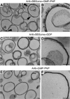
Figure 6. Morphological analysis of liposomes incubated with BBSome, Arl6 and GMP-PNP
(A) Major mix liposomes (108 µM final lipid concentration) containing 3 mol% PI(3,4)P2 were incubated with Arl6 (0.25 µM), BBSome (50 nM) and GMP-PNP (100 µM) in a 120µl reaction at 30°C for 30 min and processed for thin section electron microscopy. Distinct coated patches (arrow heads) decorate the outer layer of several liposomes. (B) Magnified view of a coated patch from (A). Repeat units are highlighted. (C) The same liposomes as in (A) were incubated with Arl6 (0.25 µM), BBSome (50 nM) and GDP (100 µM). No coated patches were seen in this preparation. (D) Magnified view of a liposome from (C). (E) The same liposomes as in (A) were incubated with Arl6 (0.25 µM) and GMP-PNP (100 µM). No coated patches were seen in this preparation. (F) Magnified view of a liposome from (E). Scale bars in (A–F), 50 nm.
Figure 7
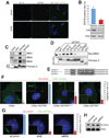
Figure 7. Direct recognition of SSTR3i3 by the BBSome imbues SSTR3i3 with ciliary targeting activity
(A) Arl6 is required for SSTR3 trafficking to cilia. Hippocampal neuron cultures were infected with shArl6-lentivirus or control lentivirus at DIV2 (2 days in vitro), and immunostained for adenylate cyclase III (ACIII), a marker of neuronal cilia, and SSTR3 at DIV8. Stacks of 10 z-sections were acquired at 0.5 µm interval with a 63x/ 1.4NA objective, deconvolved by constrained iterative and projected using sum over z-axis. Scale bars, 5 µm. (B) Top: At least 156 cilia were counted for each condition in the experiment shown in (A). Error bars represent SD between microscope fields. Bottom: Protein extracts from hippocampal neuron cultures prepared as in (A) were immunoblotted for Arl6 and actin (loading control). At least 75% of Arl6 protein was depleted upon infection with shArl6-lentivirus compared to control virus. (C) The BBSome directly recognizes the CTS of SSTR3. A MonoS-purified BBSome fraction was mixed with beads coated with GST, GST-SSTR3i3 or GST-SSTR5i3 and the material cleaved off from GST was resolved on SDS-PAGE and probed with antibodies against BBS4, BBS8 and BBS9. The bottom part of the membrane was stained with Ponceau S to show equivalent amounts of fusion moiety cleaved off from GST. 4.5 input equivalents of each eluate were loaded. (D) Generation of a point mutant in SSTR3i3 with decreased BBSome binding. Extracts of HEK cells transfected with Myc-BBS2 were applied to beads decorated with GST-SSTR3i3 variants, GST-SSTR5i3 or GST for capture assays. Total cell extracts (Input) and captured materials were immunoblotted for Myc. The bottom part of the membrane was stained with Ponceau S to show similar amounts of fusion moiety cleaved off from GST. 50 input equivalents of each eluate were loaded. Low amounts of SSTR3i3[AQ-FF] are recovered after cleavage because GST-SSTR3i3[AQ-FF] is partially degraded inside E.coli. Nonetheless, the large amounts of Myc-BBS2 recovered with SSTR3i3[AQ-FF] demonstrate that SSTR3i3[AQ-FF] binds the BBSome more tightly than SSTR3i3. (E) Sequence of select SSTR3/ 5i3 variants used in (D) and (F). The segments that differ between SSTR3i3 and SSTR5i3 are highlighted, the conserved AX[A/ S]XQ motifs are shown in bold and the mutated amino acids are red. The complete set of sequences is shown in Figure S7B. (F) The CTS activity of SSTR3i3 depends upon intact BBSome binding. CD8α, CD8α-SSTR3i3, CD8α-SSTR3i3[C-A] or CD8α-SSTR5i3 were transiently transfected into IMCD3 cells, and cells were serum starved to induce ciliation. Surface exposed CD8α chimeras were visualized by incubating cells with the OKT8 antibody directed against the extracellular domain of CD8α before fixation. Cilia were visualized by Glu-Tubulin staining. Insets show an enlargement of the CD8α channel in the region around the cilium. Images were acquired with a 63x/ 1.4NA objective and planes containing cilia are shown. At least 85 ciliated and transfected cells per experiment were counted and the percentages of CD8α chimeras targeting to cilia were plotted. Error bars represents SD between microscopic fields. Scale bars, 5 µm. (G) Ciliary targeting of CD8α-SSTR3i3 is BBSome dependent. IMCD3-[CD8α-SSTR3i3] cells were treated with control siRNA or siRNA against Arl6 or BBS4, and serum starved to induce ciliation. Surface exposed CD8α-SSTR3i3 was visualized with the OKT8 antibody and cilia were stained with Glu-Tubulin as in (F). Insets show an enlargement of the CD8α channel in the region around the cilium. Images were acquired with a 100x/ 1.4NA objective and planes containing cilia are shown. At least 110 cilia per treatment were counted and the percentages of CD8α-SSTR3i3-positive cilia were plotted. Error bars represent SD between microscopic fields. Scale bars, 5 µm. (H) A mis-sorted BBSome cargo accumulates at the plasma membrane. Surface biotinylation was performed on IMCD3-[CD8α-SSTR3i3] cells treated as in (G). Equal portions of cell lysates were either immunoprecipitated with the OKT8 antibody (Total) or captured with Neutravidin (Surface). The strips were excised from the same membrane. Arl6 and BBS4 were efficiently depleted by siRNA treatment as shown in Figure S7C. Note that total or surface exposed CD8α-SSTR3i3 protein level is not changed in siArl6- or siBBS4-treated cells.

Similar articles

Cited by

References

    1. Berbari NF, Lewis JS, Bishop GA, Askwith CC, Mykytyn K. Bardet-Biedl syndrome proteins are required for the localization of G protein-coupled receptors to primary cilia. Proc Natl Acad Sci USA. 2008a;105:4242–4246. - PMC - PubMed
    1. Berbari NF, Johnson AD, Lewis JS, Askwith CC, Mykytyn K. Identification of ciliary localization sequences within the third intracellular loop of G protein-coupled receptors. Mol Biol Cell. 2008b;19:1540–1547. - PMC - PubMed
    1. Bielas S, Silhavy J, Brancati F, Kisseleva M, Al-Gazali L, Sztriha L, Bayoumi R, Zaki M, Abdel-Aleem A, et al. Mutations in INPP5E, encoding inositol polyphosphate-5-phosphatase E, link phosphatidyl inositol signaling to the ciliopathies. Nat Genet. 2009;41:1032–1036. - PMC - PubMed
    1. Blacque OE, Reardon MJ, Li C, McCarthy J, Mahjoub MR, Ansley SJ, Badano JL, Mah AK, Beales PL, et al. Loss of C. elegans BBS-7 and BBS-8 protein function results in cilia defects and compromised intraflagellar transport. Genes Dev. 2004;18:1630–1642. - PMC - PubMed
    1. Bremser M, Nickel W, Schweikert M, Ravazzola M, Amherdt M, Hughes CA, Söllner TH, Rothman JE, Wieland FT. Coupling of coat assembly and vesicle budding to packaging of putative cargo receptors. Cell. 1999;96:495–506. - PubMed

Publication types