Subtype-specific genomic alterations define new targets for soft-tissue sarcoma therapy
- PMID: 20601955
- PMCID: PMC2911503
- DOI: 10.1038/ng.619
Subtype-specific genomic alterations define new targets for soft-tissue sarcoma therapy
Abstract
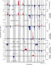
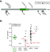
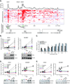
Soft-tissue sarcomas, which result in approximately 10,700 diagnoses and 3,800 deaths per year in the United States, show remarkable histologic diversity, with more than 50 recognized subtypes. However, knowledge of their genomic alterations is limited. We describe an integrative analysis of DNA sequence, copy number and mRNA expression in 207 samples encompassing seven major subtypes. Frequently mutated genes included TP53 (17% of pleomorphic liposarcomas), NF1 (10.5% of myxofibrosarcomas and 8% of pleomorphic liposarcomas) and PIK3CA (18% of myxoid/round-cell liposarcomas, or MRCs). PIK3CA mutations in MRCs were associated with Akt activation and poor clinical outcomes. In myxofibrosarcomas and pleomorphic liposarcomas, we found both point mutations and genomic deletions affecting the tumor suppressor NF1. Finally, we found that short hairpin RNA (shRNA)-based knockdown of several genes amplified in dedifferentiated liposarcoma, including CDK4 and YEATS4, decreased cell proliferation. Our study yields a detailed map of molecular alterations across diverse sarcoma subtypes and suggests potential subtype-specific targets for therapy.
Figures


Similar articles
-
Gene expression analysis of soft tissue sarcomas: characterization and reclassification of malignant fibrous histiocytoma.Mod Pathol. 2007 Jul;20(7):749-59. doi: 10.1038/modpathol.3800794. Epub 2007 Apr 27. Mod Pathol. 2007. PMID: 17464315
-
Cytogenetic analysis of 46 pleomorphic soft tissue sarcomas and correlation with morphologic and clinical features: a report of the CHAMP Study Group. Chromosomes and MorPhology.Genes Chromosomes Cancer. 1998 May;22(1):16-25. doi: 10.1002/(sici)1098-2264(199805)22:1<16::aid-gcc3>3.0.co;2-a. Genes Chromosomes Cancer. 1998. PMID: 9591630
-
Analysis of germline and tumor mutations of p53 gene in familial occurrence of soft tissue sarcomas.J Surg Oncol. 2007 Mar 15;95(4):347-50. doi: 10.1002/jso.20720. J Surg Oncol. 2007. PMID: 17192950
-
Lipomatous tumors.Monogr Pathol. 1996;38:207-39. Monogr Pathol. 1996. PMID: 8744279 Review.
-
[Pleomorphic high-grade soft tissue sarcomas: is the subclassification up to date?].Pathologe. 2011 Feb;32(1):47-56. doi: 10.1007/s00292-010-1400-4. Pathologe. 2011. PMID: 21234572 Review. German.
Cited by
-
Establishment and characterization of a new human myxoid liposarcoma cell line (DL-221) with the FUS-DDIT3 translocation.Lab Invest. 2016 Aug;96(8):885-94. doi: 10.1038/labinvest.2016.64. Epub 2016 Jun 6. Lab Invest. 2016. PMID: 27270875 Free PMC article.
-
The molecular landscape of extraskeletal osteosarcoma: A clinicopathological and molecular biomarker study.J Pathol Clin Res. 2015 Oct 29;2(1):9-20. doi: 10.1002/cjp2.29. eCollection 2016 Jan. J Pathol Clin Res. 2015. PMID: 27499911 Free PMC article.
-
Discovery of novel candidates for anti-liposarcoma therapies by medium-scale high-throughput drug screening.PLoS One. 2021 Mar 10;16(3):e0248140. doi: 10.1371/journal.pone.0248140. eCollection 2021. PLoS One. 2021. PMID: 33690666 Free PMC article.
-
Analysis of the intratumoral adaptive immune response in well differentiated and dedifferentiated retroperitoneal liposarcoma.Sarcoma. 2015;2015:547460. doi: 10.1155/2015/547460. Epub 2015 Jan 29. Sarcoma. 2015. PMID: 25705114 Free PMC article.
-
Retroperitoneal liposarcoma: current insights in diagnosis and treatment.Front Surg. 2015 Feb 10;2:4. doi: 10.3389/fsurg.2015.00004. eCollection 2015. Front Surg. 2015. PMID: 25713799 Free PMC article. Review.
References
-
- Jemal A, et al. Cancer statistics, 2009. CA Cancer J Clin. 2009;59:225–249. - PubMed
-
- Fletcher C, Unni K, Mertens F, editors. Pathology and Genetics of Tumors of Soft Tissue and Bone. Lyon: International Agency for Research on Cancer Press; 2002. p. 427.
-
- Heinrich MC, et al. PDGFRA activating mutations in gastrointestinal stromal tumors. Science. 2003;299:708–710. - PubMed
-
- Hirota S, et al. Gain-of-function mutations of c-kit in human gastrointestinal stromal tumors. Science. 1998;279:577–580. - PubMed
-
- Demetri GD, et al. Efficacy and safety of imatinib mesylate in advanced gastrointestinal stromal tumors. N Engl J Med. 2002;347:472–480. - PubMed
Publication types
MeSH terms
Grants and funding
LinkOut - more resources
Full Text Sources
Other Literature Sources
Medical
Molecular Biology Databases
Research Materials
Miscellaneous

