. 2010 Sep 3;285(36):27664-72.
doi: 10.1074/jbc.M110.131235. Epub 2010 Jul 2.

Association between the herpes simplex virus-1 DNA polymerase and uracil DNA glycosylase

Affiliations

Association between the herpes simplex virus-1 DNA polymerase and uracil DNA glycosylase

Federica Bogani et al. J Biol Chem. .

Abstract

Herpes simplex virus-1 (HSV-1) is a large dsDNA virus that encodes its own DNA replication machinery and other enzymes involved in DNA transactions. We recently reported that the HSV-1 DNA polymerase catalytic subunit (UL30) exhibits apurinic/apyrimidinic and 5'-deoxyribose phosphate lyase activities. Moreover, UL30, in conjunction with the viral uracil DNA glycosylase (UL2), cellular apurinic/apyrimidinic endonuclease, and DNA ligase IIIalpha-XRCC1, performs uracil-initiated base excision repair. Base excision repair is required to maintain genome stability as a means to counter the accumulation of unusual bases and to protect from the loss of DNA bases. Here we show that the HSV-1 UL2 associates with the viral replisome. We identified UL2 as a protein that co-purifies with the DNA polymerase through numerous chromatographic steps, an interaction that was verified by co-immunoprecipitation and direct binding studies. The interaction between UL2 and the DNA polymerase is mediated through the UL30 subunit. Moreover, UL2 co-localizes with UL30 to nuclear viral prereplicative sites. The functional consequence of this interaction is that replication of uracil-containing templates stalls at positions -1 and -2 relative to the template uracil because of the fact that these are converted into non-instructional abasic sites. These findings support the existence of a viral repair complex that may be capable of replication-coupled base excision repair and further highlight the role of DNA repair in the maintenance of the HSV-1 genome.


Figures
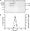
FIGURE 1.
FIGURE 1.
Co-purification of HSV-1 DNA polymerase and uracil DNA glycosylase. A, fractions eluting from the Resource Q column were resolved by 10% SDS-PAGE followed by silver staining. The positions of markers (ML and MH, Bio-Rad low and high range standards, respectively), UL30 and UL42 are as indicated. X indicates the position of an ∼36-kDa polypeptide. B, the specified fractions were assayed for pol (●) and UDG (○) activities as described under “Experimental Procedures.”
FIGURE 2.
FIGURE 2.
Co-immunoprecipitation of UL30 with UL2. Transfection, immunoprecipitation with anti-V5, and immunoblotting were performed as described under “Experimental Procedures.” A and B, co-immunoprecipitation of UL30 with UL2 in transfected cells. Aliquots of the indicated input fractions (Input; lanes 1-7) and immunoprecipitates (IP; lanes 8-14) were probed with combined anti-UL30/UL42 rabbit antisera (A) or anti-V5 (B). C and D, co-immunoprecipitation of purified UL30 with purified UL2. Aliquots of the indicated immunoprecipitates (IP; lanes 1-3) and supernatants (Supernatant; lanes 4-6) were probed with anti-UL30 rabbit serum (C) or anti-V5 (D). The combinations of proteins are indicated, as are the positions of UL30, UL42, UL2-V5, UL2-Trx-V5, and LacZ-V5. LacZ-V5 was included as a nonspecific control and used to balance the total amount of DNA in the transfections.
FIGURE 3.
FIGURE 3.
Purified UL2 binds to immobilized UL30. Purified UL2-Trx-V5 was applied to UL30 (lanes 1-4) or aldolase beads (lanes 6-9) as described under “Experimental Procedures.” Flow-through (FT), and consecutive 1 m NaCl eluates (E1, E2, and E3) were analyzed by immunoblotting using anti-V5. The position of UL2-Trx-V5 is as indicated. M (lane 5), purified UL2-Trx-V5 control.
FIGURE 4.
FIGURE 4.
Co-immunoprecipitation of pol with UL2 in HSV-1-infected cells. Transfection, infection, immunoprecipitation, and immunoblotting were performed as described under “Experimental Procedures.” A–C, immunoprecipitation with anti-V5. A, immunoprecipitates probed with a combined anti-UL30/UL42 rabbit serum. B, input fractions probed with combined anti-UL30/UL42 rabbit sera. C, input fractions probed with anti-V5. For each panel, lane 1 represents aliquots of immunoprecipitates (A) or inputs (B and C) from cells transfected with LacZ-V5 and superinfected with HSV-1. Lane 2 represents aliquots of immunoprecipitates (A) or inputs (B and C) from cells transfected with UL2-V5 and mock-infected with HSV-1. Lane 3 represents aliquots of immunoprecipitates (A) or inputs (B and C) from cells transfected with UL2-V5 and superinfected with HSV-1. D and E, immunoprecipitation with anti-UL42 serum. D, aliquots of the indicated input fractions (Input; lanes 1 and 2) and immunoprecipitates (IP; lanes 3 and 4) were probed with anti-V5. E, aliquots of HSV-1-infected (lane 1) and mock-infected (lane 2) input fractions were probed with combined anti-UL30/UL42 rabbit sera. Infection with HSV-1 is indicated with a +. The positions of UL30, UL42, LacZ-V5, and UL2-V5 are as indicated.
FIGURE 5.
FIGURE 5.
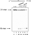
UDG activity in HSV-1-infected cell extract binds to immobilized UL30. Two hundred-μl aliquots of extract from HSV-1-infected cells were applied to aldolase (lanes 4 and 5) or UL30 (lanes 6 and 7) beads, and the input (I), flow-through (FT), and 1 m NaCl eluates (E) were assayed for UDG activity using 4 nm 5′-32P labeled PBAZ7 as described under “Experimental Procedures.” S, substrate; B, substrate with buffer. The positions of 31-mer substrate and 16-mer product are as indicated. The numbers in italics below each lane represent the percentage of cleavage of substrate. The asterisk denotes the position of the 5′ [32P] label.
FIGURE 6.
FIGURE 6.
Co-localization of UL30 and UL2 to viral prereplicative sites. Indirect immunofluorescence microscopy of transfected cells was performed as described under “Experimental Procedures.” D, DAPI staining; R, red fluorescence (UL30); G, green fluorescence (UL2); M, merged red/green fluorescence. A, cells transfected with UL30. B, cells transfected with UL2. C and D, cells transfected with UL30, the remaining six essential viral replication proteins, oriS-containing plasmid, and PAA treatment. E–H, cells transfected with UL2, UL30, the remaining six essential viral replication proteins, oriS-containing plasmid, and PAA treatment.
FIGURE 7.
FIGURE 7.
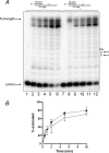
Template uracil does not affect DNA synthesis by UL30. Primer extension reactions were performed with 4 nm UL30 for the times indicated on either the T-containing (lanes 1–6) or the U-containing templates (lanes 7–12) as described under “Experimental Procedures.” A, reaction products. Lanes 1-6 and 7-12, 0, 0.5, 1, 2, 5, and 10 min of incubation. The positions of primer and full-length product and the relevant sequence of the template are as indicated. The asterisk denotes the position of the 5′ [32P] label. B, quantitation of the data from three independent experiments. ●, T-containing template; ○, U-containing template. Error bars indicate S.E.
FIGURE 8.
FIGURE 8.
UL2 causes UL30 to stall upstream of a template uracil. Primer extension reactions were performed with 4 nm UL30 and the indicated concentrations of UL2 on either the T-containing (lanes 1–7) or the U-containing templates (lanes 8–14) as described under “Experimental Procedures.” A, primer extension reactions. Lanes 1 and 8, substrate only; lanes 2-7 and 9-14, reactions with 0, 2, 4, 6, 8, and 16 nm UL2. B, UDG activity of UL2 was assayed using the 5′-32P labeled 31-mer as described under “Experimental Procedures.” Lanes 1-6, 0, 2, 4, 6, 8, and 16 nm UL2. The positions of primer, full-length product, 31-mer substrate, and 16-mer product are as indicated. The asterisk denotes the position of the 5′ [32P] label. C, quantitation of the fraction of primers stalled upstream (positions −1 and −2) of template T (●) or U (○) from the data in A. D, quantitation of the data shown in B.
FIGURE 9.
FIGURE 9.
DNA polymerase from HSV-1-infected cells stalls upstream of template uracil. Primer extension reactions were performed with the indicated concentration of HSV-1 DNA polymerase-UL2 complex purified from HSV-1-infected cells on either the T-containing (lanes 1–6) or the U-containing templates (lanes 7–12) as described under “Experimental Procedures.” A, primer extension reactions. Lanes 1-6 and 7-12, reactions with 0, 5, 10, 25, 50, and 100 nm protein. B, UDG activity of UL2 was assayed using the 5′-32P labeled 31-mer as described under “Experimental Procedures.” Lanes 1-6, 0, 5, 10, 25, 50, and 100 nm protein. The positions of primer, full-length product, 31-mer substrate, and 16-mer product are as indicated. The asterisk denotes the position of the 5′ [32P] label. C, quantitation of the data in A. Filled symbols, T-containing templates; open symbols, U-containing templates. ●, ○, total extension; ■, □, extension beyond template T or U; ▴, ▵, stalling upstream (positions −1 and −2) of template T or U. D, quantitation of the data shown in B.

Similar articles

Cited by

References

    1. Boehmer P. E., Nimonkar A. V. (2003) IUBMB Life 55, 13–22 - PubMed
    1. Boehmer P. E., Villani G. (2003) Prog. Nucleic Acid Res. Mol. Biol. 75, 139–171 - PubMed
    1. Knipe D. M., Cliffe A. (2008) Nat. Rev. Microbiol. 6, 211–221 - PubMed
    1. McGeoch D. J., Dalrymple M. A., Dolan A., McNab D., Perry L. J., Taylor P., Challberg M. D. (1988) J. Virol. 62, 444–453 - PMC - PubMed
    1. Wu C. A., Nelson N. J., McGeoch D. J., Challberg M. D. (1988) J. Virol. 62, 435–443 - PMC - PubMed

Publication types

MeSH terms

LinkOut - more resources