. 2010 Sep;84(18):9439-51.
doi: 10.1128/JVI.00533-10. Epub 2010 Jun 30.

The clinically approved proteasome inhibitor PS-341 efficiently blocks influenza A virus and vesicular stomatitis virus propagation by establishing an antiviral state

Affiliations

The clinically approved proteasome inhibitor PS-341 efficiently blocks influenza A virus and vesicular stomatitis virus propagation by establishing an antiviral state

Sabine Eva Dudek et al. J Virol. 2010 Sep.

Abstract

Recently it has been shown that the proinflammatory NF-kappaB pathway promotes efficient influenza virus propagation. Based on these findings, it was suggested that NF-kappaB blockade may be a promising approach for antiviral intervention. The classical virus-induced activation of the NF-kappaB pathway requires proteasomal degradation of the inhibitor of NF-kappaB, IkappaB. Therefore, we hypothesized that inhibition of proteasomal IkappaB degradation should impair influenza A virus (IAV) replication. We chose the specific proteasome inhibitor PS-341, which is a clinically approved anticancer drug also known as Bortezomib or Velcade. As expected, PS-341 treatment of infected A549 cells in a concentration range that was not toxic resulted in a significant reduction of progeny virus titers. However, we could not observe the proposed suppression of NF-kappaB-signaling in vitro. Rather, PS-341 treatment resulted in an induction of IkappaB degradation and activation of NF-kappaB as well as the JNK/AP-1 pathway. This coincides with enhanced expression of antiviral genes, such as interleukin-6 and, most importantly, MxA, which is a strong interferon (IFN)-induced suppressor of influenza virus replication. This suggests that PS-341 may act as an antiviral agent via induction of the type I IFN response. Accordingly, PS-341 did not affect virus titers in Vero cells, which lack type I IFN genes, but strongly inhibited replication of vesicular stomatitis virus (VSV), a highly IFN-sensitive pathogen. Thus, we conclude that PS-341 blocks IAV and VSV replication by inducing an antiviral state mediated by the NF-kappaB-dependent expression of antivirus-acting gene products.


Figures

FIG. 1.
FIG. 1.
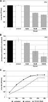
A 50 nM concentration of PS-341 is not cytotoxic or proapoptotic in cells for the indicated exposure times. A549 (A and B) and Vero, MDCK II, and HEK293 cells and HUVEC and HBEpC (all shown only in panel A) were treated with different concentrations (100 nM, 50 nM, and 10 nM) of PS-341 for the indicated times or treated with solvent or left untreated. Cells treated with 1.5 μM staurosporine (STS) were used as positive controls for metabolically inactive cells/dead cells. (A) An MTT assay was performed, and metabolic activity of cells was calculated as the percentage of the untreated control. Arrow bars represent standard deviations from four independent experiments. (B) PI staining was performed to measure membrane integrity of cells by fluorescence-activated cell sorter analysis. The diagram shows gated cells which were not stained by PI and therefore had no destruction of the cell membrane. Arrow bars represent standard deviations of three independent experiments. (C) A549 cells were treated with 50 nM PS-341 or solvent or left untreated for the indicated times. Afterwards cells were lysed and Western blot analysis was performed to detect apoptotic cells by cleavage of PARP. As a positive control cells were treated with 1 μM staurosporine. Equal amounts of protein loading were assayed by using the cellular protein ERK2. Shown is one representative blot out of three independent experiments.
FIG. 2.
FIG. 2.
PS-341 impairs FPV replication in A549 cells. (A and B) A549 cells were either pretreated for 1 h with different concentrations of PS-341 or with solvent only or were left untreated. Then, cells were infected with FPV at an MOI of 0.001 (A) or 0.05 (B). After virus inoculation cells were posttreated with different concentrations of PS-341. (A) At 24 h p.i. supernatants were obtained and progeny virus titers were measured by standard plaque assay. (B) Proteasome activity and the ability of PS-341 to inhibit the proteasome was determined 24 h p.i. (C) A549 cells were pretreated with 50 nM PS-341 or solvent or left untreated for 1 h. Afterwards cells were infected with FPV at an MOI of 0.0005. Subsequent to virus inoculation cells were posttreated with 50 nM PS-341 or solvent or left untreated. After the indicated times p.i. supernatants were obtained and progeny virus titers were determined by standard plaque assay. Arrow bars in all experiments represent standard deviations of three independent experiments.
FIG. 3.
FIG. 3.
PS-341 impairs viral propagation of different IAV strains in different cell types. A549 (A, B, and F), U937 (C), MDCK II (D), HEK293 (E), HUVEC (G), or HBEpC (H) cells were pretreated for 1 h with 50 nM PS-341 or solvent or left untreated. Then cells were infected with different human and avian IAV strains (FPV MOI of 0.001, PR8 MOI of 0.05, and KAN-1 MOI of 0.01). Subsequent to virus inoculation cells were posttreated with 50 nM PS-341 or solvent or left untreated. After the indicated times p.i. supernatants were obtained and progeny virus titers were determined by standard plaque assay. Arrow bars represent standard deviations of three independent experiments.
FIG. 4.
FIG. 4.
Early steps of viral replication within the first replication cycle are affected. (A) For time-of-addition kinetics analysis, A549 cells were either left untreated or were pretreated for 10 h or 1 h with 50 nM PS-341 before infection and additionally posttreated after infection. Cells were infected with FPV at an MOI of 0.005. After virus inoculation cells were posttreated with 50 nM PS-341. Then the proteasome inhibitor was added after virus inoculation (−10 h, −1 h, and +30 min) or it was added at the different times p.i. as indicated (+1 h, +2 h, +4 h, +6 h, and +8 h; cells were not pretreated before infection). At 9 h p.i. supernatants were obtained and progeny virus titers were determined by standard plaque assay. Shown is one representative experiment out of three independent experiments. (B) A549 cells were pretreated with 50 nM PS-341 or left untreated for 1 h. Afterwards cells were infected with avian FPV or human PR8 at an MOI of 1. Subsequent to virus inoculation cells were posttreated with 50 nM PS-341 or left untreated. After the indicated times p.i. cells were lysed and analyzed by Western blotting for accumulation of viral proteins polymerase PB1 and matrix protein M1. Cellular protein ERK2 served as a control to demonstrate equal amounts of protein loading. Shown is one representative blot out of three independent experiments.
FIG. 5.
FIG. 5.
PS-341 treatment leads to an activation of the NF-κB pathway and JNK/AP-1 pathway. (A) A549, Vero, MDCK II, HEK293, HUVEC, and HBEpC cells were treated with 50 nM PS-341 for the indicated times or left untreated. In addition, cells were stimulated with 30 ng/ml TNF-α for 15 min or left unstimulated. Cell lysates were subjected to Western blot analysis against IκBα and phosphorylated p65 (Ser536). p65 and ERK2 served as controls for equal protein amounts. (B and C) A549 cells were treated with PS-341 at 50 nM for the indicated times or left untreated. (B) Western blotting was performed with total cell lysates, using phospho-specific antibodies against JNK and the transcription factors c-Jun and ATF-2 or loading controls, respectively. (C) Native PAGE analysis was performed with total cell lysates to detect IRF-3 dimers. As a control for IRF-3 dimerization (ctr.), A549 cells were transfected for 4 h with 1 μg RNA isolated from FPV-infected A549 cells (MOI of 5; 5 h). In each case one representative blot is shown of three independent experiments.
FIG. 6.
FIG. 6.
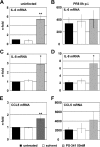
NF-κB- and AP-1-dependent gene transcripts are upregulated. A549 cells were pretreated for 1 h with 50 nM PS-341 or solvent or left untreated. Then cells were infected with PR8 at an MOI of 1 or mock infected with solvent containing no virus (uninfected). After virus inoculation cells were posttreated with 50 nM PS-341 or solvent or left untreated. At 8 h p.i. cells were lysed and RNA was subjected to reverse transcription. cDNA was analyzed by quantitative RT-PCR for amounts of transcripts of IL-6 (A and B), IL-8 (C and D), and CCL5 (E and F). Arrow bars represent standard deviations of three independent experiments and are generated as the fold change compared to untreated and mock-infected controls. *, P < 0.05; **, P < 0.01).
FIG. 7.
FIG. 7.
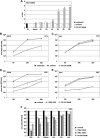
PS-341 treatment induces type I IFN response genes, leading to suppression of virus propagation. (A) A549 cells were treated for the indicated times with 50 nM PS-341 or solvent. Then cells were lysed and RNA was subjected to reverse transcription. cDNA was analyzed by quantitative RT-PCR to determine amounts of transcripts of IFN-dependent MxA. Arrow bars represent standard deviations of two independent experiments and are generated as the fold change compared to the untreated control. *, P < 0.05 compared to untreated control. (B to E) A549 cells (B and D) or Vero cells (C and E) were pretreated for 1 h with 50 nM PS-341 (B to E), for 12 h with 100 U of IFN-β (only D and E), solvent, or left untreated. After that cells were infected with avian FPV at an MOI of 0.001 (B and C) or with VSV at an MOI of 0.001 (D) or 0.0001 (E). Subsequent to virus inoculation cells were posttreated with 50 nM PS-341 (B to E), 100 U of IFN-β (D and E only), solvent, or left untreated. After the indicated times p.i. supernatants were obtained and progeny virus titers were determined by standard plaque assay. Arrow bars represent standard deviations of three independent experiments (panel E shows results of one representative experiment out of four). (F) For time-of-addition kinetics analysis, A549 cells were either pretreated for 10 h or 1 h with 50 nM PS-341 or 1,000 U of IFN-β or IFN-α before infection and were additionally posttreated after infection or left untreated. Cells were infected with VSV at an MOI of 0.001. After virus inoculation cells were posttreated with 50 nM PS-341 or 1,000 U of IFN-β or IFN-α. The proteasome inhibitor or IFNs were added after virus inoculation (−10 h, −1 h, or +30 min) or they were added at different times p.i. as indicated (+1 h, +2 h, and +4 h; cells were not pretreated before infection). At 12 h p.i. supernatants were obtained and progeny virus titers were determined by standard plaque assay. Shown is one representative experiment out of three independent experiments.

Similar articles

Cited by

References

    1. Adams, J., M. Behnke, S. Chen, A. A. Cruickshank, L. R. Dick, L. Grenier, J. M. Klunder, Y. T. Ma, L. Plamondon, and R. L. Stein. 1998. Potent and selective inhibitors of the proteasome: dipeptidyl boronic acids. Bioorg. Med. Chem. Lett. 8:333-338. - PubMed
    1. Adams, J., V. J. Palombella, E. A. Sausville, J. Johnson, A. Destree, D. D. Lazarus, J. Maas, C. S. Pien, S. Prakash, and P. J. Elliott. 1999. Proteasome inhibitors: a novel class of potent and effective antitumor agents. Cancer Res. 59:2615-2622. - PubMed
    1. An, J., Y. Sun, M. Fisher, and M. B. Rettig. 2004. Maximal apoptosis of renal cell carcinoma by the proteasome inhibitor bortezomib is nuclear factor-κB dependent. Mol. Cancer Ther. 3:727-736. - PubMed
    1. Baumeister, W., J. Walz, F. Zuhl, and E. Seemuller. 1998. The proteasome: paradigm of a self-compartmentalizing protease. Cell 92:367-380. - PubMed
    1. Belkowski, L. S., and G. C. Sen. 1987. Inhibition of vesicular stomatitis viral mRNA synthesis by interferons. J. Virol. 61:653-660. - PMC - PubMed

Publication types