. 2010 Jun 16;5(6):e11160.
doi: 10.1371/journal.pone.0011160.

Bryostatin modulates latent HIV-1 infection via PKC and AMPK signaling but inhibits acute infection in a receptor independent manner

Affiliations

Bryostatin modulates latent HIV-1 infection via PKC and AMPK signaling but inhibits acute infection in a receptor independent manner

Rajeev Mehla et al. PLoS One. .

Abstract

HIV's ability to establish long-lived latent infection is mainly due to transcriptional silencing in resting memory T lymphocytes and other non dividing cells including monocytes. Despite an undetectable viral load in patients treated with potent antiretrovirals, current therapy is unable to purge the virus from these latent reservoirs. In order to broaden the inhibitory range and effectiveness of current antiretrovirals, the potential of bryostatin was investigated as an HIV inhibitor and latent activator. Bryostatin revealed antiviral activity against R5- and X4-tropic viruses in receptor independent and partly via transient decrease in CD4/CXCR4 expression. Further, bryostatin at low nanomolar concentrations robustly reactivated latent viral infection in monocytic and lymphocytic cells via activation of Protein Kinase C (PKC) -alpha and -delta, because PKC inhibitors rottlerin and GF109203X abrogated the bryostatin effect. Bryostatin specifically modulated novel PKC (nPKC) involving stress induced AMP Kinase (AMPK) inasmuch as an inhibitor of AMPK, compound C partially ablated the viral reactivation effect. Above all, bryostatin was non-toxic in vitro and was unable to provoke T-cell activation. The dual role of bryostatin on HIV life cycle may be a beneficial adjunct to the treatment of HIV especially by purging latent virus from different cellular reservoirs such as brain and lymphoid organs.


Conflict of interest statement

Competing Interests: The authors have declared that no competing interests exist.

Figures

Figure 1
Figure 1. Bryostatin is non-toxic at its active concentration either alone or in combination with other anti-HIV compounds.
Magi and Jurkat cells were treated with various therapeutic drugs (MF-Metformin (1 mM), Min-Minocycline (5 µg/ml and 10 µg/ml), Sim-Simvastatin (2.5 µM), and AZT (100 µM), S.Sali-Sodium salicylate (1 mM) either alone or in combination with bryostatin for 48 and 72 h and cell survival was determined using WST8/CCK8 cytotoxicity assay. (A) Survival of Magi cells after treatment with bryostatin and other drugs, (B) bryostatin in combination with other drugs. (C) Jurkat cell survival after treatment with bryostatin (25 ng/ml) and other drugs, (D) bryostatin in combination with other drugs. Each treatment was done in triplicate. (n = 2).
Figure 2
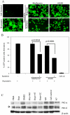
Figure 2. Bryostatin ablates R5-tropic HIV infection.
Magi cells (CD4+/CCR5+) were infected with 100 ng/ml p24 equivalent HIV recombinant NLENYU2 virus expressing YFP. (A) Productive viral infection was monitored semi-quantitatively on 3rd and 5th dpi. (B) GFP-positive cells per 10 fields were counted in HIV-infected cultures and plotted as mean. (C) Dose dependent anti-HIV effect of bryostatin was monitored by p24 assay. (D) p24 levels in the culture supernatants after treatment with bryostatin (25 ng/ml) and other compounds on 3rd and 5th day post infection are shown. Sodium salicylate (1 mM) was used as a negative control in each set of experiments. Degree of significance for bryostatin treatment was relative to virus control, * p<0.05, # p<0.001. (n = 4).
Figure 3
Figure 3. Bryostatin ablates X4-tropic HIV infection.
Jurkat cells were pretreated either with bryostatin or other drugs, infected with 50 ng/ml p24 equivalent recombinant NLENY HIV-1 expressing YFP followed by continuation in respective treatments. (A) YFP expression levels were monitored semi-quantitatively (data shown in representative pictures on 5th dpi). (B) Bryostatin treatment ablated HIV infection as demonstrated by decrease in YFP-positive Jurkat cells on 5th dpi. (C) Productive HIV replication was monitored by YFP-positive cells on 5th dpi and (D) validated by p24 levels in infected culture supernatants on 3rd and 5th dpi. Results were statistically significant (#p<0.005, * p<0.0001) compared to HIV-1 NLENY virus control. (n = 3).
Figure 4
Figure 4. Bryostatin down-regulates CD4 and CXCR4 receptor expression on Jurkat cells.
(A) Jurkat cells were treated either with bryostatin (25 nM) or left untreated for 0 to 48 h followed by immunostaining with fluorescently labeled anti-CD4 and anti-CXCR4 antibodies and monitored by flowcytometry. Representative dot plots at 3 h and 48 h of bryostatin treatment, isotype control and vehicle control (DMSO). (B) Time course of expression profile of CD4+/CXCR4+ receptors on Jurkat cells upon treatment with bryostatin (n = 2).
Figure 5
Figure 5. Bryostatin inhibits HIV-1 independent of viral receptors.
(A and B) Hela cells were pretreated with bryostatin (27 nM), vehicle, and AZT as positive control and thereafter infected with VSV pseudotyped-NLENY1 HIV. Productive HIV replication was monitored using YFP expression after 3dpi. Results were significant at p<0.05 (C) Virus production was further confirmed by p24 levels in infected culture supernatants by quantitative ELISA. (D) Schematic representation of the experimental design of virus infection in HeLa cells and infectivity assay in TZM-bl cells is shown. (E and F) Virus infectivity in the bryostatin treated HIV-infected culture supernatants was monitored by infecting TZM-bl cells either with YFP expression as a marker or (G) viral p24 production after 3dpi. (H and I) Schematic representation of single round infection experiment, bryostatin mediated HIV inhibition was monitored by p24 assay in a single round infection on Hela cells using VSV pseudotyped HIV-NLR+E- (*p<0.05, #p<0.005) (n = 2).
Figure 6
Figure 6. Bryostatin reactivates latent HIV infection in monocytic cells.
(A) THP-p89 cells were treated with bryostatin (25 ng/ml) for different durations and fluorescence was quantified by flowcytometry. (B) GFP expression was directly proportional to the level of activation in these cells as visualized by fluorescence microscopy. Quantification of GFP fluorescence in bryostatin-treated latently-infected THP-p89 cells was done by flowcytometry. (C) Bryostatin primed viral reactivation at 24 and 48 h, was followed by release of viral p24 in culture supernatants using ELISA. (D) Bryostatin induced viral reactivation was further confirmed on another latently HIV-infected lymphocytic cell model (J1.1), 48 h post treatment using viral p24 as a marker. (E and F) Comparative dose response of bryostatin, prostratin and SAHA, for viral reactivation in THP-p89 cells. Bryostatin at a 1000 fold lower concentration, EC50<0.25 nM reactivated latent HIV in THP-p89 cells more potently than SAHA and prostratin (# p<0.001, * p<0.005). (G and H) Bryostatin in absence of Tat failed to activate HIV promoter. SVGA-LTR-GFP cells were either treated with bryostatin, vehicle control or transfected with wild type Tat-expression vector (positive control) in parallel with control mutant-Tat expression vector (negative control). The LTR transactivation was measured after 48 h by GFP fluorescence. Nuclei were stained with Hoechst 33342. (G) GFP expression visualized by fluorescence microscopy; pictures and bar graph are showing level of fluorescence. (H) Dose response for Tat-expression vector with or without bryostatin (25 ng/ml) in SVGA-LTR-GFP cells (n = 3).
Figure 7
Figure 7. Bryostatin reactivates latent HIV infection via activation of classical and novel PKCs.
(A and B) THP-p89 cells were pretreated with bryostatin either alone or with broad spectrum PKC inhibitor H7 dihydrochloride or (C and D) different concentrations of classical PKC inhibitor GF109203X and novel PKC inhibitor Rottlerin as indicated and monitored for GFP fluorescence by flowcytometry. (E) Total PKCα activation at different time points post-treatment with bryostatin. (F) PKC-δ and PKC-α in replicate experiment was degraded in a time dependent manner within 72 h of treatment with bryostatin. (G) Quantitative profile of PKC-α and δ after normalization with beta actin.
Figure 8
Figure 8. AMPK mediate reactivation of latent HIV infection via PKC.
(A) GFP-expression in THP-p89 cells upon treatment with 10 µM Compound-C (CC), an AMPK inhibitor; 1 mM Metformin (MF) and AICAR (AMPK activators) either alone or in combination with bryostatin. Activation of AMPK alone does not show viral reactivation. (B) Quantification of THP-p89-reactivated GFP-positive cells by flowcytometry. (C) Western blots for phosphorylated AMPK-alpha subunit after treatment with bryostatin (25 ng/ml) were monitored at different time points. (D) Quantification of phospho-alpha and -beta subunit bands on western blots. Bryostatin treatment dephosphorylates AMPK-regulatory subunit β in a time dependent manner.
Figure 9
Figure 9. PKCs regulate reactivation of latent HIV infection downstream of AMPK.
(A and B) nPKC (novel PKC) inhibitor, rottlerin abrogated viral reactivation in THP-p89 cells after AMPK activation (metformin and AICAR). Activation of AMPK alone did not affect viral reactivation. (C) Bryostatin- and PMA-mediated activation of PKC led to complete degradation of PKC-α and PKC-δ compared to vehicle control. H7 dihydrochloride and rottlerin (positive controls) mediated inhibition of activation of classical and novel PKCs prevented the degradation of total PKCs. Metformin an AMPK activator had no effect on the PKC, however, the inhibitor of AMPK, compound-C (inhibits AMPK activation) rescued the PKC degradation or prevented PKC activation in 24 h.
Figure 10
Figure 10. Bryostatin synergizes Tat- and Nef-mediated LTR-transactivation.
SVGA-LTR-GFP reporter cells were transfected with 2 ng Tat-expression vector (suboptimal dose) and increasing concentrations of Nef-expression vector. (A) Quantification of GFP-positive cells. (B) SVGA-LTR GFP cells were transfected with 2 ng Tat-expression vector and 100 ng Nef-expression vector in the presence or absence of bryostatin (25 ng/ml) followed by GFP quantification using flowcytometry. (C) Mean fluorescence intensity (MFI) for the same experiment (B). (n = 3).
Figure 11
Figure 11. Effect of bryostatin on T-cell activation.
Human PBMCs depleted of monocytes were cultured for 3 days and either activated with PHA and IL-2 or treatment with bryostatin for 2 days, followed by immunostaining with fluorescently labeled human anti-CD25 and anti-CD69 antibodies and monitored by flowcytometry. Representative dot plots for PHA and IL-2 activation, isotype control and bryostatin treatment are shown (n = 2).
Figure 12
Figure 12. Schematic illustration of the mechanism of bryostatin action.
Bryostatin treatment transiently down-regulate CD4 and CXCR4 receptors on lymphocytes/macrophages while intracellular activation of signal transduction pathways lead to reactivation of latent HIV infection. Bryostatin primed activation of classical and novel PKCs via AMPKs triggers reactivation of latent HIV-1. Involvement of activated innate immune defense system in curtailing HIV infection is expected.

Similar articles

Cited by

References

    1. Geeraert L, Kraus G, Pomerantz J. Hide-and-seek: The challenge of viral persistence in HIV-1 infection. Annu Rev Med. 2008;59:487–450. - PubMed
    1. Chun TW, Finzi D, Margolick J, Chadwick K, Schwartz D, et al. In vivo fate of HIV-1-infected T cells: quantitative analysis of the transition to stable latency. Nat Med. 1995;1:1284–1290. - PubMed
    1. Finzi D, Hermankova M, Pierson T, Carruth LM, Buck C, et al. Identification of a reservoir for HIV-1 in patients on highly active antiretroviral therapy. Science. 1997;278:1295–1300. - PubMed
    1. Wong JK, Hezareh M, Günthard HF, Havlir DV, Ignacio CC, et al. Recovery of replication-competent HIV despite prolonged suppression of plasma viremia. Science. 1997;278:1291–1295. - PubMed
    1. Chun TW, Stuyver L, Mizell SB, Ehler LA, Mican JA, et al. Presence of an inducible HIV-1 latent reservoir during highly active antiretroviral therapy. Proc Natl Acad Sci U S A. 1997;94:13193–13197. - PMC - PubMed

Publication types