. 2010 Sep;30(17):4308-23.
doi: 10.1128/MCB.00429-10. Epub 2010 Jun 28.

Human Pat1b connects deadenylation with mRNA decapping and controls the assembly of processing bodies

Affiliations

Human Pat1b connects deadenylation with mRNA decapping and controls the assembly of processing bodies

Sevim Ozgur et al. Mol Cell Biol. 2010 Sep.

Abstract

In eukaryotic cells, degradation of many mRNAs is initiated by removal of the poly(A) tail followed by decapping and 5'-3' exonucleolytic decay. Although the order of these events is well established, we are still lacking a mechanistic understanding of how deadenylation and decapping are linked. In this report we identify human Pat1b as a protein that is tightly associated with the Ccr4-Caf1-Not deadenylation complex as well as with the Dcp1-Dcp2 decapping complex. In addition, the RNA helicase Rck and Lsm1 proteins interact with human Pat1b. These interactions are mediated via at least three independent domains within Pat1b, suggesting that Pat1b serves as a scaffold protein. By tethering Pat1b to a reporter mRNA, we further provide evidence that Pat1b is also functionally linked to both deadenylation and decapping. Finally, we report that Pat1b strongly induces the formation of processing (P) bodies, cytoplasmic foci that contain most enzymes of the RNA decay machinery. An amino-terminal region within Pat1b serves as an aggregation-prone domain that nucleates P bodies, whereas an acidic domain controls the size of P bodies. Taken together, these findings provide evidence that human Pat1b is a central component of the RNA decay machinery by physically connecting deadenylation with decapping.


Figures
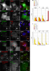
FIG. 1.
FIG. 1.
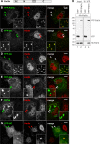
Intracellular localization of human Pat1a and Pat1b in COS7 cells. COS7 cells were transiently transfected with YFP (A), YFP-Pat1a (B), or YFP-Pat1b (C to E) and processed for immunofluorescence microscopy. P bodies in the cytoplasm were counterstained in red using an antibody against Hedls (A to C), Rck (D), or Lsm1 (E). In addition, cells were stained with Hoechst 33342 in blue to visualize nuclei. Images in panels A to E were acquired by epifluorescence microscopy. Merged images are shown on the right, and the size bar represents 10 μm. Arrowheads point toward P bodies. (F and G) Untransfected COS7 cells were stained in red for endogenous Pat1b with antibody 387 and in green for P bodies using Hedls (F) or Lsm1 (G) as a marker. Images in panels F and G were acquired by spinning-disc confocal microscopy; maximum projections of z-stacks are depicted. (H) COS7 cells transiently transfected with either YFP, YFP-Pat1a, or YFP-Pat1b were fixed and stained for P bodies using an antibody against Hedls. The number of P bodies (PBs) in transfected cells was counted, and the average distribution is represented in the graph. Error bars show the standard deviations (SD) based on three independent repeats. Untransfected COS7 cells serve as an additional control. (I) The expression of Pat1b was knocked down in U2OS cells by transfection of two different siRNAs (T2 and T3) at a concentration of 100 nM. An unspecific siRNA (D0) was transfected as a negative control. The number of PBs per cell was counted after staining for Helds, and the average distribution is represented in the graph. Error bars show the SD based on three independent repeats.
FIG. 2.
FIG. 2.
Localization of Pat1b subdomains. (A) Schematic representation of Pat1b domains. A, acidic domain; N, amino-terminal region; H, homology domain; C, carboxy-terminal region. COS7 cells were transiently transfected with YFP-Pat1b (B), YFP-dH (C), YFP-dA (D), YFP-AN (E), YFP-N (F), or YFP-HC (G) and processed for immunofluorescence microscopy. P bodies in the cytoplasm were counterstained in red using an antibody against Hedls. Images were acquired by spinning-disc confocal microscopy; maximum projections of z-stacks are depicted. Merged images are shown on the right; arrowheads point toward P bodies. Size bar, 10 μm. Localization of Pat1b subdomains is also summarized in Table S2 in the supplemental material. (H) HEK293 cells were transiently transfected with HA-Pat1b together with either YFP or YFP-Pat1b. Cytoplasmic lysates (input) were prepared for IP with GFP-binder, and Western blot analysis was carried out to visualize interaction between HA-Pat1b and YFP-Pat1b. The sizes of the molecular weight markers (in thousands) are indicated on the left.
FIG. 3.
FIG. 3.
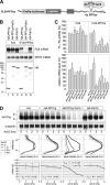
Tethering of Pat1b accelerates mRNA decay. (A) Schematic representation of the reporter containing firefly luciferase fused to β-globin (FLB). Six copies of the PP7 binding site (bs) were inserted in the 3′ UTR for the FLB-PP7bs reporter. An HA-tagged PP7 coat protein (cp) fused to Pat1b specifically binds to the PP7bs RNA. (B) HeLa cells were transiently transfected with FLB or FLB-PP7bs reporter together with either HA-PP7cp, HA-PP7cp-Pat1b, or HA-Pat1b. RNA and protein were extracted and analyzed by Northern (top two panels) and Western (bottom) blotting. The sizes of the molecular weight markers (in thousands) are indicated on the left. Ribosomal protein S7 (RPS7) mRNA serves as RNA loading control. (C) HeLa cells were transiently transfected with FLB or FLB-PP7bs reporter together with a Renilla luciferase (RL) reporter and either HA-PP7cp, HA-PP7cp-Pat1a, HA-PP7cp-Pat1b, or HA-PP7cp fused to fragments of Pat1b. Cytoplasmic lysates were prepared after 24 h to measure FL/RL activity. FLB mRNA levels were determined from the same lysates by Northern blot analysis, normalized to RPS7 mRNA, and quantified. Average values ± SD from three repeat experiments were plotted in the graph. (D) HeLa cells were transiently transfected with a T7-tagged β-globin reporter containing 6 copies of PP7bs (7B-PP7bs) together with either the HA tag alone, HA-PP7cp, HA-PP7cp-Pat1b, or HA-Pat1b. Total RNA was extracted at 1-h intervals after blocking transcription with actinomycin D (5 μg/ml). The reporter mRNA was detected by Northern blot analysis; nucleolin mRNA serves as loading control. In the middle panels, deadenylation was visualized by quantifying the signal intensity (in arbitrary units [A.U.]) of 7B-PP7bs mRNA along the length of the signal and plotting it as a function of mRNA size. In the bottom panel, the overall signal intensity of 7B-PP7bs mRNA was quantified and normalized to nucleolin mRNA. Average values ± standard errors (SE) were plotted as a percentage of the initial time point.
FIG. 4.
FIG. 4.
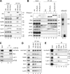
Pat1b interacts with P-body proteins. (A) HEK293 cells were transiently transfected with vector alone, HA-tagged Pat1b, or HA-tagged Pat1a. After 1 day, cytoplasmic lysates (input) were prepared for IP with anti-HA antibody. The HA-tagged proteins as well as endogenous Rck, Hedls, Xrn1, Lsm1, Lsm4, and eIF3B were detected by Western blotting. The sizes of the molecular weight markers (in thousands) are indicated on the right. (B) HEK293 cells transiently transfected with vector alone, HA-Pat1b, or HA-Pat1b-YFP were used for IP as described in the legend for panel A. Where indicated, RNase A was added to the lysates during IP. Lanes 10 and 11 show RNA extracted from the unbound fraction and stained with ethidium bromide. (C) HA-Pat1b was immunoprecipitated with HA antibody or without antibody as a control for unspecific precipitation. (D) HA-Pat1b was immunoprecipitated and subjected to increasing NaCl concentrations prior to elution of the protein complexes. *, immunoglobulin heavy chain. (E) Endogenous Xrn1, Hedls, Rck, Lsm1, and 14-3-3 were immunoprecipitated from the cytoplasmic lysate of HEK293 cells transiently transfected with HA-Pat1b. The Western blot for Rck is not shown due to an overlapping signal from the immunoglobulin heavy chain.
FIG. 5.
FIG. 5.
Separate interaction domains within Pat1b. (A) Schematic representation of Pat1b fragments. A, acidic domain; N, amino-terminal region; H, homology domain; C, carboxy-terminal region. (B) HEK293 cells were transiently transfected with vector alone, full-length HA-Pat1b, HA-AN, or HA-HC. After 1 day, cytoplasmic lysates (input) were prepared for IP with anti-HA antibody. The HA-tagged proteins as well as endogenous Rck, Lsm1, and eIF3B were detected by Western blotting. *, immunoglobulin heavy chain. The sizes of the molecular weight markers (in thousands) are indicated on the right. (C) HEK293 cells transiently transfected with vector alone, HA-Pat1b, HA-dA, HA-AN, HA-N, or HA-A were used for IP as described in the legend for panel B. The HA-tagged proteins and endogenous Rck were detected by Western blotting. (D) HEK293 cells were transiently transfected with vector alone, HA-Pat1b, HA-dH, HA-HC, HA-C, or HA-ANH and used for IP as described in the legend for panel B. The HA-tagged proteins and endogenous Lsm1 were detected by Western blotting.
FIG. 6.
FIG. 6.
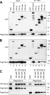
Pat1b interacts with Dcp2 and Dcp1a. (A) HEK293 cells were transiently transfected with YFP, YFP-Pat1b, YFP-A, YFP-N, YFP-AN, or YFP-HC together with Flag-Dcp2. GFP-binder was used for IP, and Western blot analysis was carried out with anti-GFP and anti-Flag antibody. The sizes of the molecular weight markers (in thousands) are indicated on the right. (B) Same analysis as described in the legend for panel A, except that Flag-Dcp1a was cotransfected. (C) HEK293 cells were transiently transfected with YFP-Pat1b together with either Flag-Dcp1a or Flag-Dcp2. IPs were carried out with GFP-binder and subjected to increasing NaCl concentrations prior to elution.
FIG. 7.
FIG. 7.
Pat1b interacts with the Ccr4-Caf1-Not complex. (A) HEK293 cells were transiently transfected with YFP or YFP-Pat1b together with Flag-Not1. GFP-binder was used for IP, and Western blot analysis was carried out with anti-GFP and anti-Flag antibody. The sizes of the molecular weight markers (in thousands) are indicated on the right. Where indicated, RNase A was added during IP. (B) IP was carried out as described in the legend for panel A using YFP or YFP-Pat1b together with myc-Ccr4; anti-myc was used for Western blot analysis. (C) IP was carried out as described in the legend for panel A using YFP or YFP-Pat1b together with HA-Caf1a or HA-Caf1b; anti-HA was used for Western blot analysis. On the right side, RNA was extracted from unbound fractions and stained with ethidium bromide. (D) HEK293 cells were transiently transfected with YFP, YFP-Pat1b, YFP-A, YFP-N, YFP-AN, or YFP-HC and processed for IP with GFP-binder. The YFP-tagged proteins and endogenous Caf1a were detected by Western blotting. (E) HEK293 cells were transiently cotransfected with YFP-Pat1b together with either Flag-Not1, myc-Ccr4, HA-Caf1a, or HA-Caf1b. IPs were carried out with GFP-binder and subjected to increasing NaCl concentrations prior to elution. (F) HEK293 cells were transiently transfected with Flag-Dcp2 together with either GFP alone, GFP-Caf1a alone, or GFP-Caf1a together with HA-Pat1b. Cytoplasmic lysates were processed for IP with GFP-binder and then subjected to Western blot analysis. In the IP samples, the Flag antibody cross-reacted with GFP-Caf1a.
FIG. 8.
FIG. 8.
Tethering of Pat1b causes mRNA deadenylation and decapping. HeLa cells were transiently transfected with the T7-tagged β-globin reporter containing 6 copies of the PP7bs (7B-PP7bs) together with HA-PP7cp-Pat1b and vector alone (A), Caf1a-wt (B), dominant negative Caf1a-AA (D40A/E42A) (C), Dcp2-wt (D), dominant negative Dcp2-AA (E147A/E148A) (E), Caf1a-wt plus Dcp2-AA (F), Caf1a-AA plus Dcp2-wt (G), and Caf1a-AA plus Dcp2-AA (H). Total RNA was extracted at 1-h intervals after blocking transcription with actinomycin D (5 μg/ml). The reporter mRNA was detected by Northern blot analysis; nucleolin mRNA served as a loading control. RNA samples marked dT were treated with oligo(dT) and RNase H and served as a size marker for deadenylated (A) reporter mRNA. In the middle panels, deadenylation was visualized by quantifying the signal intensity of 7B-PP7bs mRNA along the length of the signal and plotting it as a function of mRNA size. In the bottom panels, the overall signal intensity of 7B-PP7bs mRNA was quantified and normalized to nucleolin mRNA. Average values ± SE were plotted as a percentage of the initial time point. An asterisk indicates a significant difference in the mRNA half-life (t1/2) (P < 0.05 by two-tailed Student's t test) compared to the vector control depicted in panel A. Statistical analysis of the mRNA t1/2 is summarized in Table S3 in the supplemental material.
FIG. 9.
FIG. 9.
Tethering of Pat1b fragments. HeLa cells were transiently transfected with the 7B-PP7bs reporter and HA-tagged PP7cp (A), PP7cp-Pat1b (B), PP7cp-AN (C), PP7cp-HC (D), PP7cp-A (E), PP7cp-N (F), PP7cp-dH (G), or PP7cp-dA (H). Degradation of the reporter mRNA was analyzed and quantified as described in the legend to Fig. 8. Average values ± SE were plotted as a percentage of the initial time point. An asterisk indicates a significant difference in the mRNA t1/2 (P < 0.05 by two-tailed Student's t test) compared to tethering of PP7cp-Pat1b depicted in panel B. Statistical analysis of mRNA t1/2 is summarized in Table S4 in the supplemental material.
FIG. 10.
FIG. 10.
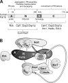
Role of Pat1b in P-body assembly and mRNA degradation. (A) Schematic representation human Pat1b subdomains, together with interacting proteins and associated functions. All the interactions depicted are RNA independent yet may be direct or indirect. (B) Hypothetical model of the Pat1b-associated complex that connects deadenylation with mRNA decapping. The acidic region (A) associates with Rck, the AN fragment with the Ccr4-Not complex, and the N fragment with Caf1 and Dcp2-Dcp1a. The homology region (H) is required for Lsm1 binding, and the HC fragment further associates with Dcp2-Dcp1a. The HC fragment also shows weak association with Edc3, Hedls, and Xrn1.

Similar articles

Cited by

References

    1. Andrei, M. A., D. Ingelfinger, R. Heintzmann, T. Achsel, R. Rivera-Pomar, and R. Luhrmann. 2005. A role for eIF4E and eIF4E-transporter in targeting mRNPs to mammalian processing bodies. RNA 11:717-727. - PMC - PubMed
    1. Bashkirov, V. I., H. Scherthan, J. A. Solinger, J. M. Buerstedde, and W. D. Heyer. 1997. A mouse cytoplasmic exoribonuclease (mXRN1p) with preference for G4 tetraplex substrates. J. Cell Biol. 136:761-773. - PMC - PubMed
    1. Bhattacharyya, S. N., R. Habermacher, U. Martine, E. I. Closs, and W. Filipowicz. 2006. Relief of microRNA-mediated translational repression in human cells subjected to stress. Cell 125:1111-1124. - PubMed
    1. Boag, P. R., A. Atalay, S. Robida, V. Reinke, and T. K. Blackwell. 2008. Protection of specific maternal messenger RNAs by the P body protein CGH-1 (Dhh1/RCK) during Caenorhabditis elegans oogenesis. J. Cell Biol. 182:543-557. - PMC - PubMed
    1. Bonnerot, C., R. Boeck, and B. Lapeyre. 2000. The two proteins Pat1p (Mrt1p) and Spb8p interact in vivo, are required for mRNA decay, and are functionally linked to Pab1p. Mol. Cell. Biol. 20:5939-5946. - PMC - PubMed

Publication types

Substances