. 2010 Sep;84(17):8888-902.
doi: 10.1128/JVI.00687-10. Epub 2010 Jun 23.

Mutagenesis of adeno-associated virus type 2 capsid protein VP1 uncovers new roles for basic amino acids in trafficking and cell-specific transduction

Affiliations

Mutagenesis of adeno-associated virus type 2 capsid protein VP1 uncovers new roles for basic amino acids in trafficking and cell-specific transduction

Jarrod S Johnson et al. J Virol. 2010 Sep.

Abstract

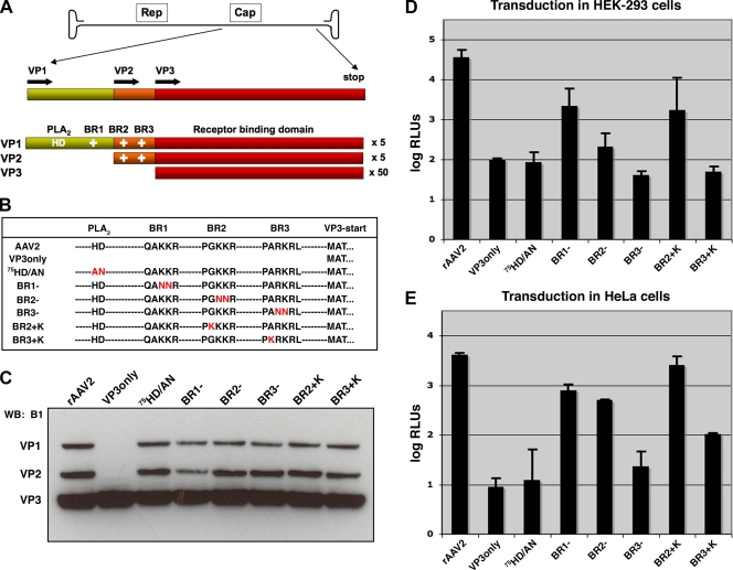
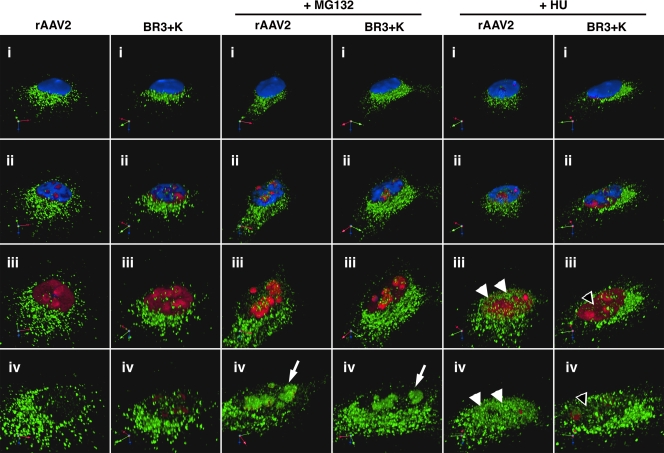
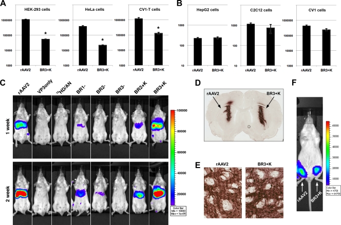
The N termini of the capsid proteins VP1 and VP2 of adeno-associated virus (AAV) play important roles in subcellular steps of infection and contain motifs that are highly homologous to a phospholipase A(2) (PLA(2)) domain and nuclear localization signals (NLSs). To more clearly understand how virion components influence infection, we have generated mutations in these regions and examined their effects on subcellular trafficking, capsid stability, transduction, and sensitivity to pharmacological enhancement. All mutants tested assembled into capsids; retained the correct ratio of VP1, VP2, and VP3; packaged DNA similarly to recombinant AAV2 (rAAV2); and displayed similar stability profiles when heat denatured. Confocal microscopy demonstrated that these mutants trafficked through a perinuclear region in the vicinity of the Golgi apparatus, with a subset of mutants displaying more-diffuse localization consistent with an NLS-deficient phenotype. When tested for viral transduction, two mutant classes emerged. Class I (BR1(-), BR2(-), and BR2+K) displayed partial transduction, whereas class II (VP3 only, (75)HD/AN, BR3(-), and BR3+K) were severely defective. Surprisingly, one class II mutant (BR3+K) trafficked identically to rAAV2 and accumulated in the nucleolus, a step recently described by our laboratory that occurs with wild-type infection. The BR3+K mutant, containing an alanine-to-lysine substitution in the third basic region of VP1, was 10- to 100-fold-less infectious than rAAV2 in transformed cell lines (such as HEK-293, HeLa, and CV1-T cells), but in contrast, it was indistinguishable from rAAV2 in several nontransformed cell lines, as well as in tissues (liver, brain, and muscle) in vivo. Complementation studies with pharmacological adjuvants or adenovirus coinfection suggested that additional positive charges in NLS regions restrict mobilization in the nucleus and limit transduction in a transformed-cell-specific fashion. Remarkably, besides displaying cell-type-specific transduction, this is the first description of a capsid mutant indicating that nuclear entry is not sufficient for AAV-mediated transduction and suggests that additional steps (i.e., subnuclear mobilization or uncoating) limit successful AAV infection.


Figures

FIG. 1.
FIG. 1.
Mutations in VP1/2 N termini affect transduction of rAAV. (A) The AAV genome contains genes that code for Rep proteins involved in replication and Cap proteins that comprise the capsid. Specific Cap proteins harbor a phospholipase domain (PLA2) (HD) and basic regions (BR) (+) that are putative nuclear localization signals. (B) Capsid mutants used in this study are listed by sequence, and amino acid changes are shown in red letters. (C) Western blot (WB) analysis of purified virus (5 × 10⁹ vg) blotted with antibody B1, which detects VP1, VP2, and VP3. (D and E) Luciferase assay of transduction in HEK-293 cells (D) and HeLa cells (E). Cells were inoculated with rAAV2 or mutants that package TR-CMV-Luc (10,000 vg/cell), and luciferase activity was quantified 24 h later. Error bars represent standard deviations from three independent samples.
FIG. 2.
FIG. 2.
Subcellular localization of capsid mutants. Volume imaging software was used to process z-stacked confocal images of HeLa cells infected with capsid mutants (10,000 vg/cell) for 16 h. Capsids (green) are shown in relation to the Golgi marker giantin (red) and nuclei (blue). All mutants, along with rAAV2, display some level of colocalization with the Golgi apparatus, with the BR1 mutant being the most intense. The BR2 and BR3 capsid mutants appear to have diffuse localization throughout the cell in addition to localizing to the Golgi apparatus compared to the localization of rAAV2 and other mutants.
FIG. 3.
FIG. 3.
Exposure of VP1 N termini. (A) Antibodies that specifically detect either intact capsids (A20), VP1 N termini (A1/αVP1), or disassembled subunits (B1) permit analysis of conformational changes in vitro in response to limited heat treatment or in cells during infection (i.e., exposure of the phospholipase and NLS motifs). (B) Immunoblot showing exposure of VP1 N termini. rAAV2 virions or empty capsids were exposed to limited heat treatment after limited heat treatment at designated temperatures for 10 min, applied onto nitrocellulose membranes, incubated with the antibodies shown, and developed after processing for chemiluminescence. Empty capsids are more resistant to exposing VP1. (C) Immunoblot comparing VP1 exposure among capsid mutants (1 × 10⁹ vg/well). Capsids disassemble prior to 75°C, as indicated by the loss of A20 reactivity and gain of B1 reactivity. An intermediate conformational change exists after heating capsids to 60°C, where VP1 epitopes are detected but capsids remain intact. (D) Immunofluorescence microscopy demonstrating VP1 exposure during infection. Capsids (green) display VP1 exposure (red) in a perinuclear region. Empty capsids, which are deficient in VP1 exposure, and VP3only particles, which lack VP1, show no significant signal from αVP1 (amino acids [aa] 2 to 16). The two mutants that are least infectious, 75HD/AN and BR3, have not lost the ability to expose VP1 during infection, implicating dysfunction in later steps during infection as the cause of the lost transduction.
FIG. 4.
FIG. 4.
Effect of proteasome inhibition on nucleolar accumulation and transduction of capsid mutants. MG132 (2 μM) was administered to HeLa cells simultaneously with virions (10,000 vg/cell), and after 16 h, samples were prepped for immunofluorescence microscopy. rAAV2, BR2+K, and BR3+K capsids (green) are found to accumulate in the nucleus (white arrows), and VP3only, 75HD/AN, BR1, BR2, and BR3 capsids are restricted mostly to extranuclear regions. (B) Subcellular fractionation of nucleoli after infection. HeLa cells were infected with mutant virions (1,000 vg/cell) for 16 h and subjected to subcellular fractionation. Capsid proteins from rAAV2 and the BR2+K and BR3+K mutants were detected in nucleolar fractions by Western blotting. (C) Luciferase assay of transduction in the presence of proteasome inhibitor. HEK-293 cells were infected (10,000 vg/cell) for 24 h in the presence of MG132 (2 μM) and then scored for transduction efficiency. Error bars represent standard deviations from three independent samples.
FIG. 5.
FIG. 5.
Stability of BR3+K capsids compared to rAAV. (A) Purified virus preparations were subjected to uranyl-acetate negative staining and applied onto discharged copper grids for electron microscopy. The scale bar represents 50 nm. (B) Immunoblotting of antibody reactivity after limited heat treatment (10 min at designated temperatures) indicates that rAAV2 and BR3+K capsids have similar macromolecular stabilities. Capsids are no longer intact at 75°C (A20), VP1 N termini become exposed after 57°C, and disassembled subunits can be detected prominently for both capsids at 65°C. (C) After heat treatment, virions were treated with DNase and submitted to qPCR to compare genome protections. rAAV2 and the BR3+K mutant have similar patterns of DNase sensitivity after heat treatment. The line graph is representative of data from one of three experiments.
FIG. 6.
FIG. 6.
Fluorescent in situ hybridization depicting that DNA from the BR3+K mutant is delivered to the nucleus during infection. HeLa cells were infected with rAAV2 or the BR3+K mutant (10,000 vg/cell) for 16 h in the absence (left) or presence (right) of MG132. Cells were prepped for FISH using an RNA probe complementary to the antisense strand of the TR-Luc genome. No staining in control samples (i and iv) indicates a lack of nonspecific binding from the probe. Both rAAV2 (v) and BR3+K (vi) genomes (red) are found to accumulate in the nucleus, specifically in nucleolar regions during proteasome inhibition.
FIG. 7.
FIG. 7.
HU treatment does not mobilize BR3+K capsids to the nucleoplasm. To visualize whether BR3+K mutant capsids could be mobilized into the nucleoplasm in response to HU, we fixed and stained infected HeLa cells for indirect immunofluorescence of capsids (green), nucleolin (red), and nuclei (blue). Roughly 40 confocal sections that span 5 μm were stacked and rendered in three dimensions by using Volocity software (i). A movie was generated to explore capsid localization in detail by digitally gating first the blue channel (DAPI, representing the nucleus) and then the red channel (nucleolin, representing the nucleolus), along with increasing the zoom. Images were captured at successive stages in the movie to reveal the nuclear interior (ii and iii) and the interior of the nucleolus (iv). Both rAAV2 and the BR3+K mutant accumulate in the nucleolus in the presence of proteasome inhibitor (white arrows). Only rAAV2 is localized to nucleoplasmic sites following HU treatment (white arrowheads, rAAV2; empty arrowheads, BR3+K mutant).
FIG. 8.
FIG. 8.
The BR3+K mutant is significantly less efficient than rAAV2 in transformed cells but is indistinguishable from rAAV2 in liver, brain, and muscle tissue in vivo. (A) Transformed cell lines such as HEK-293, HeLa, or CV1-T cells were inoculated with rAAV2 or the BR3+K mutant (1,000 vg/cell) (TR-CBA-Luc transgene) and assayed for transduction efficiency after 24 h. rAAV2 transduction was at least 10-fold-more efficient than that of the BR3+K mutant in these cell lines. (B) The normal cell lines HepG2 (human liver cells), C2C12 (mouse myoblasts), and CV1 (green monkey kidney cells) were assayed for transduction efficiency of rAAV2 or the BR3+K mutant under the same conditions as those described above (A). The transduction efficiencies of rAAV2 and the BR3+K mutant in nontransformed cells were similar (within 2-fold) (*, P value of ≤0.01 for A and B). Note that HepG2 cells are derived from an adenocarcinoma but contain no viral antigens and display many characteristics of normal hepatocytes. (C) rAAV2 or mutant virions (1010 vg) were administered by tail vein injection to female BALB/c mice, and bioluminescent luciferase activity was visualized 1 and 2 weeks later. The color scale bar represents photons/s/cm2, and data shown are representative images of a single mouse from three to four mice per vector group. (D) rAAV2 or BR3+K packaging GFP transgenes (3.8 × 10⁸ vg) was infused into rat striatum by intracerebral injection. Two weeks postinjection, brains were fixed and coronal sections were processed to detect GFP using DAB as a substrate and nickel-cobalt intensification. (E) Higher magnification of images from coronal sections similar to those in D depicting the transduction of neurons and associated processes (dark staining). (F) Bioluminescence images of transduction following intramuscular injections of rAAV2 (right hind limb) and BR3+K (left hind limb) packaging Luc transgenes acquired 2 weeks postinjection.
FIG. 9.
FIG. 9.
Model to illustrate the transformed-cell-type-specific defect of the BR3+K mutant. (A) The entry pathway of rAAV2 proceeds from binding glycoprotein receptors at the cell surface to endocytosis, trafficking to a perinuclear compartment, escape into the cytoplasm, nuclear entry, and mobilization to an unknown region of the nucleoplasm. (B) The trafficking of the BR3+K mutant is indistinguishable from that of rAAV2 except that in transformed cells, it does not mobilize to the nucleoplasm as efficiently as rAAV2 (Fig. 7, and see Movie S1 in the supplemental material). Cellular transformation modifies several nuclear processes that could influence the trafficking and transduction of the BR3+K mutant (see Discussion). These changes likely affect the processing of the BR3+K mutant after nuclear entry but prior to gene expression (subnuclear localization or uncoating).

Similar articles

Cited by

References

    1. Ahuja, D., M. T. Saenz-Robles, and J. M. Pipas. 2005. SV40 large T antigen targets multiple cellular pathways to elicit cellular transformation. Oncogene 24:7729-7745. - PubMed
    1. Bantel-Schaal, U., B. Hub, and J. Kartenbeck. 2002. Endocytosis of adeno-associated virus type 5 leads to accumulation of virus particles in the Golgi compartment. J. Virol. 76:2340-2349. - PMC - PubMed
    1. Bartlett, J. S., R. Wilcher, and R. J. Samulski. 2000. Infectious entry pathway of adeno-associated virus and adeno-associated virus vectors. J. Virol. 74:2777-2785. - PMC - PubMed
    1. Berns, K. I., and C. Giraud. 1996. Biology of adeno-associated virus. Curr. Top. Microbiol. Immunol. 218:1-23. - PubMed
    1. Bleker, S., F. Sonntag, and J. A. Kleinschmidt. 2005. Mutational analysis of narrow pores at the fivefold symmetry axes of adeno-associated virus type 2 capsids reveals a dual role in genome packaging and activation of phospholipase A2 activity. J. Virol. 79:2528-2540. - PMC - PubMed

Publication types

MeSH terms

LinkOut - more resources