. 2010 Aug 1;137(15):2527-37.
doi: 10.1242/dev.051011. Epub 2010 Jun 23.

Mib-Jag1-Notch signalling regulates patterning and structural roles of the notochord by controlling cell-fate decisions

Affiliations

Mib-Jag1-Notch signalling regulates patterning and structural roles of the notochord by controlling cell-fate decisions

Mai Yamamoto et al. Development. .

Abstract

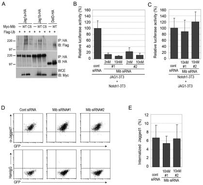
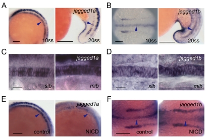
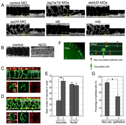
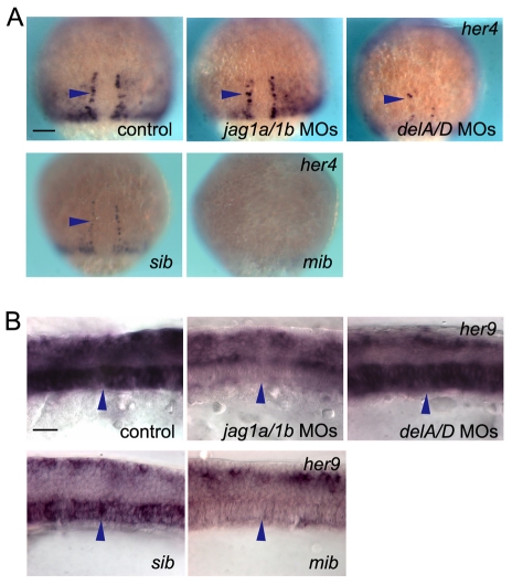
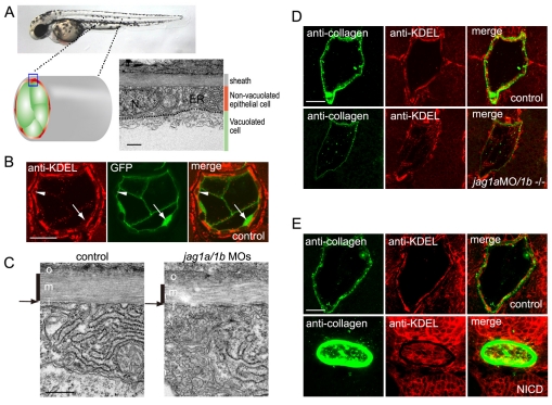
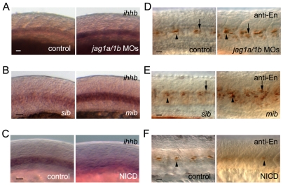
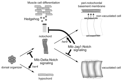
In the developing embryo, cell-cell signalling is necessary for tissue patterning and structural organization. During midline development, the notochord plays roles in the patterning of its surrounding tissues while forming the axial structure; however, how these patterning and structural roles are coordinated remains elusive. Here, we identify a mechanism by which Notch signalling regulates the patterning activities and structural integrity of the notochord. We found that Mind bomb (Mib) ubiquitylates Jagged 1 (Jag1) and is essential in the signal-emitting cells for Jag1 to activate Notch signalling. In zebrafish, loss- and gain-of-function analyses showed that Mib-Jag1-Notch signalling favours the development of non-vacuolated cells at the expense of vacuolated cells in the notochord. This leads to changes in the peri-notochordal basement membrane formation and patterning surrounding the muscle pioneer cells. These data reveal a previously unrecognized mechanism regulating the patterning and structural roles of the notochord by Mib-Jag1-Notch signalling-mediated cell-fate determination.


Figures

Fig. 1.
Fig. 1.
Mind bomb ubiquitylates Jagged 1 and is required for Jagged 1-induced Notch activation. (A) Jag1a and Jag1b were ubiquitylated by Mib. COS7 cells were transfected with HA-tagged Jag1a (Jag1a-HA), HA-tagged Jag1b (Jag1b-HA), HA-tagged DeltaD (DelD-HA), Myc-tagged wild-type Mib (WT), C1001S Mib (CS) and Flag-ubiquitin (Ub), as indicated. Cell extracts were then subjected to immunoprecipitation with an anti-HA antibody. The immunoprecipitated complexes were immunoblotted with anti-Flag (top) and anti-HA (top and middle) antibodies. Whole cell extracts (WCEs) were immunoblotted with an anti-Myc antibody (bottom). Molecular weight marker sizes in kDa are indicated on the left. (B) Mib was required for Jag1-induced Notch activation in Jag1-expressing cells. Jag1-3T3 cells were transfected with each Mib siRNA and then co-cultured with Notch1-3T3 cells. Notch activity was measured in Notch1-3T3 cells transfected with TP1, a Notch reporter plasmid. (C) Mib was not required for Jag1-induced Notch activation in Notch1-expressing cells. Notch1-3T3 cells were transfected with each Mib siRNA and the TP1 reporter plasmid, and then co-cultured with Jag1-3T3 cells. Error bars in B,C represent the mean ± s.d. of three independent experiments. (D) The expression of Jag1 on the cell surface was not altered by Mib knockdown. GFP-positive Jag1-3T3 cells were transfected with Mib siRNAs and, after 48 hours, the expression of Jag1 was examined by flow cytometry with an anti-Jag1 antibody. Hamster IgG was used as a control. (E) The Jag1 endocytosis rate was not dramatically altered by Mib knockdown. The surface proteins of Jag1-3T3 cells were labelled with Sulfo-NHS-SS-Biotin and incubated for 20 minutes at 37°C. The remaining cell-surface biotin was then stripped, and WCEs were prepared. Internalized biotinylated Jag1 and total Jag1 was measured by an ELISA. The values shown are mean ± s.d. (%).
Fig. 2.
Fig. 2.
The two zebrafish jagged 1 homologues are expressed in spatially and temporally restricted patterns in the developing notochord, and their expression is controlled by Notch signalling. (A,B) Expression of jag1a (A) and jag1b (B) in the notochord cells at 10 and 20, ss. (C,D) jag1a (C) and jag1b (D) expression was increased in the mib mutants at 10 ss. sib, sibling control. (E,F) jag1a (E) and jag1b (F) expression was decreased at 10 ss in the notochord of embryos with increased Notch activity. Control siblings (control) or double transgenic Tg(UAS:myc-Notch1a-intra);Tg(hsp70:Gal4) embryos (NICD) were heat shocked at 3 ss. Arrowheads show expression in the notochord. (A-E) Lateral view with anterior to the left, except for 10 ss in B. 10 ss in B and F is a dorsal view with anterior to the left. Scale bars: 100 μm in A,B,E,F; 20 μm in C,D.
Fig. 3.
Fig. 3.
Jagged 1a and Jagged 1b regulate cell fates during notochord development. (A) Defects in Mib-Jag1-Notch but not DeltaA/D-Notch signalling led to increased vacuoles in the notochord. Enlarged views of the yellow-boxed areas are shown in each right panel. (B) Notch activation reduced the vacuolated cells in the notochord. Control siblings (control) or double transgenic Tg(UAS:myc-Notch1a-intra);Tg(hsp70:Gal4) embryos (NICD) were heat shocked at 3 ss. A and B are side views of the BODIPY TR methyl ester-stained notochord cells in embryos at around 34 hpf. (C,D) Vacuolated cells were increased at the expense of non-vacuolated cells in jag1a/1b knockdown embryos. Nuclei were visualized by nuclear-localized mCherry (red, upper panels in C,D) or by Ntl antibody (red, lower panels in C,D). Vacuolated cells were revealed by using a transgenic fish line (214A-GFP), in which the vacuolated cells are labelled with GFP (green). Red arrowheads indicate non-vacuolated cell nuclei. Green arrows indicate vacuolated cell nuclei. (E) Quantification of the mean number of vacuoles and nuclei. The vacuoles and nuclei were counted within the trunk region of the notochord (230 μm in width, as in the upper images of C,D). Control MO (C, n=20), jag1a/1b MOs (J, n=22). (F) Vacuolated cells and non-vacuolated epithelial cells are derived from notochord precursor cells. The GFP-positive cells at 15 ss were followed until 32 hpf in an embryo injected with Flh-GFP plasmid. (G) Quantification of the proportion of non-vacuolated cells. GFP-positive vacuolated and GFP-negative non-vacuolated cells were counted in embryos co-injected with Flh-GFP plasmid and the control MO (C, n=6) or jag1a/1b MOs (J, n=8). Asterisks in F and G indicate statistically significant differences relative to the control (P<0.005). Error bars indicate s.e.m. Scale bars: 20 μm in A-D,F.
Fig. 4.
Fig. 4.
Jagged 1-Notch signalling regulates her9 but not her4 expression, independent of Delta-Notch signalling. (A) her4 expression was reduced in the hypochord precursor cells of deltaA/D (delA/D) knockdown embryos and mib mutants, but not in those of jag1a/1b (jag1a/1b) knockdown embryos. Arrowheads indicate the hypochord precursor cells. Dorsal views at 80% epibody are shown. (B) her9 expression was decreased in the notochord of jag1a/1b knockdown and mib mutant embryos, but not in that of deltaA/D knockdown embryos. Arrowheads indicate notochord cells. sib, sibling control. Side views at 10 ss are shown. Scale bars: 100 μm in A; 20 μm in B.
Fig. 5.
Fig. 5.
Jagged 1a/1b is required for notochord extracellular matrix sheath formation. (A) Non-vacuolated epithelial cells contained abundant rough ER compared with vacuolated cells. N, nuclei; ER, endoplasmic reticulum. Transmission electron micrographs (TEMs) of transverse sections of a wild-type embryo on day 2. (B) Non-vacuolated cells were rich in rough ER. ER was stained by an anti-KDEL antibody (red), and vacuolated cells were revealed by the 214A-GFP transgenic line (green). Arrowheads indicate anti-KDEL-positive non-vacuolated cells. Arrows indicate GFP-positive vacuolated cells. (C) The medial layer of the peri-notochordal basement membrane was thinner in the jag1a/1b knockdown embryos than in wild-type embryos. TEM of transverse sections. o, outer; m, medial; i, inner. Black vertical lines indicate the medial layer of the sheath. Arrows indicate the thin inner layer. (D) jag1a/1b knockdown resulted in reduced type II collagen deposition and fewer cells with abundant rough ER in the notochord. (E) Notch activation increased type II collagen deposition and ER-positive cells in the notochord. Control siblings (control) or double transgenic Tg(UAS:myc-Notch1a-intra);Tg(hsp70:Gal4) embryos (NICD) were heat shocked at 3 ss. A-E are transverse sections of day 2 embryos. Scale bars: 0.5 μm in A,C; 20 μm in B,D,E.
Fig. 6.
Fig. 6.
Mind bomb-Jagged-Notch signalling regulates muscle cell identities by altering ihhb expression. (A-C) ihhb expression was increased in jag1a/1b (jag1a/1b)-knockdown embryos and mib mutants, whereas Notch activation decreased ihhb expression at 18 ss. (D-F) Engrailed-positive (anti-En) MPs and MEFs were increased in jag1a/1b knockdown and mib mutant embryos, whereas Notch activation decreased the Engrailed-positive cells at 24 hpf. (B,E) sib, sibling control. (C,F) Control siblings (control) or double transgenic Tg(UAS:myc-Notch1a-intra);Tg(hsp70:Gal4) embryos (NICD) were heat-shocked at 3 ss. MPs and MFFs are indicated by arrowheads and arrows, respectively. Scale bars: 20 μm in A-F.
Fig. 7.
Fig. 7.
Model of how Mind bomb-Jagged 1-Notch signalling regulates the patterning and structural roles of the notochord by affecting cell-fate decisions. Mib-Delta-Notch signalling acts during the gastrula stage and determines notochord versus hypochord, possibly through her4 activation. Mib-Jag1-Notch signalling acts at later stages, and determines vacuolated versus non-vacuolated cell fate in the notochord, possibly through her9 activation. This cell-fate choice affects peri-notochordal basement membrane formation and muscle-patterning activity.

Similar articles

Cited by

References

    1. Appel B., Fritz A., Westerfield M., Grunwald D. J., Eisen J. S., Riley B. B. (1999). Delta-mediated specification of midline cell fates in zebrafish embryos. Curr. Biol. 9, 247-247 - PubMed
    1. Artavanis-Tsakonas S., Rand M. D., Lake R. J. (1999). Notch signaling: cell fate control and signal integration in development. Science 284, 770-770 - PubMed
    1. Asakawa K., Suster M. L., Mizusawa K., Nagayoshi S., Kotani T., Urasaki A., Kishimoto Y., Hibi M., Kawakami K. (2008). Genetic dissection of neural circuits by Tol2 transposon-mediated Gal4 gene and enhancer trapping in zebrafish. Proc. Natl. Acad. Sci. USA 105, 1255-1255 - PMC - PubMed
    1. Broder Y. C., Katz S., Aronheim A. (1998). The ras recruitment system, a novel approach to the study of protein-protein interactions. Curr. Biol. 8, 1121-1121 - PubMed
    1. Coutinho P., Parsons M. J., Thomas K. A., Hirst E. M., Saude L., Campos I., Williams P. H., Stemple D. L. (2004). Differential requirements for COPI transport during vertebrate early development. Dev. Cell 7, 547-547 - PubMed

Publication types

MeSH terms

LinkOut - more resources