

TRPV4 channels augment macrophage activation and ventilator-induced lung injury
- PMID: 20562229
- PMCID: PMC2951075
- DOI: 10.1152/ajplung.00315.2009
TRPV4 channels augment macrophage activation and ventilator-induced lung injury
Abstract
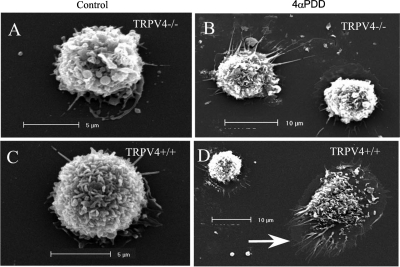
We have previously implicated transient receptor potential vanilloid 4 (TRPV4) channels and alveolar macrophages in initiating the permeability increase in response to high peak inflation pressure (PIP) ventilation. Alveolar macrophages were harvested from TRPV4(-/-) and TRPV4(+/+) mice and instilled in the lungs of mice of the opposite genotype. Filtration coefficients (K(f)) measured in isolated perfused lungs after ventilation with successive 30-min periods of 9, 25, and 35 cmH(2)O PIP did not significantly increase in lungs from TRPV4(-/-) mice but increased >2.2-fold in TRPV4(+/+) lungs, TRPV4(+/+) lungs instilled with TRPV4(-/-) macrophages, and TRPV4(-/-) lungs instilled with TRPV4(+/+) macrophages after ventilation with 35 cmH(2)O PIP. Activation of TRPV4 with 4-alpha-phorbol didecanoate (4alphaPDD) significantly increased intracellular calcium, superoxide, and nitric oxide production in TRPV4(+/+) macrophages but not TRPV4(-/-) macrophages. Cross-sectional areas increased nearly 3-fold in TRPV4(+/+) macrophages compared with TRPV4(-/-) macrophages after 4alphaPDD. Immunohistochemistry staining of lung tissue for nitrotyrosine revealed increased amounts in high PIP ventilated TRPV4(+/+) lungs compared with low PIP ventilated TRPV4(+/+) or high PIP ventilated TRPV4(-/-) lungs. Thus TRPV4(+/+) macrophages restored susceptibility of TRPV4(-/-) lungs to mechanical injury. A TRPV4 agonist increased intracellular calcium and reactive oxygen and nitrogen species in harvested TRPV4(+/+) macrophages but not TRPV4(-/-) macrophages. K(f) increases correlated with tissue nitrotyrosine, a marker of peroxynitrite production.
Figures


Similar articles
-
TRPV4 initiates the acute calcium-dependent permeability increase during ventilator-induced lung injury in isolated mouse lungs.Am J Physiol Lung Cell Mol Physiol. 2007 Oct;293(4):L923-32. doi: 10.1152/ajplung.00221.2007. Epub 2007 Jul 27. Am J Physiol Lung Cell Mol Physiol. 2007. PMID: 17660328
-
Prevention of ventilator-induced lung edema by inhalation of nanoparticles releasing ruthenium red.Am J Respir Cell Mol Biol. 2014 Jun;50(6):1107-17. doi: 10.1165/rcmb.2013-0163OC. Am J Respir Cell Mol Biol. 2014. PMID: 24405281 Free PMC article.
-
Phosphoinositide 3-kinase, Src, and Akt modulate acute ventilation-induced vascular permeability increases in mouse lungs.Am J Physiol Lung Cell Mol Physiol. 2007 Jul;293(1):L11-21. doi: 10.1152/ajplung.00279.2005. Epub 2007 Feb 23. Am J Physiol Lung Cell Mol Physiol. 2007. PMID: 17322282
-
Emerging roles of calcium-activated K channels and TRPV4 channels in lung oedema and pulmonary circulatory collapse.Acta Physiol (Oxf). 2017 Jan;219(1):176-187. doi: 10.1111/apha.12768. Epub 2016 Sep 16. Acta Physiol (Oxf). 2017. PMID: 27497091 Review.
-
Nitric oxide and peroxynitrite in ozone-induced lung injury.Adv Exp Med Biol. 2001;500:183-90. doi: 10.1007/978-1-4615-0667-6_24. Adv Exp Med Biol. 2001. PMID: 11764933 Review.
Cited by
-
Pharmacological inhibitors of TRPV4 channels reduce cytokine production, restore endothelial function and increase survival in septic mice.Sci Rep. 2016 Sep 22;6:33841. doi: 10.1038/srep33841. Sci Rep. 2016. PMID: 27653046 Free PMC article.
-
Emerging pharmacological therapies for ARDS: COVID-19 and beyond.Intensive Care Med. 2020 Dec;46(12):2265-2283. doi: 10.1007/s00134-020-06141-z. Epub 2020 Jul 11. Intensive Care Med. 2020. PMID: 32654006 Free PMC article.
-
Caveolar peroxynitrite formation impairs endothelial TRPV4 channels and elevates pulmonary arterial pressure in pulmonary hypertension.Proc Natl Acad Sci U S A. 2021 Apr 27;118(17):e2023130118. doi: 10.1073/pnas.2023130118. Proc Natl Acad Sci U S A. 2021. PMID: 33879616 Free PMC article.
-
Distribution and expression of non-neuronal transient receptor potential (TRPV) ion channels in rosacea.J Invest Dermatol. 2012 Apr;132(4):1253-62. doi: 10.1038/jid.2011.424. Epub 2011 Dec 22. J Invest Dermatol. 2012. PMID: 22189789 Free PMC article.
-
Urgent reconsideration of lung edema as a preventable outcome in COVID-19: inhibition of TRPV4 represents a promising and feasible approach.Am J Physiol Lung Cell Mol Physiol. 2020 Jun 1;318(6):L1239-L1243. doi: 10.1152/ajplung.00161.2020. Epub 2020 May 13. Am J Physiol Lung Cell Mol Physiol. 2020. PMID: 32401673 Free PMC article.
References
-
- Akei H, Whitsett JA, Buroker M, Ninomiya T, Tatsumi H, Weaver TE, Ikegami M. Surface tension influences cell shape and phagocytosis in alveolar macrophages. Am J Physiol Lung Cell Mol Physiol 291: L572–L579, 2006 - PubMed
-
- Broeke RT, Leusink-Muis T, Hilberdink R, Van Ark I, van den Worm E, Villain M, De Clerck F, Blalock JE, Nijkamp FP, Folkerts G. Specific modulation of calmodulin activity induces a dramatic production of superoxide by alveolar macrophages. Lab Invest 84: 29–40, 2003 - PubMed
-
- Brower RG, Matthay MA, Morris A, Schoenfeld D, Thompson BT. Ventilation with lower tidal volumes as compared with traditional tidal volumes for acute lung injury and respiratory distress syndrome. N Engl J Med 342: 1301–1308, 2000 - PubMed
-
- Dos Santos CC, Slutsky AS. Invited review: mechanisms of ventilator-induced lung injury: a perspective. J Appl Physiol 89: 1645–1655, 2000 - PubMed
Publication types
MeSH terms
Substances
Grants and funding
LinkOut - more resources
Full Text Sources
Molecular Biology Databases