High expression of macrophage colony-stimulating factor-1 receptor in peritumoral liver tissue is associated with poor outcome in hepatocellular carcinoma after curative resection
- PMID: 20551429
- PMCID: PMC3228006
- DOI: 10.1634/theoncologist.2009-0170
High expression of macrophage colony-stimulating factor-1 receptor in peritumoral liver tissue is associated with poor outcome in hepatocellular carcinoma after curative resection
Abstract
Background: Macrophage colony-stimulating factor 1 receptor (CSF-1R) expression in hepatocellular carcinoma (HCC) and its prognostic values are unclear. This study evaluated the prognostic values of the intratumoral and peritumoral expression of CSF-1R in HCC patients after curative resection.
Methods: Tissue microarrays containing material from cohort 1 (105 patients) and cohort 2 (32 patients) were constructed. Immunohistochemistry was performed and prognostic values of these and other clinicopathological data were evaluated. The CSF-1R mRNA level was assessed by quantitative real-time polymerase chain reaction in cohort 3 (52 patients).
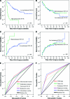
Results: Both the CSF-1R density and its mRNA level were significantly higher in peritumoral liver tissue than in the corresponding tumor tissue. CSF-1R was distributed in a gradient in the long-distance peritumoral tissue microarray, with its density decreasing as the distance from the tumor margin increased. High peritumoral CSF-1R was significantly associated with more intrahepatic metastases and poorer survival. Peritumoral CSF-1R was an independent prognostic factor for both overall survival and time to recurrence and affected the incidence of early recurrence. However, intratumoral CSF-1R did not correlate with any clinicopathological feature. Peritumoral CSF-1R was also associated with both overall survival and time to recurrence in a subgroup with small HCCs (< or =5 cm).
Conclusions: Peritumoral CSF-1R is associated with intrahepatic metastasis, tumor recurrence, and patient survival after hepatectomy, highlighting the critical role of the peritumoral liver milieu in HCC progression. CSF-1R may become a potential therapeutic target for postoperative adjuvant treatment.
Conflict of interest statement
The content of this article has been reviewed by independent peer reviewers to ensure that it is balanced, objective, and free from commercial bias. No financial relationships relevant to the content of this article have been disclosed by the authors or independent peer reviewers.
Figures


Similar articles
-
High expression of macrophage colony-stimulating factor in peritumoral liver tissue is associated with poor survival after curative resection of hepatocellular carcinoma.J Clin Oncol. 2008 Jun 1;26(16):2707-16. doi: 10.1200/JCO.2007.15.6521. J Clin Oncol. 2008. PMID: 18509183
-
Prognostic roles of cross-talk between peritumoral hepatocytes and stromal cells in hepatocellular carcinoma involving peritumoral VEGF-C, VEGFR-1 and VEGFR-3.PLoS One. 2013 May 30;8(5):e64598. doi: 10.1371/journal.pone.0064598. Print 2013. PLoS One. 2013. PMID: 23737988 Free PMC article.
-
Expression and prognostic significance of placental growth factor in hepatocellular carcinoma and peritumoral liver tissue.Int J Cancer. 2011 Apr 1;128(7):1559-69. doi: 10.1002/ijc.25492. Epub 2010 Jun 2. Int J Cancer. 2011. PMID: 20521248
-
High-mobility group protein box1 expression correlates with peritumoral macrophage infiltration and unfavorable prognosis in patients with hepatocellular carcinoma and cirrhosis.BMC Cancer. 2016 Nov 11;16(1):880. doi: 10.1186/s12885-016-2883-z. BMC Cancer. 2016. PMID: 27836008 Free PMC article.
-
Colony Stimulating Factor-1 and its Receptor in Gastrointestinal Malignant Tumors.J Cancer. 2021 Oct 17;12(23):7111-7119. doi: 10.7150/jca.60379. eCollection 2021. J Cancer. 2021. PMID: 34729112 Free PMC article. Review.
Cited by
-
microRNA-26a suppresses recruitment of macrophages by down-regulating macrophage colony-stimulating factor expression through the PI3K/Akt pathway in hepatocellular carcinoma.J Hematol Oncol. 2015 May 29;8:56. doi: 10.1186/s13045-015-0150-4. J Hematol Oncol. 2015. PMID: 26021873 Free PMC article.
-
The Histologic Cut-off Point for Adjacent and Remote Non-neoplastic Liver Parenchyma of Hepatocellular Carcinoma in Chronic Hepatitis B Patients.Korean J Pathol. 2012 Aug;46(4):349-58. doi: 10.4132/KoreanJPathol.2012.46.4.349. Epub 2012 Aug 23. Korean J Pathol. 2012. PMID: 23110027 Free PMC article.
-
Mechanisms driving macrophage diversity and specialization in distinct tumor microenvironments and parallelisms with other tissues.Front Immunol. 2014 Mar 26;5:127. doi: 10.3389/fimmu.2014.00127. eCollection 2014. Front Immunol. 2014. PMID: 24723924 Free PMC article. Review.
-
The combination of HTATIP2 expression and microvessel density predicts converse survival of hepatocellular carcinoma with or without sorafenib.Oncotarget. 2014 Jun 15;5(11):3895-906. doi: 10.18632/oncotarget.2019. Oncotarget. 2014. PMID: 25008315 Free PMC article.
-
MicroRNA-26a inhibits angiogenesis by down-regulating VEGFA through the PIK3C2α/Akt/HIF-1α pathway in hepatocellular carcinoma.PLoS One. 2013 Oct 23;8(10):e77957. doi: 10.1371/journal.pone.0077957. eCollection 2013. PLoS One. 2013. PMID: 24194905 Free PMC article.
References
-
- Parkin DM, Bray F, Ferlay J, et al. Global cancer statistics, 2002. CA Cancer J Clin. 2005;55:74–108. - PubMed
-
- Llovet JM, Burroughs A, Bruix J. Hepatocellular carcinoma. Lancet. 2003;362:1907–1917. - PubMed
-
- Tang ZY, Ye SL, Liu YK, et al. A decade's studies on metastasis of hepatocellular carcinoma. J Cancer Res Clin Oncol. 2004;130:187–196. - PubMed
-
- Ding Y, Chen B, Wang S, et al. Overexpression of Tiam1 in hepatocellular carcinomas predicts poor prognosis of HCC patients. Int J Cancer. 2009;124:653–658. - PubMed
Publication types
MeSH terms
Substances
LinkOut - more resources
Full Text Sources
Other Literature Sources
Medical
Research Materials
Miscellaneous

