. 2010 Jun 10;6(6):e1000951.
doi: 10.1371/journal.ppat.1000951.

Epigenetic repression of p16(INK4A) by latent Epstein-Barr virus requires the interaction of EBNA3A and EBNA3C with CtBP

Affiliations

Epigenetic repression of p16(INK4A) by latent Epstein-Barr virus requires the interaction of EBNA3A and EBNA3C with CtBP

Lenka Skalska et al. PLoS Pathog. .

Abstract

As an inhibitor of cyclin-dependent kinases, p16(INK4A) is an important tumour suppressor and inducer of cellular senescence that is often inactivated during the development of cancer by promoter DNA methylation. Using newly established lymphoblastoid cell lines (LCLs) expressing a conditional EBNA3C from recombinant EBV, we demonstrate that EBNA3C inactivation initiates chromatin remodelling that resets the epigenetic status of p16(INK4A) to permit transcriptional activation: the polycomb-associated repressive H3K27me3 histone modification is substantially reduced, while the activation-related mark H3K4me3 is modestly increased. Activation of EBNA3C reverses the distribution of these epigenetic marks, represses p16(INK4A) transcription and allows proliferation. LCLs lacking EBNA3A express relatively high levels of p16(INK4A) and have a similar pattern of histone modifications on p16(INK4A) as produced by the inactivation of EBNA3C. Since binding to the co-repressor of transcription CtBP has been linked to the oncogenic activity of EBNA3A and EBNA3C, we established LCLs with recombinant viruses encoding EBNA3A- and/or EBNA3C-mutants that no longer bind CtBP. These novel LCLs have revealed that the chromatin remodelling and epigenetic repression of p16(INK4A) requires the interaction of both EBNA3A and EBNA3C with CtBP. The repression of p16(INK4A) by latent EBV will not only overcome senescence in infected B cells, but may also pave the way for p16(INK4A) DNA methylation during B cell lymphomagenesis.


Conflict of interest statement

The authors have declared that no competing interests exist.

Figures

Figure 1
Figure 1. EBNA3C inactivation and reactivation in LCL 3CHT.
Cells that had been grown in culture medium containing HT were washed and re-suspended in medium with HT omitted. After culturing for the number of days indicated, protein extracts were western blotted and probed with an anti-EBNA3C MAb to reveal EBNA3C-HT. After 17 days some cells were transferred to medium containing HT and after 1, 5 or 8 days samples were again taken for western blotting. The western blot was re-probed with anti-γ-tubulin to ensure equal loading of the proteins.
Figure 2
Figure 2. Proliferation of LCL 3CHT after inactivation and reactivation of EBNA3C.
(A) LCL 3CHT cells were cultured either with HT (+HT) or for 33 days after the removal of HT (-HT) from the growth medium. Cells were pulsed for 1 hour with BrdU, harvested, fixed and stained with anti-BrdU-FITC and propidium iodide. The cells were analysed by flow cytometry. The gated BrdU-positive population is reduced in the absence of HT. (B) Two LCL 3CHT (-A and -C) generated from independent 3CHT-BACs using PBL from a single donor were analysed as in (A) after 14 and 33 days without HT. Histograms show BrdU incorporation relative to the control, cycling population. The control (ctrl) was a proliferating population of LCL 3CHT grown in medium with HT. (C) LCLs 3CHT-A and -C were cultured for 14 days in the absence of HT (0) and BrdU-incorporation was assayed as in (A). The incorporation of BrdU was also assayed as above after 4, 7 and 12 days after re-adding HT. (D) A similar experiment was performed on cells cultured without HT for 33 days.
Figure 3
Figure 3. Repression of p16INK4A following reactivation of EBNA3C.
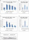
(A) After 14 days without HT (0), total RNA was extracted from aliquots from two LCL 3CHT populations (-A and -C). HT was re-added to the remaining cells and further RNA samples were taken at the times indicated. Real time quantitative RT-PCR (qRT-PCR) was performed to quantify CDKN2A transcripts. The histogram corresponds to CDKN2A mRNA relative to that in control cycling populations of each LCL 3CHT. (B) As in (A) but using a p16INK4A-specific qPCR. (C) & (D) Similar assays to those described in (A) and (B) after 33 days without HT. (E) Western blots – probed with a p16INK4A-specific MAb – of protein extracts from LCL 3CHT-C cells to which HT was re-added after 14 or 33 days in its absence (014 and 033 respectively). The control (ctrl) is LCL 3CHT-C cells continuously cultured in medium with HT.
Figure 4
Figure 4. Inactivation of EBNA3C and accumulation of p16INK4A leads to de-phosphorylation of Rb, reduced expression of p107 and an increase in p130; activation of EBNA3C reverses these processes.
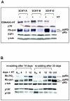
(A) Western blot analyses of three LCL 3CHT lines performed on whole cell lysates from cells cultured with HT (+) without HT for 26 days (−). As the amount of EBNA3C protein reduces (and is inactivated) and p16INK4A accumulates, so hyperphosphorylated Rb (ppRb) protein disappears and only hypophosphorylated Rb (pRb) is detected. The levels of E2F1 and γ-tubulin (γ-tub) remain unchanged irrespective of the culture conditions. (B) Western blot analysis of extracts from cells after re-addition of HT to cultures starved of HT for either 14 or 33 days (014 and 033 respectively). A pan-specific anti-Rb MAb and phospho-specific MAb (Ser 807–811) both show an increase in hyperphosphorylated Rb (ppRb) after HT was added. After 12–16 days the degree of phosphorylation is equivalent to that in the proliferating control LCL 3CHT population (ctrl). As Rb becomes phosphorylated, expression of p107 increases and expression of p130 decreases. The level of γ-tubulin (γ-tub) did not alter throughout the experiments.
Figure 5
Figure 5. ChIP analysis to quantify the H3K27me3 and H3K4me3 marks on p16INK4A exon 1 when EBNA3C is inactivated and re-activated in LCL 3CHT.
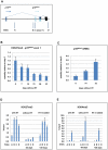
(A) Schematic of the human p16INK4A-ARF locus showing the location of coding exons (boxes) and transcription start sites (horizontal arrows) – not drawn to scale. The vertical (A-D) arrows refer to the approximate locations of primer pairs used for qPCR analysis of precipitated chromatin (as described in Materials and Methods). (B) ChIP analysis of H3K27me3 distribution on exon 1 (site C) of p16INK4A. The histogram shows a decline in H3K27me3 relative to a cycling LCL 3CHT (day 0). (C) Corresponding changes in p16INK4A mRNA quantified by qRT-PCR. (D) ChIP analysis of H3K27me3 distribution across the p16INK4A-ARF locus in LCL 3CHT proliferating in the presence of HT, after 30 days without HT and 20 days after re-adding HT. (E) ChIP analysis of H3K4me3 the p16INK4A-ARF locus in LCL 3CHT proliferating in the presence of HT, after 30 days without HT and 20 days after re-adding HT.
Figure 6
Figure 6. EBNA3C-mediated regulation of p16INK4A does not require Rb.
(A) Western blot analysis of whole cell lysates from two LCL 3CHT (-D and -E) cultured with (+) or without (-) HT for 26 days. Although Rb is undetectable in LCL 3CHT-E, when HT is removed from the growth medium, EBNA3C decreases and p16INK4A (p16) increases. E2F1 and γ-tubulin (γ-tub) levels do not alter. (B) Steady-state levels of Rb mRNA in LCL 3CHT-E relative to 3 other LCL 3CHT and a WT-BAC LCL all cultured with HT, quantified by qRT-PCR. (C) ChIP analysis of H3K27Me3 distribution across the p16INK4A-ARF locus in LCL 3CHT-E cells (expressing little or no Rb) with HT or without HT in the growth medium. (D) ChIP analysis of H3K4me3 distribution across the p16INK4-ARF locus essentially as described in (C).
Figure 7
Figure 7. EBNA3A contributes to the regulation of p16INK4A.
(A) Western blot analysis of three LCLs established with virus derived from B95.8-EBV BAC (WT) and two established with EBNA3A-KO recombinants. The steady state levels of p16INK4A (p16) are elevated in the EBNA3A-KO cells relative to the WT-EBV infected cells. It should be noted that similar results were reported using a larger panel of EBNA3A-KO LCLs . An LCL 3CHT (3CHT) with (+) or without (-) HT is shown for comparison. (B) & (C) ChIP analysis of H3K27me3 and H3K4me3 distribution on exon 1 (site C) and site A in the p16INK4A-ARF locus from an EBNA3A-KO LCL (3AKO) and a WT-EBV LCL (WT).
Figure 8
Figure 8. LCLs expressing CtBP-binding mutants of EBNA3A and EBNA3C grow relatively poorly.
(A) Schematic showing mutations of CtBP-binding sites in EBNA3A and EBNA3C that were introduced into the CtBPM recombinant viruses. EBNA3C includes a single consensus PLDLS site that was mutated to ALDAS . EBNA3A includes two non-canonical CtBP-binding sites that synergise to produce very efficient binding to CtBP; the ALDLS site was mutated to ALDAA and the VLDLS site was mutated to VLDAA . (B) Schematic showing the generation of a series of CtBP-binding mutant EBVs, by serially mutating the EBNA3 CtBP binding sites, and then reverting them, creating both single and double CtBP mutant EBVs, and a revertant. (C) A plate of cells 40 days after infection of PBLs with 1000 rgu of virus per well. Difference in the colour of the medium and the visible density/size of cell clumps in the wells are clear indicators that CtBP-mutant LCLs grow out more slowly than wild-type (not shown) and revertant LCLs. This appears to be more striking in 3CCtBP and E3CtBP LCLs. (D) Established LCLs (approximately 3 months post-infection) were seeded at 5×10⁴ cells/ml and counted each day for a week. CtBP-mutants (dashed lines) grow more slowly, and/or have a lower maximum cell density than wild-type and revertant LCLs (solid lines). Two cell lines were tested for each mutant.
Figure 9
Figure 9. Expression of p16INK4A is increased in CtBPM LCLs relative to revertant and WT LCLs.
(A) E3CtBP, 3ACtBP, 3CCtBP and two revCtBP LCLs were harvested 28 days after the infection of primary B cells with the recombinant EBVs. RNA was extracted and the relative levels of CDKN2A transcripts were quantified by qRT-PCR. (B) RNA was extracted from two established E3CtBP LCLs, two 3ACtBP LCLs, 3CCtBP LCL, a revertant (revCtBP) and a WT-EBV LCL. The relative levels of p16INK4A transcripts were quantified by qRT-PCR. (C) Western blot analysis of protein extracts from two E3CtBP LCLs, two 3ACtBP LCLs and a 3CCtBP LCL all established from a single donor. A revertant (revCtBP) and a WT-EBV LCL are shown for comparison. Levels of p16INK4A (p16) are shown relative to γ-tubulin (γ-tub).
Figure 10
Figure 10. The interaction of EBNA3A and EBNA3C with CtBP is necessary for the chromatin remodelling associated with the repression of p16INK4A.
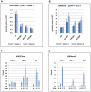
(A) & (B) ChIP analysis of H3K27me3 and H3K4me3 in p16INK4A exon 1 (site C) performed in duplicate (assays i & ii) on an E3CtBP LCL, a 3ACtBP and a 3CCtBP. Similar analysis of a WT-EBV LCL is shown for comparison. (C) & (D) ChIP anaysis of H3K27me3 and H4Kme3 distribution across the p16INK4A-ARF locus in an E3CtBP LCL. Similar assays performed on revertant (revCtBP) and WT-EBV LCLs are shown for comparison. The sites correspond to those shown in Figure 5A.

Similar articles

Cited by

References

    1. Bornkamm GW, Hammerschmidt W. Molecular virology of Epstein-Barr virus. Philos Trans R Soc Lond B Biol Sci. 2001;356:437–459. - PMC - PubMed
    1. Thorley-Lawson DA, Gross A. Persistence of the Epstein-Barr Virus and the Origins of Associated Lymphomas. The New England journal of medicine. 2004;350:1328–1337. - PubMed
    1. Roughan JE, Thorley-Lawson DA. The intersection of Epstein-Barr virus with the germinal center. J Virol. 2009;83:3968–3976. - PMC - PubMed
    1. Crawford DH. Biology and disease associations of Epstein-Barr virus. Philos Trans R Soc Lond B Biol Sci. 2001;356:461–473. - PMC - PubMed
    1. Touitou R, O'Nions J, Heaney J, Allday MJ. Epstein-Barr virus EBNA3 proteins bind to the C8/alpha7 subunit of the 20S proteasome and are degraded by 20S proteasomes in vitro, but are very stable in latently infected B cells. J Gen Virol. 2005;86:1269–1277. - PubMed

Publication types

MeSH terms