Merkel cell polyomavirus and two previously unknown polyomaviruses are chronically shed from human skin
- PMID: 20542254
- PMCID: PMC2919322
- DOI: 10.1016/j.chom.2010.05.006
Merkel cell polyomavirus and two previously unknown polyomaviruses are chronically shed from human skin
Abstract
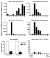
Mounting evidence indicates that Merkel cell polyomavirus (MCV), a circular double-stranded DNA virus, is a causal factor underlying a highly lethal form of skin cancer known as Merkel cell carcinoma. To explore the possibility that MCV and other polyomaviruses commonly inhabit healthy human skin, we developed an improved rolling circle amplification (RCA) technique to isolate circular DNA viral genomes from human skin swabs. Complete MCV genomes were recovered from 40% of healthy adult volunteers tested, providing full-length, apparently wild-type cloned MCV genomes. RCA analysis also identified two previously unknown polyomavirus species that we name human polyomavirus-6 (HPyV6) and HPyV7. Biochemical experiments show that polyomavirus DNA is shed from the skin in the form of assembled virions. A pilot serological study indicates that infection or coinfection with these three skin-tropic polyomaviruses is very common. Thus, at least three polyomavirus species are constituents of the human skin microbiome.
Copyright (c) 2010 Elsevier Inc. All rights reserved.
Figures


Similar articles
-
Specific rolling circle amplification of low-copy human polyomaviruses BKV, HPyV6, HPyV7, TSPyV, and STLPyV.J Virol Methods. 2015 Apr;215-216:17-21. doi: 10.1016/j.jviromet.2015.02.004. Epub 2015 Feb 16. J Virol Methods. 2015. PMID: 25698464
-
Prevalence and Genetic Variability of Human Polyomaviruses 6 and 7 in Healthy Skin Among Asymptomatic Individuals.J Infect Dis. 2018 Jan 17;217(3):483-493. doi: 10.1093/infdis/jix516. J Infect Dis. 2018. PMID: 29161422
-
Genome analysis of the new human polyomaviruses.Rev Med Virol. 2012 Nov;22(6):354-77. doi: 10.1002/rmv.1711. Epub 2012 Mar 28. Rev Med Virol. 2012. PMID: 22461085 Review.
-
Characterization of two novel polyomaviruses of birds by using multiply primed rolling-circle amplification of their genomes.J Virol. 2006 Apr;80(7):3523-31. doi: 10.1128/JVI.80.7.3523-3531.2006. J Virol. 2006. PMID: 16537620 Free PMC article.
-
[New, newer, newest human polyomaviruses: how far?].Mikrobiyol Bul. 2013 Apr;47(2):362-81. doi: 10.5578/mb.5377. Mikrobiyol Bul. 2013. PMID: 23621738 Review. Turkish.
Cited by
-
Viral pathogen discovery.Curr Opin Microbiol. 2013 Aug;16(4):468-78. doi: 10.1016/j.mib.2013.05.001. Epub 2013 May 29. Curr Opin Microbiol. 2013. PMID: 23725672 Free PMC article. Review.
-
Prevalence of Merkel Cell Polyomavirus in Tehran: An Age-Specific Serological Study.Iran Red Crescent Med J. 2016 Feb 14;18(5):e26097. doi: 10.5812/ircmj.26097. eCollection 2016 May. Iran Red Crescent Med J. 2016. PMID: 27437129 Free PMC article.
-
Agnoprotein of polyomavirus BK interacts with proliferating cell nuclear antigen and inhibits DNA replication.Virol J. 2015 Feb 1;12:7. doi: 10.1186/s12985-014-0220-1. Virol J. 2015. PMID: 25638270 Free PMC article.
-
Review on the relationship between human polyomaviruses-associated tumors and host immune system.Clin Dev Immunol. 2012;2012:542092. doi: 10.1155/2012/542092. Epub 2012 Mar 25. Clin Dev Immunol. 2012. PMID: 22489251 Free PMC article. Review.
-
Identification of MW polyomavirus, a novel polyomavirus in human stool.J Virol. 2012 Oct;86(19):10321-6. doi: 10.1128/JVI.01210-12. Epub 2012 Jun 27. J Virol. 2012. PMID: 22740408 Free PMC article.
References
-
- Antonsson A, Erfurt C, Hazard K, Holmgren V, Simon M, Kataoka A, Hossain S, Hakangard C, Hansson BG. Prevalence and type spectrum of human papillomaviruses in healthy skin samples collected in three continents. J Gen Virol. 2003;84:1881–1886. - PubMed
-
- Boldorini R, Veggiani C, Barco D, Monga G. Kidney and urinary tract polyomavirus infection and distribution: molecular biology investigation of 10 consecutive autopsies. Arch Pathol Lab Med. 2005;129:69–73. - PubMed
Publication types
MeSH terms
Substances
Associated data
- Actions
- Actions
- Actions
- Actions
- Actions
- Actions
- Actions
- Actions
- Actions
- Actions
- Actions
- Actions
- Actions
- Actions
- Actions
- Actions
- Actions
- Actions
- Actions
- Actions
- Actions
- Actions
- Actions
- Actions
- Actions
- Actions
- Actions
- Actions
- Actions
- Actions
- Actions
- Actions
Grants and funding
LinkOut - more resources
Full Text Sources
Other Literature Sources
Research Materials

