. 2010 Aug 20;285(34):26135-48.
doi: 10.1074/jbc.M110.146423. Epub 2010 Jun 10.

Postischemic deactivation of cardiac aldose reductase: role of glutathione S-transferase P and glutaredoxin in regeneration of reduced thiols from sulfenic acids

Affiliations

Postischemic deactivation of cardiac aldose reductase: role of glutathione S-transferase P and glutaredoxin in regeneration of reduced thiols from sulfenic acids

Karin Wetzelberger et al. J Biol Chem. .

Abstract

Aldose reductase (AR) is a multifunctional enzyme that catalyzes the reduction of glucose and lipid peroxidation-derived aldehydes. During myocardial ischemia, the activity of AR is increased due to the oxidation of its cysteine residues to sulfenic acids. It is not known, however, whether the activated, sulfenic form of the protein (AR-SOH) is converted back to its reduced, unactivated state (AR-SH). We report here that in perfused mouse hearts activation of AR during 15 min of global ischemia is completely reversed by 30 min of reperfusion. During reperfusion, AR-SOH was converted to a mixed disulfide (AR-SSG). Deactivation of AR and the appearance of AR-SSG during reperfusion were delayed in hearts of mice lacking glutathione S-transferase P (GSTP). In vitro, GSTP accelerated glutathiolation and inactivation of AR-SOH. Reduction of AR-SSG to AR-SH was facilitated by glutaredoxin (GRX). Ischemic activation of AR was increased in GRX-null hearts but was attenuated in the hearts of cardiospecific GRX transgenic mice. Incubation of AR-SSG with GRX led to the regeneration of the reduced form of the enzyme. In ischemic cardiospecific AR transgenic hearts, AR was co-immunoprecipitated with GSTP, whereas in reperfused hearts, the association of AR with GRX was increased. These findings suggest that upon reperfusion of the ischemic heart AR-SOH is converted to AR-SSG via GSTP-assisted glutathiolation. AR-SSG is then reduced by GRX to AR-SH. Sequential catalysis by GSTP and GRX may be a general redox switching mechanism that regulates the reduction of protein sulfenic acids to cysteines.


Figures
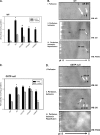
FIGURE 1.
FIGURE 1.
Ischemia-induced activation of AR. Hearts from WT (A and B) and GSTP-null (C and D) mice were excised and perfused ex vivo with either buffer alone for 75 min (P), subjected to global ischemia (I) for 15 min, or subjected to reperfusion (R) for the indicated times after 15 min of ischemia. After the indicated treatments, the hearts were snap frozen, and the AR activity was measured as described under “Experimental Procedures” in the absence or presence of 1 μm sorbinil (A). ′, minutes. Two-dimensional Western blot analyses of AR in homogenates prepared from WT (B) and GSTP-null (D) hearts subjected either to perfusion for 75 min (i), 15 min of perfusion followed by 15 min of ischemia (ii), or 15 min of perfusion and 15 min of ischemia followed by 30 min of reperfusion (iii) are shown. At the end of the protocol, hearts were homogenized, and proteins were resolved by two-dimensional gel electrophoresis. Western blots (WB) were developed using anti-AR antibody, biotin-labeled DD and horseradish peroxidase-tagged streptavidin, or anti-PSSG antibody as indicated. Arrows indicate reduced AR (AR-SH), sulfenic acid-modified AR (AR-SOH), and glutathiolated AR (AR-SSG) forms of the AR protein. “?” indicates an unidentified form of AR. Values are mean ± S.D. *, p < 0.05 versus 75 min of perfusion; #, p < 0.05 versus 15 min of ischemia (n = 3 hearts/group).
FIGURE 2.
FIGURE 2.
Distribution of AR during myocardial ischemia-reperfusion. A, percentage of AR activity attributable to AR-SOH formation in WT and GSTP-null hearts subjected to 75 min of perfusion (P), 15 min of global ischemia (I), or 15, 30, and 60 min of reperfusion (R) after 15 min of ischemia as indicated. The extent of AR-SOH formation was calculated from sorbinil inhibition data shown in Fig. 1 and was based on the assumption that 1 μm sorbinil inhibits 90% AR-SH and 20% AR-SOH activity (Equation 1). ′, minutes. B, inhibition of AR by dimedone. AR activity was measured in lysates of hearts subjected to 75 min of perfusion (75′P), 15 min of ischemia (15′I), or 15 min of ischemia followed by 30 min of reperfusion (15′I 30′R) with or without 0.5 mm dimedone. C, AR catalytic activity in WT and GSTP-null hearts. Measured values of AR activity are from Fig. 1 and are normalized to AR activity in perfused heart. Predicted values were calculated from the extent of AR-SOH formation (shown in A) and are based on the assumption that the catalytic activity of AR-SOH is 4-fold higher than that of AR-SH. The mismatch between the predicted and the measured values (highlighted in yellow) was used to calculate the percentage of AR in the inactive AR-SSG form. The calculated distribution of different AR forms during ischemia-reperfusion in WT (D) and GSTP-null (E) hearts is shown. Values in B are mean ± S.D. *, p < 0.05 versus perfused hearts; #, p < 0.05 versus ischemic hearts without dimedone treatment; +, p < 0.05 versus ischemic hearts (n = 3).
FIGURE 3.
FIGURE 3.
Role of GSTP in glutathiolation of activated AR. Human recombinant AR was treated with 0.1 mm peroxynitrite for 1 h as described under “Experimental Procedures.” The peroxynitrite-modified enzyme was then incubated with 2 mm GSH (A) or GSH and human recombinant GSTP (B) in 50 mm phosphate buffer, pH 7.0. Aliquots were withdrawn from the reaction mixture at the indicated time for measurement of AR activity with 10 mm dl-glyceraldehyde as substrate without and with sorbinil (1 μm). ′, minutes. C, effect of DD on reduced and oxidized AR. Reduced or peroxynitrite-treated AR was incubated with 0.5 mm DD; after 60 min of incubation, aliquots were withdrawn from the reaction mixture; and AR activity was measured without and with sorbinil (1 μm). Incubation with GSTP or GSH alone did not affect AR activity. Values are mean ± S.D. *, p < 0.05 versus untreated AR; #, p < 0.05 versus ONOO-treated AR (n = 3).
FIGURE 4.
FIGURE 4.
GSTP-dependent AR glutathiolation. Recombinant human AR was reduced with 0.1 m DTT and desalted by passing through a PD-10 column. The sulfhydryl group of AR was converted to sulfenic acid by treatment with peroxynitrite. The oxidized enzyme was then incubated with GSH (A) or GSH + GSTP (B). Aliquots of the reaction mixture were withdrawn at the indicated times, and the extent of glutathiolation was measured. The upper panels show Western blots (WB) of the aliquots developed using anti-AR and anti-PSSG antibodies, and measurements of band intensity are shown in the lower panels. Values are mean ± S.D. *, p < 0.05 versus time 0; #, p < 0.05 versus 15 min (n = 3 experiments). A.U., arbitrary units.
FIGURE 5.
FIGURE 5.
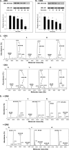
Glutathiolation of AR protein by GSTP. Reduced recombinant AR treated with ONOO was desalted and treated with GSH (A) or GSH + GSTP (B). Aliquots were withdrawn at the indicated times and analyzed by ESI+/MS. A, deconvoluted spectra of unmodified, reduced AR protein (37,883 Da) and a minor unidentified peak (37,924 Da) (i), AR incubated with ONOO showing a 16-Da increase in mass (AR-SOH, 37,899 Da) (ii), and AR after incubation with 2 mm GSH for 15 min showing the AR-SOH (37,899 Da) and the AR-SOSG (38,206 Da) forms of the protein (iii). B, AR glutathiolation in the presence of GSTP showing unmodified AR (37,882 Da) (i), reduced AR incubated with 0.1 mm ONOO showing AR-SOH (37,899 Da) (ii), and AR-SOH incubated for 15 min with 2 mm GSH and GSTP showing both AR-SH (37,885 Da) and the AR-SSG (38,192 Da) forms (iii).
FIGURE 6.
FIGURE 6.
Reduction of AR-SSG by GRX. Reduced AR was incubated with 0.1 mm ONOO. After incubation, the mixture was desalted to remove ONOO. Catalytic activity of the reduced (C) and modified (M) enzyme with or without 1 μm sorbinil is shown. The modified enzyme was then incubated with 2 mm GSH alone for 15 min; after that, glutathione reductase (GR) and NADPH were added to GSH-containing modified enzyme, and the mixture was then incubated at 25 °C in the absence (A) or presence (B) of GRX as indicated. Aliquots from the reaction mixture were withdrawn at the indicated time intervals and assayed for AR activity with and without sorbinil (1 μm). Data are expressed as mean ± S.D. *, p < 0.05 versus AR (C) alone; #, p < 0.05 versus AR + ONOO (M); +, p < 0.05 versus AR + ONOO + GSH (GSH); ▴, p < 0.05 versus modified AR + glutathione reductase + GSH (0 min) or modified AR + glutathione reductase + GRX + GSH (0 min); n = 3 experiments at each time point.
FIGURE 7.
FIGURE 7.
Deglutathiolation of AR-SSG by GRX. AR reduced with 0.1 m DTT and incubated with 0.1 mm ONOO was desalted and then incubated with 2 mm GSH, glutathione reductase (GR), and NADPH at 25 °C in the absence (A) or presence (B) of GRX. Aliquots from the reaction mixture were withdrawn at the indicated times, and glutathiolated AR was assayed by Western blot (WB) using anti-PSSG antibody. Values are mean ± S.D. *, p < 0.05 versus 0-min time point (n = 3 experiments at each time point). Aliquots of the reaction mixture withdrawn at regular time intervals were also analyzed by ESI/MS. C (i–vi) shows ESI/MS spectra of AR incubated with ONOO, GSH, and GSTP showing both the native (37,883 Da; minor peak) and the AR-SSG form (38,190 Da; major peak) (i). Incubation of the AR-SSG form with glutathione reductase for 0 (ii), 15 (iii), 30 (iv), 60 (v), and 90 (vi) min shows the major form AR-SSG (38,190 Da) and a minor form corresponding to native AR (37,883 Da). Incubation of AR-SSG, generated by incubating AR with ONOO (D, i), with GSH, GSTP, and GRX (D, ii) for 0 min shows both the AR-SSG (38,190 Da; major peak) and the native forms (37,883 Da; minor peak), whereas incubation for 15 min (D, iii) shows two equal peaks of the native (37,883 Da) and AR-SSG (38,190 Da) forms of the protein. Incubation of AR-SSG with 2 mm GSH and GRX for 30 (D, iv), 60 (D, v), and 90 min (D, vi) shows the major reduced AR-SH form (37,883 Da) and the minor AR-SSG form (38,190 Da) of the protein. A.U., arbitrary units.
FIGURE 8.
FIGURE 8.
Regulation of reperfusion-induced AR deactivation by GRX in heart. Isolated perfused hearts from WT and GRX-TG (A) and WT and GRX-null (B) mice were subjected to 15 min of global ischemia alone (15′I) or 15 min of ischemia followed by 30 min of reperfusion (15′I/30′R). 45′P, 45 min of perfusion. WT hearts subjected to the same treatment were used as controls. AR activity was measured as described under “Experimental Procedures.” Values are mean ± S.D. (n = 3 experiments). *, p < 0.05 compared with perfusion only; ▴, p < 0.05 compared with 15 min of ischemia in WT; #, p < 0.05 compared with 15 min of ischemia.
FIGURE 9.
FIGURE 9.
Association of AR with GSTP and GRX during myocardial ischemia-reperfusion. A, AR activity was measured in WT and AR-TG heart homogenates that were subjected to 15 min of ischemia alone (15′I) or 15 min of ischemia followed by 30 min of reperfusion (15′I/30′R). The 75 min of perfusion (75′P) group data served as controls. B, the extent of AR glutathiolation and interaction with GRX and GSTP was assessed by immunoprecipitating AR from perfused (P), ischemic (I), or ischemic-reperfused hearts (I/R). Western blots (WB) of the immunoprecipitate (IP) were developed using anti-PSSG, anti-GSTP, and anti-GRX antibodies. C, levels of each of these proteins (i–iii) were normalized to the amount of AR detected in the immunoprecipitate measured by Western blotting using anti-His antibodies that recognize the AR transgene in the heart. No AR-His protein, GRX, or GSTP was immunoprecipitated with an unrelated antibody. Values are mean ± S.D. (n = 3 experiments). *, p < 0.05 versus 75 min of perfusion; ▴, p < 0.05 versus 75 min of perfusion; #, p < 0.05 versus 15 min of ischemia. A.U., arbitrary units.
SCHEME 1.
SCHEME 1.
Mechanism for glutathiolation and deglutathiolation of AR. Reduced AR (AR-SH) is oxidized by peroxynitrite (ONOO generated during ischemia) to AR-SOH (activated form). The sulfenic acid form of AR (AR-SOH) could be irreversibly oxidized to sulfinic acid (AR-SO2H) and sulfonic acid (AR-SO3H) forms. The reversible, sulfenic acid form of AR is deactivated to an inactive form by glutathiolation (AR-SSG) of the sulfenic acid residue via a reaction catalyzed by GSTP in the presence of GSH. The AR-SSG is recycled to the native, reduced form (AR-SH) by GRX in the presence of GSH. *, sorbinil-sensitive AR with basal activity; #, dimedone-sensitive activated AR; 1, catalytic activity unknown; 2, enzymatically inactive AR.

Similar articles

Cited by

References

    1. Bolli R., Patel B. S., Jeroudi M. O., Lai E. K., McCay P. B. (1988) J. Clin. Investig. 82, 476–485 - PMC - PubMed
    1. Bolli R., Jeroudi M. O., Patel B. S., Aruoma O. I., Halliwell B., Lai E. K., McCay P. B. (1989) Circ. Res. 65, 607–622 - PubMed
    1. Tortolani A. J., Powell S. R., Misík V., Weglicki W. B., Pogo G. J., Kramer J. H. (1993) Free Radic. Biol. Med. 14, 421–426 - PubMed
    1. Li X. Y., McCay P. B., Zughaib M., Jeroudi M. O., Triana J. F., Bolli R. (1993) J. Clin. Investig. 92, 1025–1041 - PMC - PubMed
    1. Bolli R., Zughaib M., Li X. Y., Tang X. L., Sun J. Z., Triana J. F., McCay P. B. (1995) J. Clin. Investig. 96, 1066–1084 - PMC - PubMed

Publication types