Effect of NKG2D ligand expression on host immune responses
- PMID: 20536569
- PMCID: PMC2885032
- DOI: 10.1111/j.0105-2896.2010.00893.x
Effect of NKG2D ligand expression on host immune responses
Abstract
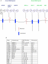
Natural killer group 2, member D (NKG2D) is an activating receptor present on the surface of natural killer (NK) cells, some NKT cells, CD8(+) cytotoxic T cells, gammadelta T cells, and under certain conditions CD4(+) T cells. Present in both humans and mice, this highly conserved receptor binds to a surprisingly diverse family of ligands that are distant relatives of major histocompatibility complex class I molecules. There is increasing evidence that ligand expression can result in both immune activation (tumor clearance, viral immunity, autoimmunity, and transplantation) and immune silencing (tumor evasion). In this review, we describe this family of NKG2D ligands and the various mechanisms that control their expression in stressed and normal cells. We also discuss the host response to both membrane-bound and secreted NKG2D ligands and summarize the models proposed to explain the consequences of this differential expression.
Figures


Similar articles
-
Natural killer group 2D receptor and its ligands in cancer immune escape.Mol Cancer. 2019 Feb 27;18(1):29. doi: 10.1186/s12943-019-0956-8. Mol Cancer. 2019. PMID: 30813924 Free PMC article. Review.
-
Soluble ligands for the NKG2D receptor are released during HIV-1 infection and impair NKG2D expression and cytotoxicity of NK cells.FASEB J. 2013 Jun;27(6):2440-50. doi: 10.1096/fj.12-223057. Epub 2013 Feb 8. FASEB J. 2013. PMID: 23395909
-
Generation of soluble NKG2D ligands: proteolytic cleavage, exosome secretion and functional implications.Scand J Immunol. 2013 Aug;78(2):120-9. doi: 10.1111/sji.12072. Scand J Immunol. 2013. PMID: 23679194 Review.
-
Importance of NKG2D-NKG2D ligands interaction for cytolytic activity of natural killer cell.Cell Immunol. 2012 Mar-Apr;276(1-2):122-7. doi: 10.1016/j.cellimm.2012.04.011. Epub 2012 May 3. Cell Immunol. 2012. PMID: 22613008
-
Immunobiology and conflicting roles of the human NKG2D lymphocyte receptor and its ligands in cancer.J Immunol. 2013 Aug 15;191(4):1509-15. doi: 10.4049/jimmunol.1301071. J Immunol. 2013. PMID: 23913973 Free PMC article. Review.
Cited by
-
Innate lymphoid cells in early tumor development.Front Immunol. 2022 Aug 12;13:948358. doi: 10.3389/fimmu.2022.948358. eCollection 2022. Front Immunol. 2022. PMID: 36032129 Free PMC article. Review.
-
Transcriptome analysis reveals markers of aberrantly activated innate immunity in vitiligo lesional and non-lesional skin.PLoS One. 2012;7(12):e51040. doi: 10.1371/journal.pone.0051040. Epub 2012 Dec 10. PLoS One. 2012. PMID: 23251420 Free PMC article.
-
NKG2D-NKG2D Ligand Interaction Inhibits the Outgrowth of Naturally Arising Low-Grade B Cell Lymphoma In Vivo.J Immunol. 2016 Jun 1;196(11):4805-13. doi: 10.4049/jimmunol.1501982. Epub 2016 May 2. J Immunol. 2016. PMID: 27183590 Free PMC article.
-
An ENU mutagenesis approach to dissect "self"-induced immune responses: Unraveling the genetic footprint of immunosurveillance.Oncoimmunology. 2012 Sep 1;1(6):856-862. doi: 10.4161/onci.20580. Oncoimmunology. 2012. PMID: 23162753 Free PMC article.
-
The requirement for NKG2D in NK cell-mediated rejection of parental bone marrow grafts is determined by MHC class I expressed by the graft recipient.Blood. 2010 Dec 9;116(24):5208-16. doi: 10.1182/blood-2010-05-285031. Epub 2010 Aug 24. Blood. 2010. PMID: 20736452 Free PMC article.
References
Publication types
MeSH terms
Substances
Grants and funding
LinkOut - more resources
Full Text Sources
Other Literature Sources
Research Materials

