. 2010 Aug 1;21(15):2578-88.
doi: 10.1091/mbc.e10-03-0227. Epub 2010 Jun 9.

The laforin-malin complex, involved in Lafora disease, promotes the incorporation of K63-linked ubiquitin chains into AMP-activated protein kinase beta subunits

Affiliations

The laforin-malin complex, involved in Lafora disease, promotes the incorporation of K63-linked ubiquitin chains into AMP-activated protein kinase beta subunits

Daniel Moreno et al. Mol Biol Cell. .

Abstract

Lafora progressive myoclonus epilepsy is a fatal neurodegenerative disorder caused by defects in the function of at least two proteins: laforin, a dual-specificity protein phosphatase, and malin, an E3-ubiquitin ligase. In this study, we report that a functional laforin-malin complex promotes the ubiquitination of AMP-activated protein kinase (AMPK), a serine/threonine protein kinase that acts as a sensor of cellular energy status. This reaction occurs when any of the three AMPK subunits (alpha, beta, and gamma) are expressed individually in the cell, and it also occurs on AMPK beta when it is part of a heterotrimeric complex. We also report that the laforin-malin complex promotes the formation of K63-linked ubiquitin chains, which are not involved in proteasome degradation. On the contrary, this modification increases the steady-state levels of at least AMPK beta subunit, possibly because it leads to the accumulation of this protein into inclusion bodies. These results suggest that the modification introduced by the laforin-malin complex could affect the subcellular distribution of AMPK beta subunits.


Figures
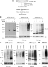
Figure 1.
Figure 1.
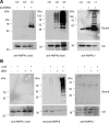
The laforin–malin complex is able to promote ubiquitination of individual AMPK subunits. (A) Diagram of the protocol used to determine the presence of ubiquitinated proteins, based on the use of a 6xHis-tagged version of ubiquitin (Kaiser and Tagwerker, 2005). (B) HEK293 cells were transfected with plasmid pCMV-His6xUbiq and the indicated combination of plasmids (pLaforin, pCMV-HA-laforin; and pMalin, pcDNA3-HA-malin). Cell extracts were then obtained as described in Materials and Methods and the clarified extract (CE; 40 μg), and the material bound to the metal-affinity chromatography column (bound; 40 μl) was analyzed by SDS-PAGE and Western blotting using anti-myc antibodies. Molecular mass markers are indicated on the left of each panel. For AMPKα2 and AMPKγ1, we also detected a minor modification when only laforin was overexpressed, perhaps because it can force endogenous malin to carry out the corresponding ubiquitination. (C) HEK293 cells were transfected with plasmid pCMV-His6xUbiq and the indicated combination of plasmids (Mdm2, pCMV-Mdm2; Laf/Mal, pCMV-HA-laforin/pcDNA3-HA-malin; and empty, pCMV-HA). Cell extracts were analyzed as described above using anti-myc antibodies. (D) HEK293 cells were transfected with plasmid pCMV-His6xUbiq and plasmids pCMV-p53 and pCMV-Mdm2 or pCMV-HA (empty). Cell extracts were analyzed as described above using anti-p53 antibodies.
Figure 2.
Figure 2.
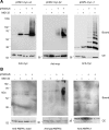
Laforin–malin-dependent ubiquitination of the heterotrimeric AMPK complex. (A) Analysis of overexpressed AMPK complex. HEK293 cells were transfected with plasmid pCMV-His6xUbiq and the indicated combination of plasmids (αβγ, pCMVmyc-AMPKα2 + pCMVmyc-AMPKβ2 + pCMVmyc-AMPKγ1; α2, pCMVmyc-AMPKα2; β2, pCMVmyc-AMPKβ2; γ1, pCMVmyc-AMPKγ1; and pLaf/pMal, pCMV-HA-laforin/pcDNA3-HA-malin). Cell extracts were analyzed as described in Figure 1 by using anti-AMPKα total, anti-AMPKβ total, or anti-AMPKγ1 antibodies. (B) Analysis of endogenous AMPK complex. HEK293 cells were transfected with plasmid pCMV-His6xUbiq and plasmids pCMV-HA-laforin (pLaf) and pcDNA3-HA-malin (pMal), when indicated. Cell extracts were analyzed as described in A but using anti-AMPKα total, anti-panAMPKβ, and anti-AMPKγ1 antibodies to determine the components of the endogenous AMPK complex (*, nonspecific band). Molecular mass markers are indicated on the left of each panel.
Figure 3.
Figure 3.
AMPK subunits accumulate as ubiquitinated proteins upon proteasome inhibition. (A) Analysis of the expression of individual AMPK subunits. HEK293 cells were transfected with plasmid pCMV-His6xUbiq (when indicated) and either pCMVmyc-AMPKα2, pCMVmyc-AMPKβ2, or pCMVmyc-AMPKγ1. Eighteen hours after transfection, cells were treated with or without 50 μM MG132 and incubated 18 h further. Cell extracts were then analyzed as described in Figure 1 by using anti-myc antibodies. (B) Analysis of the subunits of the endogenous AMPK complex. HEK293 cells were transfected only with plasmid pCMV-His6xUbiq (when indicated) and analyzed as described in A but using anti-AMPKα total, anti-panAMPKβ, and anti-AMPKγ1 antibodies to determine the components of the endogenous AMPK complex (*, nonspecific band). Molecular mass markers are indicated on the left of each panel.
Figure 4.
Figure 4.
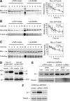
Degradation rates of individual AMPK subunits in the presence or absence of the laforin–malin complex. HEK293 cells were transfected with plasmid pCMVmyc-AMPKα2 (A), pCMVmyc-AMKβ2 (B), or pCMVmyc-AMPKγ1 (C) without or with a combination of plasmids pCMV-HA-laforin and pcDNA3-HA-malin. Twenty-four hours after transfection, cells were treated with 355 μM CHX, and at the indicated times, aliquots were taken from the cultures and cell extracts (25 μg) were analyzed by SDS-PAGE and Western blotting using anti-myc antibodies. The same blots were analyzed using anti-tubulin antibodies as loading controls. A representative blot is shown in A, B, and C. M, molecular mass standards lane. The right of each panel shows a plot of the levels of the corresponding myc-AMPK subunit with respect to the levels of tubulin of each time point, expressed as a percentage of the value at time 0. Plots are the mean of three different experiments (bars are SD). (D) The laforin–malin complex increases the steady-state levels of AMPKβ subunits. HEK293 cells were cotransfected with plasmid pCMV-GFP, and pCMVmyc-AMPKβ1 or pCMVmyc-AMPKβ2 and a combination or not of plasmids pCMV-HA-laforin and pcDNA3-HA-malin (L/M; empty: pCMV-HA). After 24 h of transfection, cell extracts (30 μg) were analyzed by SDS-PAGE and Western blotting using anti-AMPKβ (top) and anti-GFP (bottom) antibodies. (E) HEK293 cells cotransfected with plasmid pCMVmyc-AMPKβ2 and a combination or not of plasmids pCMV-HA-laforin and pcDNA3-HA-malin (pLaforin + pMalin). After 18 h of transfection, cells were treated for 5 h with either 20 mM ammonium chloride and 100 μM leupeptin (to inhibit lysosomes) or with 5 μM lactacystin (to inhibit proteasomes) or left untreated (Untr). Cell extracts (30 μg) were then analyzed by SDS-PAGE and Western blotting using anti-myc (top) and anti-tubulin (bottom) antibodies. (F) CIDEA and the laforin–malin complex have opposite effects on AMPKβ2 accumulation. HEK293 cells were transfected with plasmid pCMVmyc-AMPKβ2 and with plasmids pFLAG-CIDEA or pCMV-HA-laforin and pcDNA3-HA-malin (Laf/Mal) or with an empty plasmid (pCMV-HA). Then, cell extracts (30 μg) were analyzed by SDS-PAGE and Western blotting using anti-myc (top) and anti-tubulin (bottom) antibodies.
Figure 5.
Figure 5.
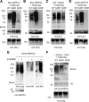
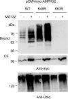
The laforin–malin complex promotes the formation of K63-linked ubiquitin chains into its corresponding substrates. HEK293 cells were transfected with plasmids pCMVmyc-AMPKα2 (A), pCMVmyc-AMPKβ2 (B), or pCMVmyc-R5/PTG (D), and pCMV-HA-laforin, pcDNA3-HA-malin and either pCMV-His6xUbiq (WT), pCMV-His6xUbiq K48R (K48R), or pCMV-His6xUbiq K63R (K63R). Cell extracts were analyzed as described in Figure 1 using anti-myc antibodies. (C) HEK293 cells were transfected with plasmids pCMV-p53, pCMV-Mdm2, and either pCMV-His6xUbiq (WT), pCMV-His6xUbiq K48R (K48R), or pCMV-His6xUbiq K63R (K63R). Cell extracts were analyzed as described in Figure 1 by using anti-p53 antibodies. Molecular mass markers are indicated on the left of each panel. Total ubiquitinated proteins were analyzed in the crude extracts (40 μg) of each panel by using anti-ubiquitin conjugates antibodies. (E) His-AMPKβ2 is also modified with K63-ubiquitin linkages by the laforin–malin complex. HEK293 cells were transfected with plasmid pcDNA4/His-AMPKβ2 (6xHis-AMPKβ2) with or without plasmids pCMV-HA-laforin and pcDNA3-HA-malin (pLaf/pMal). Cell extracts were prepared as described in Figure 1, and the bound material was analyzed using antibodies against conjugated ubiquitin (anti-ubiq), K48-linked poly-ubiquitin (anti-K48), or K63-linked poly-ubiquitin (anti-K63) chains. CEs (40 μg) also were analyzed using anti-AMPKβ antibodies. (F) Ubiquitination of endogenous AMPKβ subunits. HEK293 cells were transfected with plasmids pCMV-HA-laforin, pcDNA3-HA-malin, and either pCMV-Ubiqx6His (WT), pCMV-Ubiqx6His K48R (K48R), or pCMV-Ubiqx6His K63R (K63R). Cell extracts were analyzed as described in A: the bound material and CE (40 μg) were analyzed using anti-pan AMPKβ antibodies.
Figure 6.
Figure 6.
Endogenous E3-ubiquitin ligases also promote the K63-linked ubiquitination of AMPKβ2. HEK293 cells were transfected with plasmid pCMVmyc-AMPKβ2 and either pCMV-His6xUbiq (WT), pCMV-His6xUbiq K48R (K48R), or pCMV-His6xUbiq K63R (K63R). Eighteen hours after transfection, cells were treated with or without 50 μM MG132 and incubated 18 h further. Cell extracts were then analyzed as described in Figure 1 by using anti-myc antibodies. Total ubiquitinated proteins were analyzed in the crude extracts (40 μg) of each panel by using anti-ubiquitin conjugates antibodies.
Figure 7.
Figure 7.
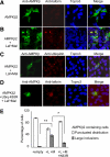
The laforin–malin complex promotes the aggregation of coexpressed AMPKβ subunits. (A–D) HEK293 cells were transfected with plasmid pCMVmyc-AMPKβ2 with (B and C) or without (A) plasmids pCMV-HA-laforin and pcDNA3-HA-malin (Laf+Mal). When indicated, cells were also transfected with plasmid pCMV-His6xUbiq K63R (D). The subcellular localization of AMPKβ2 subunit was carried out as described in Materials and Methods by using anti-AMPKβ total as primary and anti-rabbit Alexa-Fluor 488 as secondary antibodies. The same samples were treated with Topro3 to stain the nucleus and with anti-laforin or anti-ubiquitin as primary and anti-mouse Texas Red as secondary antibodies to determine the localization of laforin and ubiquitin conjugates. The three images were subjected to a merge analysis. (E) Quantification of cells expressing AMPKβ2 and showing either a punctuated distribution or large inclusion bodies. One hundred cells expressing AMPKβ2 from each of the above conditions were used to estimate the proportion of cells with or without inclusions. Bars indicate SD; statistical significance was considered at *p < 0.05 and **p < 0.01.

Similar articles

Cited by

References

    1. Bateman A. The structure of a domain common to archaebacteria and the homocystinuria disease protein. Trends Biochem. Sci. 1997;22:12–13. - PubMed
    1. Brady M., Vlatkovic N., Boyd M. T. Regulation of p53 and MDM2 activity by MTBP. Mol. Cell. Biol. 2005;25:545–553. - PMC - PubMed
    1. Crute B. E., Seefeld K., Gamble J., Kemp B. E., Witters L. A. Functional domains of the alpha1 catalytic subunit of the AMP-activated protein kinase. J. Biol. Chem. 1998;273:35347–35354. - PubMed
    1. Chan E. M., Omer S., Ahmed M., Bridges L. R., Bennett C., Scherer S. W., Minassian B. A. Progressive myoclonus epilepsy with polyglucosans (Lafora disease): evidence for a third locus. Neurology. 2004;63:565–567. - PubMed
    1. Chan E. M., et al. Mutations in NHLRC1 cause progressive myoclonus epilepsy. Nat. Genet. 2003;35:125–127. - PubMed

Publication types

MeSH terms