Systemically dispersed innate IL-13-expressing cells in type 2 immunity
- PMID: 20534524
- PMCID: PMC2895098
- DOI: 10.1073/pnas.1003988107
Systemically dispersed innate IL-13-expressing cells in type 2 immunity
Abstract
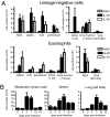
Type 2 immunity is a stereotyped host response to allergens and parasitic helminths that is sustained in large part by the cytokines IL-4 and IL-13. Recent advances have called attention to the contributions by innate cells in initiating adaptive immunity, including a novel lineage-negative population of cells that secretes IL-13 and IL-5 in response to the epithelial cytokines IL-25 and IL-33. Here, we use IL-4 and IL-13 reporter mice to track lineage-negative innate cells that arise during type 2 immunity or in response to IL-25 and IL-33 in vivo. Unexpectedly, lineage-negative IL-25 (and IL-33) responsive cells are widely distributed in tissues of the mouse and are particularly prevalent in mesenteric lymph nodes, spleen, and liver. These cells expand robustly in response to exogenous IL-25 or IL-33 and after infection with the helminth Nippostrongylus brasiliensis, and they are the major innate IL-13-expressing cells under these conditions. Activation of these cells using IL-25 is sufficient for worm clearance, even in the absence of adaptive immunity. Widely dispersed innate type 2 helper cells, which we designate Ih2 cells, play an integral role in type 2 immune responses.
Conflict of interest statement
The authors declare no conflict of interest.
Figures


Similar articles
-
Type 2 immunity is controlled by IL-4/IL-13 expression in hematopoietic non-eosinophil cells of the innate immune system.J Exp Med. 2006 Jun 12;203(6):1435-46. doi: 10.1084/jem.20052448. Epub 2006 May 15. J Exp Med. 2006. PMID: 16702603 Free PMC article.
-
Epithelial cell-specific Act1 adaptor mediates interleukin-25-dependent helminth expulsion through expansion of Lin(-)c-Kit(+) innate cell population.Immunity. 2012 May 25;36(5):821-33. doi: 10.1016/j.immuni.2012.03.021. Epub 2012 May 17. Immunity. 2012. PMID: 22608496 Free PMC article.
-
Up-regulation of gasdermin C in mouse small intestine is associated with lytic cell death in enterocytes in worm-induced type 2 immunity.Proc Natl Acad Sci U S A. 2021 Jul 27;118(30):e2026307118. doi: 10.1073/pnas.2026307118. Proc Natl Acad Sci U S A. 2021. PMID: 34290141 Free PMC article.
-
The role of rare innate immune cells in Type 2 immune activation against parasitic helminths.Parasitology. 2017 Sep;144(10):1288-1301. doi: 10.1017/S0031182017000488. Epub 2017 Jun 6. Parasitology. 2017. PMID: 28583216 Free PMC article. Review.
-
Spatial regulation of IL-4 signalling in vivo.Cytokine. 2015 Sep;75(1):51-6. doi: 10.1016/j.cyto.2015.02.026. Epub 2015 Mar 25. Cytokine. 2015. PMID: 25819429 Review.
Cited by
-
Waves of layered immunity over innate lymphoid cells.Front Immunol. 2022 Oct 4;13:957711. doi: 10.3389/fimmu.2022.957711. eCollection 2022. Front Immunol. 2022. PMID: 36268032 Free PMC article. Review.
-
Asthma as a chronic disease of the innate and adaptive immune systems responding to viruses and allergens.J Clin Invest. 2012 Aug;122(8):2741-8. doi: 10.1172/JCI60325. Epub 2012 Aug 1. J Clin Invest. 2012. PMID: 22850884 Free PMC article. Review.
-
Type 2 innate immunity in helminth infection is induced redundantly and acts autonomously following CD11c(+) cell depletion.Infect Immun. 2012 Oct;80(10):3481-9. doi: 10.1128/IAI.00436-12. Epub 2012 Jul 30. Infect Immun. 2012. PMID: 22851746 Free PMC article.
-
Macrophages as IL-25/IL-33-responsive cells play an important role in the induction of type 2 immunity.PLoS One. 2013;8(3):e59441. doi: 10.1371/journal.pone.0059441. Epub 2013 Mar 25. PLoS One. 2013. PMID: 23536877 Free PMC article.
-
Mast cells orchestrate type 2 immunity to helminths through regulation of tissue-derived cytokines.Proc Natl Acad Sci U S A. 2012 Apr 24;109(17):6644-9. doi: 10.1073/pnas.1112268109. Epub 2012 Apr 9. Proc Natl Acad Sci U S A. 2012. PMID: 22493240 Free PMC article.
References
-
- Finkelman FD, et al. Interleukin-4- and interleukin-13–mediated host protection against intestinal nematode parasites. Immunol Rev. 2004;201:139–155. - PubMed
-
- Voehringer D, Shinkai K, Locksley RM. Type 2 immunity reflects orchestrated recruitment of cells committed to IL-4 production. Immunity. 2004;20:267–277. - PubMed
-
- Gessner A, Mohrs K, Mohrs M. Mast cells, basophils, and eosinophils acquire constitutive IL-4 and IL-13 transcripts during lineage differentiation that are sufficient for rapid cytokine production. J Immunol. 2005;174:1063–1072. - PubMed
Publication types
MeSH terms
Substances
Grants and funding
LinkOut - more resources
Full Text Sources
Other Literature Sources
Molecular Biology Databases
Research Materials

