. 2010 Jul 5;207(7):1513-23.
doi: 10.1084/jem.20092474. Epub 2010 Jun 7.

Enterocyte-specific A20 deficiency sensitizes to tumor necrosis factor-induced toxicity and experimental colitis

Affiliations

Enterocyte-specific A20 deficiency sensitizes to tumor necrosis factor-induced toxicity and experimental colitis

Lars Vereecke et al. J Exp Med. .

Abstract

A20 is a nuclear factor kappaB (NF-kappaB) target gene that encodes a ubiquitin-editing enzyme that is essential for the termination of NF-kappaB activation after tumor necrosis factor (TNF) or microbial product stimulation and for the prevention of TNF-induced apoptosis. Mice lacking A20 succumb to inflammation in several organs, including the intestine, and A20 mutations have been associated with Crohn's disease. However, ablation of NF-kappaB activity, specifically in intestinal epithelial cells (IECs), promotes intestinal inflammation. As A20 deficiency sensitizes cells to TNF-induced apoptosis yet also promotes NF-kappaB activity, it is not clear if A20 deficiency in IECs would exacerbate or ameliorate intestinal inflammation. We generated mice lacking A20 specifically in IECs. These mice did not show spontaneous intestinal inflammation but exhibited increased susceptibility to experimental colitis, and their IECs were hypersensitive to TNF-induced apoptosis. The resulting TNF-driven breakdown of the intestinal barrier permitted commensal bacterial infiltration and led to systemic inflammation. These studies define A20 as a major antiapoptotic protein in the intestinal epithelium and further indicate that defects in A20 might contribute to inflammatory bowel disease in humans.


Figures
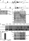
Figure 1.
Figure 1.
Generation and molecular analysis of A20IEC-KO mice. (A) Targeting scheme. The diagram shows the loxP-flanked (floxed) and deleted A20 alleles. The boxes indicate exons 1–9 (E1–E9). Restriction enzyme sites and the location of the probe used for Southern blot analysis are depicted. B, BamH1; V, EcoRV. LoxP and Frt sites are indicated by arrowheads. (B) Southern blot analysis on DNA from WT (+/+) and homologous recombinant (NFL/+) ES cells. (C) Western blot analysis for A20 expression in WT (+/+), heterozygous (+/−), and A20 knockout (−/−) primary MEFs either stimulated or not for 5 h with recombinant mouse TNF. (D) DNA isolated from various tissues of a A20FL/FL/Villin-Cre+ mouse and a control littermate (A20FL/FL/Villin-Cre) was subjected to Southern blot analysis. Δ, deleted allele; FL, floxed allele; SI, small intestine. (E) Quantitative PCR measurement of A20 mRNA expression in purified IECs from A20IEC-KO (n = 2) and control WT littermate mice (n = 2) 0 or 30 min after mouse TNF injection. Error bars represent SEM. (F) Western blot analysis for A20 expression in colonic epithelial cells from two individual A20IEC-KO and control WT littermate mice. *, unspecific. Data are representative of two independent experiments.
Figure 2.
Figure 2.
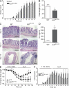
Enterocyte expression of A20 is required for recovery of mice from acute DSS-induced inflammation. (A) Clinical score of A20IEC-KO mice (n = 18) and control littermates (WT, n = 20) treated with 1.5% DSS. (B) Colon length of A20IEC-KO mice (n = 18) and control littermates (WT, n = 20) after 10 d of 1.5% DSS treatment. (C) Hematoxylin and eosin (H&E) histology on DSS-treated A20IEC-KO mice and control littermates (WT). Bars: (days 2 and 4) 100 µm; (day 10) 250 µm. (D) Serum IL-6 levels after 1.5% DSS treatment for 4 d. (E and F) Clinical score and body weight of A20IEC-KO mice (n = 6) and control littermates (WT, n = 5) treated for 6 d with 1.5% DSS followed by normal drinking water. Data in A–D are representative of three independent experiments. Experiments in E and F were performed two times. Error bars represent SEM. *, P < 0.05.
Figure 3.
Figure 3.
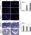
A20 deficiency sensitizes IECs to DSS-induced apoptosis. (A) TUNEL staining on distal colon sections of A20IEC-KO mice and control littermates (WT) after 2 and 6 d of 1.5% DSS treatment, and after 9 d during recovery (6 d of 1.5% DSS followed by normal drinking water). Bars, 150 µm. (B) Quantification of the number of TUNEL-positive cells/field from untreated and DSS-treated mice. Error bars represent SEM. *, P < 0.05. (C) Detection of BrdU incorporation on distal colon sections of A20IEC-KO mice and control littermates (WT) after 6 d of 1.5% DSS treatment, and after 9 d during recovery (6 d of 1.5% DSS followed by normal drinking water). Bars: (day 4) 50 µm; (day 9) 250 µm. (D) Quantification of the number of BrdU-positive cells/field from untreated and DSS-treated mice. Error bars represent SEM. Data are representative of three independent experiments.
Figure 4.
Figure 4.
MyD88 deficiency sensitizes and TNFR1 deficiency reduces experimental colitis in A20IEC-KO mice. (A) Clinical score of A20IEC-KOMyD88-deficient mice (n = 4) and control A20IEC-KOMyD88 heterozygous mice (n = 4) treated with 1.5% DSS. (B) Clinical score of double A20IEC-KOMyD88-deficient mice (n = 4) and single MyD88 knockout mice (n = 4) treated with 1.5% DSS. (C) Clinical score of A20IEC-KOTNFR1-deficient mice (n = 10) and control A20IEC-KOTNFR1 heterozygous mice (n = 5) treated with 1.5% DSS. (D) Clinical score of double A20IEC-KOTNFR1-deficient mice (n = 10) and single TNFR1 knockout mice (n = 7) treated with 1.5% DSS. Error bars represent SEM. *, P < 0.05. Data are representative of two independent experiments.
Figure 5.
Figure 5.
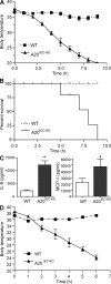
A20 deficiency in IECs sensitizes mice to TNF-induced toxicity. (A and B) Mice were injected i.p. with 5 µg of recombinant mouse TNF. Body temperature (A) and survival (B) of A20IEC-KO mice (n = 8) and littermate control mice (WT; n = 6). (C) Serum IL-6 and MCP-1 levels 4 h after mouse TNF injection. (D) Body temperature of A20IEC-KO mice (n = 6) and control littermate mice (n = 6) after injection with 50 µg of recombinant human TNF. Data are representative of three independent experiments. Error bars represent SEM. *, P < 0.05.
Figure 6.
Figure 6.
A20 deficiency in IECs sensitizes to TNF-induced damage of the small intestine and liver. (A) H&E histology on a section of the small intestine from A20IEC-KO mice and control littermates (WT) 0 and 6 h after mouse TNF injection. Bars, 100 µm. (B) TUNEL staining on sections from small intestine 0 and 6 h after mouse TNF injection, staining apoptotic cells in red. Bars, 50 µm. (C) Caspase activity assayed on tissue homogenates of terminal ileum of A20IEC-KO mice and WT littermates at 0 (control) and 90 min after mouse TNF injection. Error bars represent SEM. (D) H&E histology on liver samples from control (WT) and A20IEC-KO mice 0 and 6 h after mouse TNF injection. Bars, 50 µm. Data are representative of two independent experiments.
Figure 7.
Figure 7.
Commensal intestinal microbes contribute to TNF-induced toxicity in A20IEC-KO mice. (A) Body temperature after mouse TNF challenge of A20IEC-KO mice and control littermate mice (WT) either untreated (IEC-KO, n = 6; WT, n = 5) or treated (IEC-KO, n = 6; WT, n = 8) with broad-spectrum antibiotics. (B) H&E histology on sections from distal ileum 0 and 7 h after i.p. injection of mouse TNF in control (WT) and A20IEC-KO mice treated with antibiotics. Bottom images are magnifications of rectangles in top images. Bars: (top) 100 µm; (bottom) 75 µm. (C) H&E histology on liver samples 0 and 7 h after TNF injection in control (WT) and A20IEC-KO mice either untreated or treated with antibiotics. Bars, 75 µm. Data are representative of two independent experiments. Error bars represent SEM.

Similar articles

Cited by

References

    1. Ameloot P., Takahashi N., Everaerdt B., Hostens J., Eugster H.P., Fiers W., Brouckaert P. 2002. Bioavailability of recombinant tumor necrosis factor determines its lethality in mice. Eur. J. Immunol. 32:2759–2765 10.1002/1521-4141(2002010)32:10<2759::AID-IMMU2759>3.0.CO;2-L - DOI - PubMed
    1. Arsenescu R., Bruno M.E., Rogier E.W., Stefka A.T., McMahan A.E., Wright T.B., Nasser M.S., de Villiers W.J., Kaetzel C.S. 2008. Signature biomarkers in Crohn’s disease: toward a molecular classification. Mucosal Immunol. 1:399–411 10.1038/mi.2008.32 - DOI - PubMed
    1. Artis D. 2008. Epithelial-cell recognition of commensal bacteria and maintenance of immune homeostasis in the gut. Nat. Rev. Immunol. 8:411–420 10.1038/nri2316 - DOI - PubMed
    1. Barmada M.M., Brant S.R., Nicolae D.L., Achkar J.P., Panhuysen C.I., Bayless T.M., Cho J.H., Duerr R.H. 2004. A genome scan in 260 inflammatory bowel disease-affected relative pairs. Inflamm. Bowel Dis. 10:15–22 10.1097/00054725-200401000-00002 - DOI - PubMed
    1. Baumgart D.C., Carding S.R. 2007. Inflammatory bowel disease: cause and immunobiology. Lancet. 369:1627–1640 10.1016/S0140-6736(07)60750-8 - DOI - PubMed

Publication types

MeSH terms

Substances