Breast cancer cells in three-dimensional culture display an enhanced radioresponse after coordinate targeting of integrin alpha5beta1 and fibronectin
- PMID: 20516121
- PMCID: PMC2933183
- DOI: 10.1158/0008-5472.CAN-09-2319
Breast cancer cells in three-dimensional culture display an enhanced radioresponse after coordinate targeting of integrin alpha5beta1 and fibronectin
Abstract
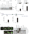
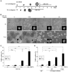
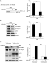
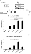
Tactics to selectively enhance cancer radioresponse are of great interest. Cancer cells actively elaborate and remodel their extracellular matrix (ECM) to aid in survival and progression. Previous work has shown that beta1-integrin inhibitory antibodies can enhance the growth-inhibitory and apoptotic responses of human breast cancer cell lines to ionizing radiation, either when cells are cultured in three-dimensional laminin-rich ECM (3D lrECM) or grown as xenografts in mice. Here, we show that a specific alpha heterodimer of beta1-integrin preferentially mediates a prosurvival signal in human breast cancer cells that can be specifically targeted for therapy. 3D lrECM culture conditions were used to compare alpha-integrin heterodimer expression in malignant and nonmalignant cell lines. Under these conditions, we found that expression of alpha5beta1-integrin was upregulated in malignant cells compared with nonmalignant breast cells. Similarly, we found that normal and oncofetal splice variants of fibronectin, the primary ECM ligand of alpha5beta1-integrin, were also strikingly upregulated in malignant cell lines compared with nonmalignant acini. Cell treatment with a peptide that disrupts the interactions of alpha5beta1-integrin with fibronectin promoted apoptosis in malignant cells and further heightened the apoptotic effects of radiation. In support of these results, an analysis of gene expression array data from breast cancer patients revealed an association of high levels of alpha5-integrin expression with decreased survival. Our findings offer preclinical validation of fibronectin and alpha5beta1-integrin as targets for breast cancer therapy.
Copyright 2010 AACR.
Conflict of interest statement
Figures


Similar articles
-
β1-Integrin via NF-κB signaling is essential for acquisition of invasiveness in a model of radiation treated in situ breast cancer.Breast Cancer Res. 2013;15(4):R60. doi: 10.1186/bcr3454. Breast Cancer Res. 2013. PMID: 23883667 Free PMC article.
-
Regulation of ionizing radiation-induced adhesion of breast cancer cells to fibronectin by alpha5beta1 integrin.Radiat Res. 2014 Jun;181(6):650-8. doi: 10.1667/RR13543.1. Epub 2014 May 1. Radiat Res. 2014. PMID: 24785587 Free PMC article.
-
ERBB2-mediated transcriptional up-regulation of the alpha5beta1 integrin fibronectin receptor promotes tumor cell survival under adverse conditions.Cancer Res. 2006 Apr 1;66(7):3715-25. doi: 10.1158/0008-5472.CAN-05-2823. Cancer Res. 2006. PMID: 16585198
-
beta1 integrin targeting to enhance radiation therapy.Int J Radiat Biol. 2009 Nov;85(11):923-8. doi: 10.3109/09553000903232876. Int J Radiat Biol. 2009. PMID: 19895268 Review.
-
Targeting integrin α5β1 in urological tumors: opportunities and challenges.Front Oncol. 2023 Jul 6;13:1165073. doi: 10.3389/fonc.2023.1165073. eCollection 2023. Front Oncol. 2023. PMID: 37483505 Free PMC article. Review.
Cited by
-
Identifications of novel mechanisms in breast cancer cells involving duct-like multicellular spheroid formation after exposure to the Random Positioning Machine.Sci Rep. 2016 May 27;6:26887. doi: 10.1038/srep26887. Sci Rep. 2016. PMID: 27230828 Free PMC article.
-
β1-Integrin via NF-κB signaling is essential for acquisition of invasiveness in a model of radiation treated in situ breast cancer.Breast Cancer Res. 2013;15(4):R60. doi: 10.1186/bcr3454. Breast Cancer Res. 2013. PMID: 23883667 Free PMC article.
-
ITGA5 inhibition in pancreatic stellate cells attenuates desmoplasia and potentiates efficacy of chemotherapy in pancreatic cancer.Sci Adv. 2019 Sep 4;5(9):eaax2770. doi: 10.1126/sciadv.aax2770. eCollection 2019 Sep. Sci Adv. 2019. PMID: 31517053 Free PMC article.
-
Integrin α5β1, the Fibronectin Receptor, as a Pertinent Therapeutic Target in Solid Tumors.Cancers (Basel). 2013 Jan 15;5(1):27-47. doi: 10.3390/cancers5010027. Cancers (Basel). 2013. PMID: 24216697 Free PMC article.
-
Caveolin-1-negative head and neck squamous cell carcinoma primary tumors display increased epithelial to mesenchymal transition and prometastatic properties.Oncotarget. 2015 Dec 8;6(39):41884-901. doi: 10.18632/oncotarget.6099. Oncotarget. 2015. PMID: 26474461 Free PMC article.
References
-
- Yao ES, Zhang H, Chen YY, et al. Increased β1 integrin is associated with decreased survival in invasive breast cancer. Cancer Res. 2007;67:659–64. - PubMed
-
- Hynes RO. Integrins: bidirectional, allosteric signaling machines. Cell. 2002;110:673–87. - PubMed
-
- Ruoslahti E, Pierschbacher MD. New perspectives in cell adhesion: RGD and integrins. Science. 1987;238:491–7. - PubMed
Publication types
MeSH terms
Substances
Grants and funding
- R37 CA064786/CA/NCI NIH HHS/United States
- U54 CA126552/CA/NCI NIH HHS/United States
- R37CA064786/CA/NCI NIH HHS/United States
- U54 CA143836-01/CA/NCI NIH HHS/United States
- U01CA143233/CA/NCI NIH HHS/United States
- R01 CA057621/CA/NCI NIH HHS/United States
- R01CA057621/CA/NCI NIH HHS/United States
- U01 CA143233/CA/NCI NIH HHS/United States
- U54CA126552/CA/NCI NIH HHS/United States
- U54CA112970/CA/NCI NIH HHS/United States
- U01 CA143233-01/CA/NCI NIH HHS/United States
- R01 CA057621-10/CA/NCI NIH HHS/United States
- U54 CA143836/CA/NCI NIH HHS/United States
- U54CA143836/CA/NCI NIH HHS/United States
- R01 CA124891/CA/NCI NIH HHS/United States
- U54 CA112970-01/CA/NCI NIH HHS/United States
- U54 CA112970/CA/NCI NIH HHS/United States
- 1R01CA124891/CA/NCI NIH HHS/United States
- U54 CA126552-04/CA/NCI NIH HHS/United States
- R37 CA064786-14/CA/NCI NIH HHS/United States
LinkOut - more resources
Full Text Sources
Other Literature Sources
Medical

