. 2010 May 28;141(5):786-98.
doi: 10.1016/j.cell.2010.03.049.

Actin-bundling protein TRIOBP forms resilient rootlets of hair cell stereocilia essential for hearing

Affiliations

Actin-bundling protein TRIOBP forms resilient rootlets of hair cell stereocilia essential for hearing

Shin-ichiro Kitajiri et al. Cell. .

Abstract

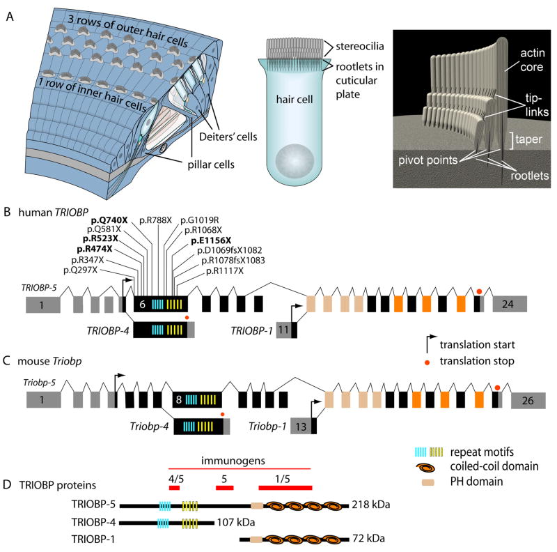
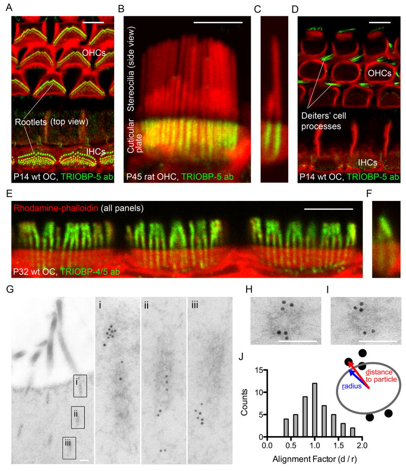
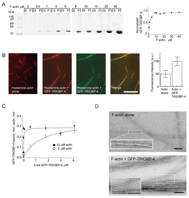
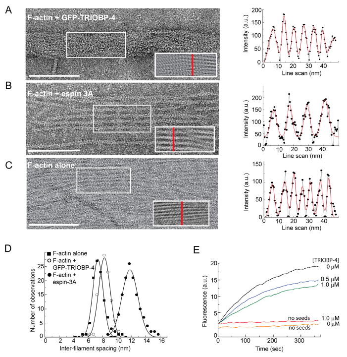
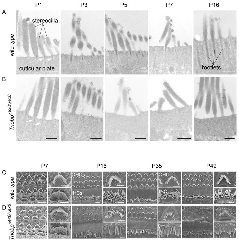
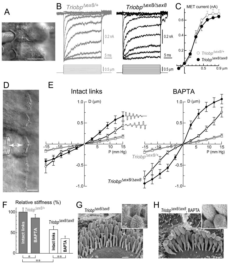
Inner ear hair cells detect sound through deflection of mechanosensory stereocilia. Each stereocilium is supported by a paracrystalline array of parallel actin filaments that are packed more densely at the base, forming a rootlet extending into the cell body. The function of rootlets and the molecules responsible for their formation are unknown. We found that TRIOBP, a cytoskeleton-associated protein mutated in human hereditary deafness DFNB28, is localized to rootlets. In vitro, purified TRIOBP isoform 4 protein organizes actin filaments into uniquely dense bundles reminiscent of rootlets but distinct from bundles formed by espin, an actin crosslinker in stereocilia. We generated mutant Triobp mice (Triobp(Deltaex8/Deltaex8)) that are profoundly deaf. Stereocilia of Triobp(Deltaex8/Deltaex8) mice develop normally but fail to form rootlets and are easier to deflect and damage. Thus, F-actin bundling by TRIOBP provides durability and rigidity for normal mechanosensitivity of stereocilia and may contribute to resilient cytoskeletal structures elsewhere.


Figures

Figure 1
Figure 1. Stereocilia Rootlets within the Organ of Corti and TRIOBP Structure, Isoforms and Immunogens
(A) Organ of Corti schematic showing three rows of outer hair cells (OHCs) and one row of inner hair cells (IHCs) supported by non-sensory pillar cells, Deiter’s cells and other supporting cells (left panel). Mechanosensitive stereocilia are arranged into three rows of increasing heights at the apical surface of each hair cell and anchored to the cuticular plate by rootlets protruding into the cell body (middle panel). Unidirectional actin filaments form a paracrystalline core of the stereocilium and become denser at the taper and within the cuticular plate, forming the rootlet (right panel). When stereocilia are deflected, rootlets are bent at the pivot points. (B) Human TRIOBP gene structure showing the three TRIOBP transcript classes (TRIOBP-5, TRIOBP-4 and TRIOBP-1), alternative promoters upstream of exons 1 and 11, and thirteen mutations causing DFNB28 deafness that are all located in exon 6 (Riazuddin et al., 2006; Shahin et al., 2006; four novel mutations are shown in bold). TRIOBP-4 has a translation stop codon and 3’ UTR in exon 6. Exon 11 includes the 5’ UTR and translation start codon of TRIOBP-1. Cassette exons 4 and 10 provide additional variation. Transcripts encoding TRIOBP-1 and TRIOBP-4 share no exons or protein sequence similarity. (C) Mouse Triobp gene structure is similar to human TRIOBP. Mouse exon 8 corresponds in sequence to human exon 6. (D) Three deduced protein isoforms encoded by mouse Triobp and predicted domains. Immunogens labeled 4/5, 5 and 1/5 were used to generate antibodies recognizing both TRIOBP-4 and TRIOBP-5, TRIOBP-5 only, and both TRIOBP-1 and TRIOBP-5, respectively.
Figure 2
Figure 2. TRIOBP Is Localized to Stereocilia Rootlets
(A) TRIOBP-5 localizes to stereocilia rootlets of wild type (wt) mouse inner (IHCs) and outer (OHCs) hair cells. In all panels A–F, green represents TRIOBP antibody staining (ab), while red is rhodamine-phalloidin staining of the F-actin cytoskeleton. Scale bars in panels A–F, 5 μm. (B and C) Side view of stereocilia shows rootlets penetrating into the cuticular plate. (D) TRIOBP-5 labeling in non-sensory Deiters’ and pillar cells of the organ of Corti. (E and F) TRIOBP-4/5 labeling in the rootlets and stereocilia core of IHCs (E) and OHCs (F). (G) TEM image of post-embedded immunogold labeled transverse sections of stereocilia rootlets in wild type mouse OHCs using TRIOBP-5 antiserum. Insets (i, ii, iii) show magnified views of rootlets. Scale bars, 200 nm. (H and I) TRIOBP-5 immunogold labeling at the periphery of the stereocilia rootlets in horizontal cross sections. Scale bars, 100 nm. (J) Gold particles (n = 47, 10 rootlets) are distributed with a modal alignment factor (d/r) close to 1 suggesting predominant localization at the periphery of the rootlet. Age of the specimens: A and D, postnatal day 14 (P14); B and C, P45; E and F, P32; G–I, P6. See also Figure S1.
Figure 3
Figure 3. TRIOBP-4 Binds and Bundles Actin Filaments
(A) Binding affinity of GFP-TRIOBP-4 for F-actin measured using high-speed co-sedimentation. Coomassie-stained SDS PAGE analysis (left panel) of 2 μM GFP-TRIOBP-4 mixed with increasing amounts of F-actin (0 to 40 μM, lower band). Supernatants (S) and pellets (P) are shown after 385,000 x gmax for 15 min. GFP-TRIOBP-4 (upper band) did not pellet in the absence of F-actin. Bound GFP-TRIOBP-4 was calculated from the amount depleted from supernatants (right panel). The density of each band was measured and normalized to GFP-TRIOBP-4 alone. (B) GFP-TRIOBP-4 binding to actin (right 3 panels). Left panel shows TIRF imaging of 20% rhodamine-labeled actin filaments without GFP-TRIOBP-4 (red). The next three panels show 20% rhodamine-labeled filaments (red) incubated with 2 μM GFP-TRIOBP-4 (green) and the merge of red and green channels. Scale bar, 10 μm. Right panel shows average fluorescent intensity of rhodamine-labeled F-actin with and without GFP-TRIOBP-4. (C) Evaluation of the binding of GFP-TRIOBP-4 to F-actin from four independent experiments. The molar ratio of GFP-TRIOBP-4 sedimented at 22,000 x gmax for 20 min with total actin was plotted against free GFP-TRIOBP-4 in the supernatant. Proteins were separated by SDS-PAGE, and the amount of free and bound GFP-TRIOBP-4 quantified using densitometry, plotted and fitted to a hyperbola. (D) TEM of negative stained 2-D rafts of F-actin alone (left panel) and F-actin bundled with GFP-TRIOBP-4 (right). Insets show the actin filaments at 3X higher magnification. Scale bars, 1 μm.
Figure 4
Figure 4. TRIOBP-4 Assembles Actin into the Bundles of Unusually High Density That Resemble Stereocilia Rootlets
(A–C) TEM images of negative stained actin filaments that were formed on a monolayer lipid membrane at a 4:1 molar ratio of actin to GFP-TRIOBP-4 (A), at a 4:1 molar ratio of actin to espin 3A (B), and actin filaments alone at a density that allows stochastic parallel arrangement of filaments (C). Scale bars, 100 nm. Right insets show spatial periodicity of the data within the left inserts that was revealed by FFT filtering. Right panels show the intensity profiles along red vertical scans perpendicular to actin filaments (grey lines, solid circles) and the fit to a sum of Gaussian distributions (red lines). (D) Histograms of distances between the centers of actin filaments from panels A–C (right) is shown with actin alone (square), actin plus GFP-TRIOBP-4 (open circle) or espin 3A (closed circle). The histograms were fit to a Gaussian distribution. (E) Purified recombinant TRIOBP-4 inhibits the rate of actin polymerization but does not cause a marked nucleation effect. The plots show solution-based actin polymerization using 10% pyrene-labeled actin (2 μM total actin) with the designated concentration of TRIOBP (μM) in the presence or absence of 0.4 nM actin seeds. Similar results were obtained with GFP-tagged TRIOBP-4. See also Figure S2.
Figure 5
Figure 5. Dynamics of F-actin Bundle Formation with GFP-TRIOBP-4 Observed by Time-lapse TIRF Microscopy
(A) Two F-actin structures coalesce in a zipper-like fashion (green arrows). (B) Coalescence of F-actin structures that become attached to one another (red arrows). (C) Bipolar bundles are elongating from both ends (white arrow heads, top row). Scale bar, 5 μm. Movie S1 showing a larger field of view and more examples of bundling behavior is available as supporting information.
Figure 6
Figure 6. TriobpΔex8/Δex8 Stereocilia Fail to Develop Rootlets, Fuse Together, and Degenerate
(A and B) TEM images of stereocilia insertions into the cuticular plate in Triobp+/+ (A) and TriobpΔex8/Δex8 (B) cochlear hair cells. Scale bars, 500 nm. (C and D) SEM images of stereocilia bundles of Triobp+/+ (C) and TriobpΔex8/Δex8 (D) cochlear hair cells. A set of images is shown for each developmental stage (P7–P49). The left image of each set is a surface view of the organ of Corti showing one row of IHCs (bottom) and three rows of OHCs (upper rows). The upper right image illustrates an OHC at higher magnification, while the right lower image illustrates an IHC. Scale bars, left overview 5 μm; right panels of individual cells, 2 μm. See also Figures S3 and S4.
Figure 7
Figure 7. TRIOBP-4/5 Deficiency Does Not Affect Mechano-Electrical Transduction (MET) But Reduces Rigidity of the Stereocilia Bundle
(A) An OHC with a piezo-driven probe (left) and a patch pipette (right). Scale bar, 10 μm. (B) MET responses (top traces) evoked by graded deflections of stereocilia (bottom traces) in TriobpΔex8/+ (left) and TriobpΔex8/Δex8 (right) OHCs. Age of the cells: P2+3 days and P2+2 days in vitro, respectively. Holding potential: −80 mV. (C) Relationship between peak transduction current and probe displacement in TriobpΔex8/+ (open circles, n=4) and TriobpΔex8/Δex8 (closed circles, n=3) OHCs. Average data are shown as mean ± SE in all panels. (D) Deflection of an IHC bundle by fluid-jet. Pressure steps from −15 to +15 mm Hg produced fluid flow (arrow) from the puff pipette (contour on the left) that deflects stereocilia in a positive or negative direction. Scale bar, 5 μm. For quantification of fluid-jet stimuli see Figure S5. (E) Displacement of tallest stereocilia rank (D, μm) as a function of fluid-jet pressure (P, mm Hg) in IHCs of TriobpΔex8/+ (grey symbols) and TriobpΔex8/Δex8 (black symbols) littermates with intact stereocilia links (left panel) and after application of Ca2+-free solution with BAPTA (right). A single initial large deflection of TriobpΔex8/Δex8 stereocilia (but not TriobpΔex8/+) resulted in a very flexible bundle. Thus, stimuli were presented in increasing (open symbols) and decreasing intensity order (solid symbols). Number of cells: TriobpΔex8/+ (no BAPTA) n = 19 (increasing stimuli), n = 5 (decreasing stimuli); TriobpΔex8/+ (BAPTA) n = 19 (increasing stimuli), n = 5 (decreasing stimuli); TriobpΔex8/Δex8 n = 16 (each of four groups). Age of the cells: P4 + 2–4 days in vitro. (F) Relative changes of hair bundle stiffness. Stiffness was assumed to be inversely proportional to the slope (D/P) of displacement-pressure relationships at low stimuli intensities (from −6 to +6 mm Hg) and expressed as percentage of average stiffness of TriobpΔex8/+ bundles with intact links. Each bundle was deflected by the same fluid-jet before and after BAPTA application. Stiffness data for TriobpΔex8/+ IHCs are combined for two different stimuli presentation regimes, as no difference was observed to increasing and decreasing stimuli. Stiffness data for TriobpΔex8/Δex8 IHCs were collected only for increasing stimuli. Significance was assessed by t-test: TriobpΔex8/Δex8 IHCs vs. control TriobpΔex8/+ IHCs, independent t-test; control vs. BAPTA-treated, paired t-test (* - p<0.05; ** - p<0.01). Number of cells: TriobpΔex8/+ n = 23; TriobpΔex8/Δex8 n = 16. (G and H) SEM images of TriobpΔex8/Δex8 IHCs incubated in standard HBSS (G) and in Ca2+-free HBSS with 10 mM BAPTA for 5 min (H). Insets show magnified images of stereocilia links. Links between stereocilia were eliminated by BAPTA. Age of the cells: (G) P2+2 days in vitro; (H) P3+7 days in vitro. Scale bars, 0.5 μm (main panels), 200 nm (insets). See also Figure S5.

Comment in

  • Wrapping up stereocilia rootlets.
    Boutet de Monvel J, Petit C. Boutet de Monvel J, et al. Cell. 2010 May 28;141(5):748-50. doi: 10.1016/j.cell.2010.05.022. Cell. 2010. PMID: 20510920

Similar articles

Cited by

References

    1. Bartles JR, Zheng L, Li A, Wierda A, Chen B. Small espin: a third actin-bundling protein and potential forked protein ortholog in brush border microvilli. J Cell Biol. 1998;143:107–119. - PMC - PubMed
    1. Belyantseva IA, Boger ET, Naz S, Frolenkov GI, Sellers JR, Ahmed ZM, Griffith AJ, Friedman TB. Myosin-XVa is required for tip localization of whirlin and differential elongation of hair-cell stereocilia. Nat Cell Biol. 2005;7:148–156. - PubMed
    1. Belyantseva IA, Perrin BJ, Sonnemann KJ, Zhu M, Stepanyan R, McGee J, Frolenkov GI, Walsh EJ, Friderici KH, Friedman TB, et al. Gamma-actin is required for cytoskeletal maintenance but not development. Proc Natl Acad Sci USA. 2009;106:9703–9708. - PMC - PubMed
    1. Beurg M, Nam JH, Crawford A, Fettiplace R. The actions of calcium on hair bundle mechanics in mammalian cochlear hair cells. Biophys J. 2008;94:2639–2653. - PMC - PubMed
    1. Beurg M, Fettiplace R, Nam JH, Ricci AJ. Localization of inner hair cell mechanotransducer channels using high-speed calcium imaging. Nat Neurosci. 2009;12:553–558. - PMC - PubMed

Publication types

Substances

Associated data