Immunological basis for the gender differences in murine Paracoccidioides brasiliensis infection
- PMID: 20505765
- PMCID: PMC2873977
- DOI: 10.1371/journal.pone.0010757
Immunological basis for the gender differences in murine Paracoccidioides brasiliensis infection
Abstract
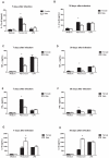
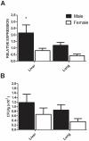
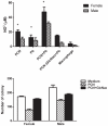
This study aimed to investigate the immunological mechanisms involved in the gender distinct incidence of paracoccidioidomycosis (pcm), an endemic systemic mycosis in Latin America, which is at least 10 times more frequent in men than in women. Then, we compared the immune response of male and female mice to Paracoccidioides brasiliensis infection, as well as the influence in the gender differences exerted by paracoccin, a P. brasiliensis component with carbohydrate recognition property. High production of Th1 cytokines and T-bet expression have been detected in the paracoccin stimulated cultures of spleen cells from infected female mice. In contrast, in similar experimental conditions, cells from infected males produced higher levels of the Th2 cytokines and expressed GATA-3. Macrophages from male and female mice when stimulated with paracoccin displayed similar phagocytic capability, while fungicidal activity was two times more efficiently performed by macrophages from female mice, a fact that was associated with 50% higher levels of nitric oxide production. In order to evaluate the role of sexual hormones in the observed gender distinction, we have utilized mice that have been submitted to gonadectomy followed by inverse hormonal reconstitution. Spleen cells derived from castrated males reconstituted with estradiol have produced higher levels of IFN-gamma (1291+/-15 pg/mL) and lower levels of IL-10 (494+/-38 pg/mL), than normal male in response to paracoccin stimulus. In contrast, spleen cells from castrated female mice that had been treated with testosterone produced more IL-10 (1284+/-36 pg/mL) and less IFN-gamma (587+/-14 pg/mL) than cells from normal female. In conclusion, our results reveal that the sexual hormones had a profound effect on the biology of immune cells, and estradiol favours protective responses to P. brasiliensis infection. In addition, fungal components, such as paracoccin, may provide additional support to the gender dimorphic immunity that marks P. brasiliensis infection.
Conflict of interest statement
Figures


Similar articles
-
Recombinant paracoccin reproduces the biological properties of the native protein and induces protective Th1 immunity against Paracoccidioides brasiliensis infection.PLoS Negl Trop Dis. 2014 Apr 17;8(4):e2788. doi: 10.1371/journal.pntd.0002788. eCollection 2014 Apr. PLoS Negl Trop Dis. 2014. PMID: 24743161 Free PMC article.
-
Impact of Paracoccin Gene Silencing on Paracoccidioides brasiliensis Virulence.mBio. 2017 Jul 18;8(4):e00537-17. doi: 10.1128/mBio.00537-17. mBio. 2017. PMID: 28720727 Free PMC article.
-
In pulmonary paracoccidioidomycosis IL-10 deficiency leads to increased immunity and regressive infection without enhancing tissue pathology.PLoS Negl Trop Dis. 2013 Oct 24;7(10):e2512. doi: 10.1371/journal.pntd.0002512. eCollection 2013. PLoS Negl Trop Dis. 2013. PMID: 24205424 Free PMC article.
-
[The Research Encouragement Award. Effects of sex hormones on sexual difference of experimental paracoccidioidomycosis].Nihon Ishinkin Gakkai Zasshi. 1999;40(1):1-8. doi: 10.3314/jjmm.40.1. Nihon Ishinkin Gakkai Zasshi. 1999. PMID: 9929575 Review. Japanese.
-
Cytokines produced by susceptible and resistant mice in the course of Paracoccidioides brasiliensis infection.Braz J Med Biol Res. 1998 May;31(5):615-23. doi: 10.1590/s0100-879x1998000500003. Braz J Med Biol Res. 1998. PMID: 9698765 Review.
Cited by
-
Immunity to fungal infections.Nat Rev Immunol. 2011 Apr;11(4):275-88. doi: 10.1038/nri2939. Epub 2011 Mar 11. Nat Rev Immunol. 2011. PMID: 21394104 Review.
-
Single-Cell RNA Sequencing Reveals the Heterogeneity of Tumor-Associated Macrophage in Non-Small Cell Lung Cancer and Differences Between Sexes.Front Immunol. 2021 Nov 5;12:756722. doi: 10.3389/fimmu.2021.756722. eCollection 2021. Front Immunol. 2021. PMID: 34804043 Free PMC article.
-
SeXX Matters in Multiple Sclerosis.Front Neurol. 2020 Jul 3;11:616. doi: 10.3389/fneur.2020.00616. eCollection 2020. Front Neurol. 2020. PMID: 32719651 Free PMC article. Review.
-
Role of interleukin 4 and its receptor in clinical presentation of chronic extrinsic allergic alveolitis: a pilot study.Multidiscip Respir Med. 2013 May 30;8(1):35. doi: 10.1186/2049-6958-8-35. Multidiscip Respir Med. 2013. PMID: 23721656 Free PMC article.
-
Epidemiology of Invasive Fungal Infections in Latin America.Curr Fungal Infect Rep. 2012 Mar;6(1):23-34. doi: 10.1007/s12281-011-0081-7. Epub 2012 Jan 5. Curr Fungal Infect Rep. 2012. PMID: 22363832 Free PMC article.
References
-
- Restrepo A, Benard G, de Castro CC, Agudelo CA, Tobon AM. Pulmonary paracoccidioidomycosis. Semin Respir Crit Care Med. 2008;29:182–197. - PubMed
-
- McEwen JG, Bedoya V, Patino MM, Salazar ME, Restrepo A. Experimental murine paracoccidiodomycosis induced by the inhalation of conidia. J Med Vet Mycol. 1987;25:165–175. - PubMed
-
- Ramos ESM, Saraiva Ldo E. Paracoccidioidomycosis. Dermatol Clin. 2008;26:257–269, vii. - PubMed
-
- Tobon AM, Agudelo CA, Osorio ML, Alvarez DL, Arango M, et al. Residual pulmonary abnormalities in adult patients with chronic paracoccidioidomycosis: prolonged follow-up after itraconazole therapy. Clin Infect Dis. 2003;37:898–904. - PubMed
Publication types
MeSH terms
Substances
LinkOut - more resources
Full Text Sources

