. 2010 Jun 7;207(6):1321-32.
doi: 10.1084/jem.20092253. Epub 2010 May 24.

Microbiota innate stimulation is a prerequisite for T cell spontaneous proliferation and induction of experimental colitis

Affiliations

Microbiota innate stimulation is a prerequisite for T cell spontaneous proliferation and induction of experimental colitis

Ting Feng et al. J Exp Med. .

Erratum in

  • J Exp Med. 2010 Jul 5;207(7):1569

Abstract

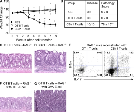
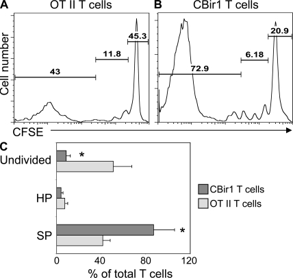
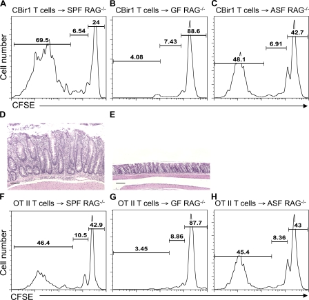
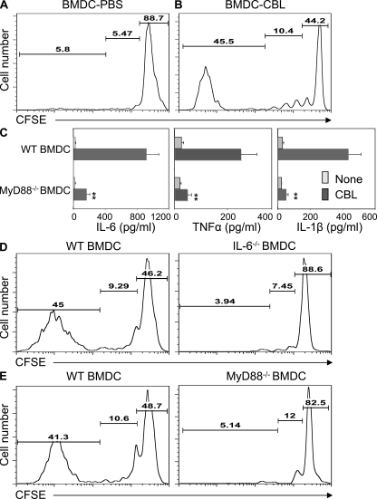
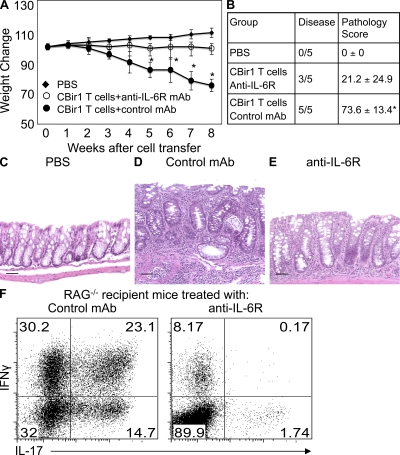
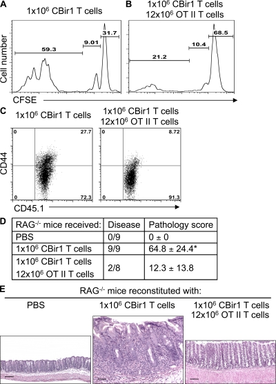
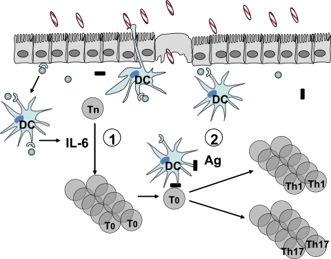
Little is known about how the microbiota regulates T cell proliferation and whether spontaneous T cell proliferation is involved in the pathogenesis of inflammatory bowel disease. In this study, we show that stimulation of innate pathways by microbiota-derived ligands and antigen-specific T cell stimulation are both required for intestinal inflammation. Microbiota-derived ligands promoted spontaneous T cell proliferation by activating dendritic cells (DCs) to produce IL-6 via Myd88, as shown by the spontaneous proliferation of T cells adoptively transferred into specific pathogen-free (SPF) RAG-/- mice, but not in germfree RAG-/- mice. Reconstitution of germfree RAG-/- mice with cecal bacterial lysate-pulsed DCs, but not with IL-6-/- or Myd88-/- DCs, restored spontaneous T cell proliferation. CBir1 TCR transgenic (CBir1 Tg) T cells, which are specific for an immunodominant microbiota antigen, induced colitis in SPF RAG-/- mice. Blocking the spontaneous proliferation of CBir1 Tg T cells by co-transferring bulk OT II CD4+ T cells abrogated colitis development. Although transferred OT II T cells underwent spontaneous proliferation in RAG-/- mice, the recipients failed to develop colitis because of the lack of cognate antigen in the intestinal lumen. Collectively, our data demonstrate that induction of colitis requires both spontaneous proliferation of T cells driven by microbiota-derived innate signals and antigen-specific T cell proliferation.


Figures

Figure 1.
Figure 1.
Adoptive transfer of CBir1 Tg CD4+ T cells but not OT II CD4+ T cells induces colitis in RAG−/− mice. Groups of 5 B6.RAG−/− mice were injected with 106 CBir1 Tg CD4+ T cells, 106 OT II CD4+ T cells, or PBS as control. Recipient mice were weighed weekly. 8 wk after transfer, the mice were sacrificed and assessed for histopathology. LP CD4+ T cell cytokine production was determined by flow cytometry. (A) Weight changes of the recipient mice. *, P < 0.05 compared with the PBS group. (B) Pathological scores of the recipient mice. **, P < 0.01 compared with the PBS group. (C and D) Colonic histopathology of the recipient mice. (E) Intestinal LP CD4+ T cell IL-17 and IFN-γ production. (F and G) Groups of 5 B6.RAG−/− mice were injected with 5 × 10⁶ OT II CD4+ T cells and then intrarectally administered with TET-E. coli (F) or with OVA-E. coli (G) every week. Data are from one of two independent experiments with similar results. Bars: (C, D, and F) 50 µm; (E) 100 µm.
Figure 2.
Figure 2.
More CBir1 Tg CD4+ T cells undergo spontaneous proliferation than do OT II CD4+ T cells in RAG−/− mice. 106 CFSE-CBir1 Tg CD4+ T cells or 106 CFSE-OT II CD4+ T cells were transferred into RAG−/− mice, and the recipient mice were sacrificed 10 d later. CD4+ T cell proliferation in the spleen was analyzed by CFSE dilution by flow cytometry. (A) OT II CD4+ T cell proliferation in RAG−/− mice. (B) CBir1 Tg CD4+ T cell proliferation in RAG−/− mice. Numbers in each plot represent the percentage of donor cells undergoing spontaneous proliferation, homeostatic proliferation, and staying undivided, respectively. Data are representative of at least four individual mice of each group from three independent experiments. (C) Percentage of transferred T cells in spontaneous proliferation (SP), homeostatic proliferation (HP), and undivided from three independent experiments. *, P < 0.05 for CBir1 Tg T cells against OT II T cells.
Figure 3.
Figure 3.
CD4+ T cells do not undergo spontaneous proliferation in germfree RAG−/− mice. 106 CFSE-CBir1 Tg CD4+ T cells (A–C) or 106 CFSE-OT II CD4+ T cells (F–H) were transferred into RAG−/− mice housed under SPF conditions (A and F), germfree (GF) conditions (B and G), or ASF conditions (C and H). 10 d after the transfer, division of the CFSE-labeled CD4+ T cells was determined by CFSE dilution. Numbers in each plot represent the percentage of donor cells undergoing spontaneous proliferation, homeostatic proliferation, and staying undivided, respectively. Data are representative of at least three individual mice of each group from three independent experiments. Histopathology of the colon of CBir1 Tg T cell recipients at 5 mice/group under SPF conditions (D) and germfree conditions (E) was assessed 8 wk after cell transfer. Bars, 100 µm. Data are representative of three independent experiments with similar results.
Figure 4.
Figure 4.
Microbiota stimulation of DC IL-6 production via Myd88 pathway promotes T cell spontaneous proliferation. BMDCs generated from B6 mice were pulsed with PBS or 100 µg/ml of CBLs for 24 h. 106 CFSE-RAG−/−.OT II CD4+ T cells were transferred together with 106 PBS-pulsed BMDCs (A) or with CBL-pulsed BMDCs (B) into germfree RAG−/− mice. 10 d after the transfer, division of the CFSE-labeled CD4+ T cells was determined by flow cytometry. Numbers in each plot represent the percentage of donor cells undergoing spontaneous proliferation, homeostatic proliferation, and staying undivided, respectively. (C) BMDCs generated from B6 mice or B6.Myd88−/− mice were stimulated with 100 µg/ml of CBL for 24 h. To determine IL-1β production, 2 mM ATP was added in the last 10 min of the culture. IL-6, TNF, and IL-1β production in the supernatant was determined by ELISA. **, P < 0.01 compared with WT DCs. (D) 106 CFSE-RAG−/−.OT II CD4+ T cells were transferred together with 106 CBL-pulsed B6.BMDCs or CBL-pulsed B6.IL-6−/− BMDCs into germfree RAG−/− mice. (E) 106 CFSE-RAG−/−.OT II CD4+ T cells were transferred together with 106 CBL-pulsed B6.BMDCs or CBL-pulsed B6.Myd88−/− BMDCs into germfree RAG−/− mice. 10 d after the transfer, division of the CFSE-labeled CD4+ T cells was determined by flow cytometry. Numbers in each plot represent the percentage of donor cells undergoing spontaneous proliferation, homeostatic proliferation, and undivided, respectively. Data are representative of at least four mice of each group from two independent experiments.
Figure 5.
Figure 5.
Administration of anti–IL-6R mAb inhibits colitis development induced by CBir1 Tg CD4+ T cells in RAG−/− mice. RAG−/− mice were reconstituted with 106 CBir1 Tg CD4+ T cells. Groups of 5 recipient mice were injected with 100 µg anti–IL-6R mAb or control mAb i.p. weekly. A group of five recipient mice were injected i.p. with PBS as control. Recipient mice were weighed weekly. 8 wk after cell transfer, the mice were sacrificed and assessed for histopathology. LP CD4+ T cell cytokine production was determined by flow cytometry. (A) Weight changes of the recipient mice. *, P < 0.05 compared with anti–IL-6R mAb–treated mice. (B) Colonic pathology scores of the recipient mice. *, P < 0.05 compared with anti–IL-6R mAb–treated mice (nonparametric Mann-Whitney test). (C–E) Histopathology of the recipient mice. Bars, 50 µm. (F) Intestinal LP CD4+ T cell IL-17 and IFN-γ production. The representative experiment shown was performed twice with similar data.
Figure 6.
Figure 6.
Co-transfer of large numbers of OT II CD4+ T cells restrains CBir1 Tg T cell spontaneous proliferation and inhibits colitis induction. 106 CFSE-CD45.1 CBir1 Tg CD4+ T cells were transferred into RAG−/− mice alone (A) or together with 12 × 106 CD45.2 RAG−/−.OT II CD4 T cells (B). 10 d after the transfer, division of the CFSE-labeled CBir1 Tg T cells was determined by CFSE dilution by gating on CD45.1+ cells. Numbers in each plot represent the percentage of donor cells undergoing spontaneous proliferation, homeostatic proliferation, and staying undivided, respectively. (C) CD44 expression of CBir1 Tg T cells was determined by flow cytometry by gating on CD45.1+ cells. Data are from one of two independent experiments with similar results. (D) The recipient mice were sacrificed 8 wk after cell transfer and the total pathology score of cecum and large intestine was assessed. *, P < 0.05 compared with the group that received CBir1 Tg T cells and bulk OT II T cells (nonparametric Mann-Whitney test). Data are sum of two independent experiments. (E) Histopathology of colon in the recipient mice. Bars: (E, left) 100 µm; (E, middle and right) 50 µm. The representative experiment shown was performed twice with similar data.
Figure 7.
Figure 7.
Two-hit model of the role of microbiota in the pathogenesis of colitis. (1) Microbiota stimulates intestinal innate cell IL-6 production through TLR–TLR ligand interactions. IL-6, and probably other factors, promotes spontaneous proliferation of T cells, which are in T0 status and do not produce effector cytokines. (2) Once T0 cells encounter their cognate microbiota antigen, they will proliferate in a TCR-dependent matter and differentiate into effector T cells, which produce effector cytokines IFN-γ and IL-17 locally in the colon and induce inflammation.

Similar articles

Cited by

References

    1. Akira S., Uematsu S., Takeuchi O. 2006. Pathogen recognition and innate immunity. Cell. 124:783–801 10.1016/j.cell.2006.02.015 - DOI - PubMed
    1. Barthlott T., Kassiotis G., Stockinger B. 2003. T cell regulation as a side effect of homeostasis and competition. J. Exp. Med. 197:451–460 10.1084/jem.20021387 - DOI - PMC - PubMed
    1. Beutler B. 2004. Inferences, questions and possibilities in Toll-like receptor signalling. Nature. 430:257–263 10.1038/nature02761 - DOI - PubMed
    1. Blumberg R.S., Saubermann L.J., Strober W. 1999. Animal models of mucosal inflammation and their relation to human inflammatory bowel disease. Curr. Opin. Immunol. 11:648–656 10.1016/S0952-7915(99)00032-1 - DOI - PubMed
    1. Bourgeois C., Stockinger B. 2006. CD25+CD4+ regulatory T cells and memory T cells prevent lymphopenia-induced proliferation of naive T cells in transient states of lymphopenia. J. Immunol. 177:4558–4566 - PubMed

Publication types

MeSH terms