. 2010 Sep 2;116(9):1528-38.
doi: 10.1182/blood-2009-12-259747. Epub 2010 May 20.

JAK2 V617F impairs hematopoietic stem cell function in a conditional knock-in mouse model of JAK2 V617F-positive essential thrombocythemia

Affiliations

JAK2 V617F impairs hematopoietic stem cell function in a conditional knock-in mouse model of JAK2 V617F-positive essential thrombocythemia

Juan Li et al. Blood. .

Abstract

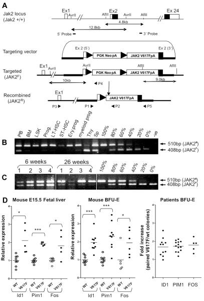
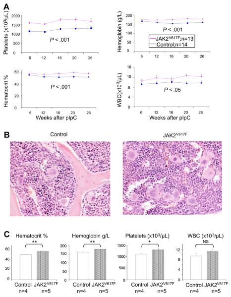
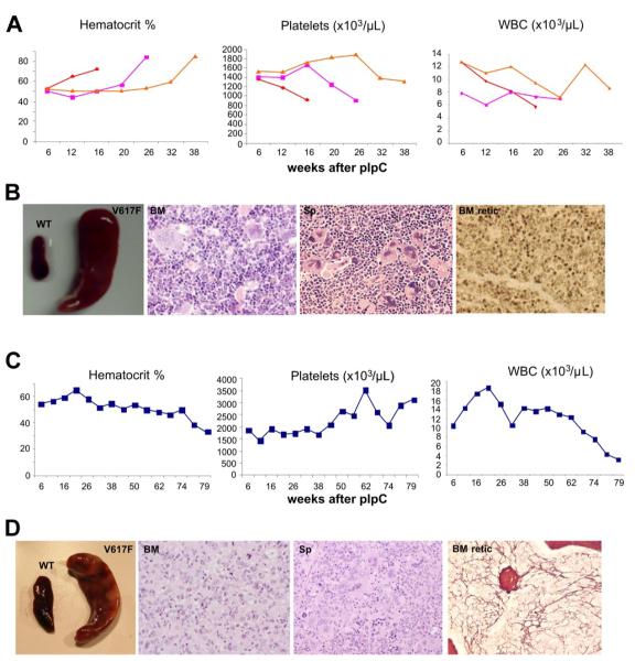
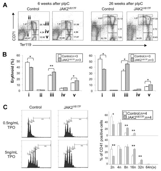
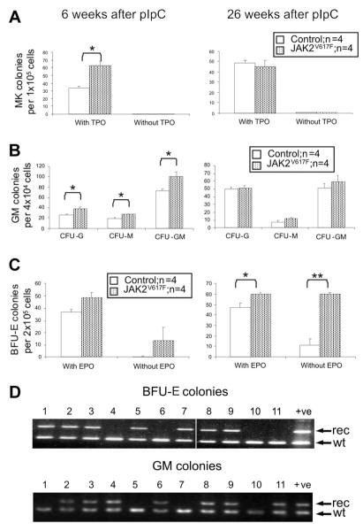
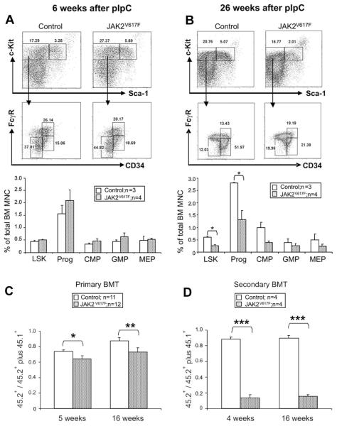
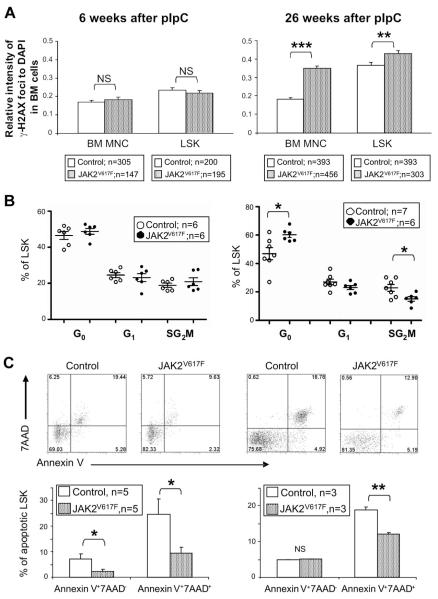
The JAK2 V617F mutation is found in most patients with a myeloproliferative neoplasm and is sufficient to produce a myeloproliferative phenotype in murine retroviral transplantation or transgenic models. However, several lines of evidence suggest that disease phenotype is influenced by the level of mutant JAK2 signaling, and we have therefore generated a conditional knock-in mouse in which a human JAK2 V617F is expressed under the control of the mouse Jak2 locus. Human and murine Jak2 transcripts are expressed at similar levels, and mice develop modest increases in hemoglobin and platelet levels reminiscent of human JAK2 V617F-positive essential thrombocythemia. The phenotype is transplantable and accompanied by increased terminal erythroid and megakaryocyte differentiation together with increased numbers of clonogenic progenitors, including erythropoietin-independent erythroid colonies. Unexpectedly, JAK2(V617F) mice develop reduced numbers of lineage(-)Sca-1(+)c-Kit(+) cells, which exhibit increased DNA damage, reduced apoptosis, and reduced cell cycling. Moreover, competitive bone marrow transplantation studies demonstrated impaired hematopoietic stem cell function in JAK2(V617F) mice. These results suggest that the chronicity of human myeloproliferative neoplasms may reflect a balance between impaired hematopoietic stem cell function and the accumulation of additional mutations.


Figures

Figure 1
Figure 1. Generation of a conditional human JAK2 V617F knock-in allele
(A) Diagram showing the endogenous mouse Jak2 allele, the targeting vector, the knock-in allele resulting from homologous recombination, and the activated recombined allele after excision of the PGKNeo-poly (A) cassette. (B) PCR showing high levels of recombination in BM, stem cells, and progenitors and relatively lower levels in peripheral blood, spleen, and thymus. PCR was performed using P1 + P2 + P4 (A) on genomic DNA from peripheral blood (PB), BM, LSK, total progenitors (Prog), LT-HSC (lineageSca-1+c-Kit+CD34), ST-HSC (lineageSca-1+c-Kit+CD34+), myeloid progenitors (lineageSca-lc-Kit+CD34+ CD71), and erythroid progenitors (lineageSca-lc-Kit+CD34 CD71+), thymus (Thy), and spleen cells (Sp). Serial dilutions were made by mixing the corresponding amount of genomic DNA from JAK2F/+ and JAK2R/+ ES cells. Top (P1 + P2) and bottom (P1 + P4) bands represent the recombined and floxed allele, respectively. (C) PCR (performed as in panel B) showing that the proportion of recombined allele in peripheral blood samples from JAK2V617F mice increases with time. Vertical lines have been inserted to indicate removal of gel lanes. (D) Real-time quantitative PCR analysis showing comparable up-regulation of Stat5 and Erk1/2 target genes in erythroid cells (both in fetal and adult erythroid cells) from JAK2V617F mice compared with results obtained from ET patients. In ET patient samples, individual BFU-E were genotyped, pooled according to genotypes, and transcript levels of target genes in JAK2 V617F mutant and wild-type colonies were compared (far right panel). The fold increase represents the ratio of gene expression in V617F and WT pools with each data point representing an individual patient. Transcript levels of the same target genes were increased by a similar amount in both fetal liver and adult BM BFU-E from JAK2V617F mice (V617F) compared with littermate controls (WT). BFU-E colonies were derived from 3 JAK2V617F and 3 wild-type control mice. Individual colonies from JAK2V617F mice were genotyped to distinguish those carrying the active recombined allele. Colonies were pooled according to the genotype (4-6 colonies/pool). Expression of target genes in a pooled BFU-E sample was calculated relative to the mean of the wild-type pooled samples, which was defined as 1. *P < .05. ***P < .001.
Figure 2
Figure 2. JAK2V617F mice develop a myeloproliferative disease
(A) Time course of blood parameters of JAK2V617F and control mice showing significantly increased platelets, hematocrit, and hemoglobin, and moderately elevated white blood cell counts (JAK2V617F, JAK2F/+Mx1Cre+; controls, JAK2+/+Mx1Cre+). Data are mean ± SEM. (B) Hematoxylin and eosin staining of BM with megakaryocytic hyperplasia with increased clustering and hyperlobated nuclei. (C) Myeloproliferative phenotype is transplantable. Histograms show blood counts of recipient mice 12 weeks after transplantation of 1 × 10⁶ BM cells from either JAK2V617F or control mice. *P < .05. **P < .01. NS indicates not significant. Data are mean ± SEM.
Figure 3
Figure 3. JAK2V617F mice develop PV and myelofibrosis
(A) Blood parameters of mice with a PV-like phenotype displaying a marked increase in hematocrit and a fall in their platelet counts. (B) Mice with a PV-like phenotype show splenomegaly; BM hematoxylin and eosin (BM) showing erythroid and megakaryocytic hyperplasia with clustering and highly pleomorphic morphology; spleen hematoxylin and eosin (Sp) showing megakaryocytic and erythroid hyperplasia; reticulin stain showing no fibrosis in BM (BM retic) and spleen (not shown). (C) Blood parameters of a mouse with BM fibrosis displaying a gradual decline of blood count parameters, including hematocrit and white cells, and an increase in its platelet counts. (D) Splenomegaly in mouse with BM fibrosis; BM hematoxylin and eosin (BM) showing granulocytic hyperplasia with reduced megakaryocytic and erythroid cells; spleen hematoxylin and eosin (Sp) showing megakaryocytic, erythroid, and granulocytic expansion; reticulin stain showing fibrosis in BM (BM retic) but not in spleen (not shown).
Figure 4
Figure 4. JAK2V617F mice exhibit enhanced erythroid and megakaryocyte differentiation
(A) Representative FACS profiles stained with CD71 and Ter119 antibodies are shown; i through v correspond to progressive stages of erythroid differentiation. (B) Histograms show increased terminal erythroid differentiation in JAK2V617F mice BM at 6 and 26 weeks after pIpC. *P < .05. **P < .01. Data are mean ± SEM. (C) Increased megakaryocyte ploidy in JAK2V617F mice. FACS profiles for megakaryocytes grown in liquid culture are shown. Histograms represent results from 2 independent experiments. *P < .05. **P < .01. Data are mean ± SEM.
Figure 5
Figure 5. Increased clonogenic progenitors in JAK2V617F mice
Colony assays were performed 6 and 26 weeks after pIpC. (A) Megakaryocyte colonies (CFU-MK). (B) Granulocyte-macrophage colonies (CFU-GM). (C) Erythroid colonies (BFU-E). *P < .05. **P < .01. Data are mean ± SEM. (D) PCR analysis of individual BFU-E and CFU-GM colonies from mice 26 weeks after pIpC using primers P1 + P2 + P5 (see Figure 1A). No colonies were homozygous for the recombined allele. Vertical line has been inserted to indicate removal of a gel lane. Lanes 1 to 11 indicate individual colonies; +ve, ES cell clone (JAK2R/+); rec, recombined allele (P1 + P2); and wt, wild type allele (P1 + P5).
Figure 6
Figure 6. Reduced numbers and impaired function of hematopoietic stem and progenitor cells in JAK2V617F mice
(A-B) Representative FACS plots and histograms summarizing the frequencies of LSK and progenitor cells (lineageSca-1c-Kit+), common myeloid progenitors (lineagec-Kit+CD34+FcγR), megakaryocyte-erythroid progenitor (lineagec-Kit+CD34FcγR), and granulocyte-monocyte progenitor (lineagec-Kit+CD34+FcγR+) at 6 and 26 weeks after pIpC. (C) BM cells from JAK2V617F mice display reduced repopulating capacity in competitive BM transplantation analysis. BM cells from 2 individual donors (6 weeks after pIpC) for each test genotype (ie, JAK2V617F or JAK2wt littermate controls) were injected into recipients F1 C57Bl6/129SvEvBrd (CD45.1/CD45.2). Histograms show the proportion of peripheral blood-nucleated cells derived from test cells (CD45.2+) compared with repopulation from total donor cells (ie, test plus competitor; CD45.2+ plus CD45.1+). (D) Marked HSC repopulation defect in secondary transplantation recipients. BM cells from primary transplantation recipients were injected into secondary recipients F1 C57Bl6/129SvEvBrd (CD45.1/CD45.2), which were analyzed 4 and 16 weeks after transplantation. Histograms represent the proportion of peripheral blood nucleated cells derived from the test cells (CD45.2+) compared with repopulation from the test plus competitor cells (CD45.2+ plus CD45.1+). *P < .05. **P < .01. ***P < .001.
Figure 7
Figure 7. LSK cells from JAK2V617F mice exhibit increased DNA damage, reduced cell cycling, and reduced apoptosis
(A) Intensities of γ-H2AX foci relative to DAPI obtained for BM cells (70-138 nuclei per mouse) and LSK cells (33-112 nuclei per mouse) from JAK2V617F mice at both 6 and 26 weeks after pIpC. Histograms show significantly higher relative intensities of γ-H2AX foci in cells from JAK2V617F mice compared with the cells from the control mice at 26 weeks, but not at 6 weeks after pIpC. (B) LSK cell-cycle analysis showing increased quiescence in JAK2V617F 26 weeks after pIpC. LSK cells were subjected to 2-parameter analysis with DNA content versus Ki-67 expression, and the percentages of LSK cells in each of the cell-cycle phases were obtained (G0, Ki-67lowPIlow; G1, Ki-67hiPIlow; S/G2/M, Ki-67hiPIhi). *P < .05. (C) Representative FACS plots and histograms showing reduced apoptosis in LSK cells from JAK2V617F mice. Annexin V and 7AAD staining was analyzed on gated LSK cell populations from JAK2V617F mice at both 6 and 26 weeks after pIpC. *P < .05. **P < .01. ***P < .001. NS indicates not significant.

Comment in

  • JAK2 impairs stem cell function?
    Skoda RC. Skoda RC. Blood. 2010 Sep 2;116(9):1392-3. doi: 10.1182/blood-2010-06-287318. Blood. 2010. PMID: 20813904 No abstract available.

Similar articles

Cited by

References

    1. James C, Ugo V, Le Couedic JP, et al. A unique clonal JAK2 mutation leading to constitutive signalling causes polycythaemia vera. Nature. 2005;434(7037):1144–1148. - PubMed
    1. Baxter EJ, Scott LM, Campbell PJ, et al. Acquired mutation of the tyrosine kinase JAK2 in human myeloproliferative disorders. Lancet. 2005;365(9464):1054–1061. - PubMed
    1. Levine RL, Wadleigh M, Cools J, et al. Activating mutation in the tyrosine kinase JAK2 in polycythemia vera, essential thrombocythemia, and myeloid metaplasia with myelofibrosis. Cancer Cell. 2005;7(4):387–397. - PubMed
    1. Kralovics R, Passamonti F, Teo SS, et al. A gain of function mutation in Jak2 is frequently found in patients with myeloproliferative disorders. N Engl J Med. 2005;352(17):1779–1790. - PubMed
    1. Fialkow P, Gartler SM, Yoshida A. Clonal origin of chronic myelocytic leukemia in man. Proc Natl Acad Sci U S A. 1967;58(4):1468–1471. - PMC - PubMed

Publication types

MeSH terms