. 2010 Jul;30(14):3519-30.
doi: 10.1128/MCB.00394-10. Epub 2010 May 17.

Ric-8A and Gi alpha recruit LGN, NuMA, and dynein to the cell cortex to help orient the mitotic spindle

Affiliations

Ric-8A and Gi alpha recruit LGN, NuMA, and dynein to the cell cortex to help orient the mitotic spindle

Geoffrey E Woodard et al. Mol Cell Biol. 2010 Jul.

Abstract

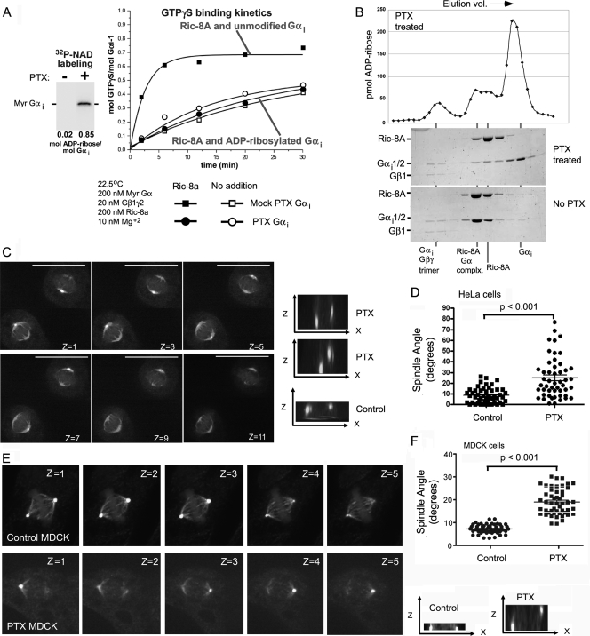
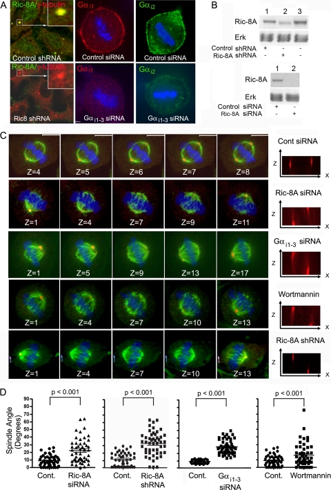
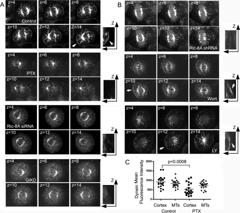
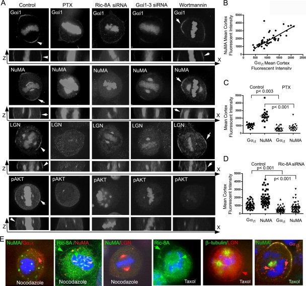
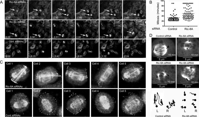
In model organisms, resistance to inhibitors of cholinesterase 8 (Ric-8), a G protein alpha (G alpha) subunit guanine nucleotide exchange factor (GEF), functions to orient mitotic spindles during asymmetric cell divisions; however, whether Ric-8A has any role in mammalian cell division is unknown. We show here that Ric-8A and G alpha(i) function to orient the metaphase mitotic spindle of mammalian adherent cells. During mitosis, Ric-8A localized at the cell cortex, spindle poles, centromeres, central spindle, and midbody. Pertussis toxin proved to be a useful tool in these studies since it blocked the binding of Ric-8A to G alpha(i), thus preventing its GEF activity for G alpha(i). Linking Ric-8A signaling to mammalian cell division, treatment of cells with pertussis toxin, reduction of Ric-8A expression, or decreased G alpha(i) expression similarly affected metaphase cells. Each treatment impaired the localization of LGN (GSPM2), NuMA (microtubule binding nuclear mitotic apparatus protein), and dynein at the metaphase cell cortex and disturbed integrin-dependent mitotic spindle orientation. Live cell imaging of HeLa cells expressing green fluorescent protein-tubulin also revealed that reduced Ric-8A expression prolonged mitosis, caused occasional mitotic arrest, and decreased mitotic spindle movements. These data indicate that Ric-8A signaling leads to assembly of a cortical signaling complex that functions to orient the mitotic spindle.


Figures

FIG. 1.
FIG. 1.
Localization of Ric-8A in HeLa cells during the cell cycle. (A) Immunoblot of Ric-8A expression. HeLa cell lysates from cells transfected with GFP or GFP-Ric-8A were immunoblotted with a Ric-8A antibody. Endogenous Ric-8A and GFP-Ric-8A had estimated molecular masses of 60 and 87 kDa, respectively. (B) Confocal images of HeLa Cells immunostained as indicated. Ric-8A and γ-tubulin colocalize (arrow in first panel) in the centrosomes of interphase cells (the inset shows a ×4 electronic zoom). The second panel shows Ric-8A immunostaining in prophase cells in a pattern consistent with centromere staining (the inset is a ×4 electronic zoom of region overlapping condensing chromosomes). Centrosome staining (third panel) is indicated by arrows. Midbody region staining and cell cortex in a telophase cell is shown with arrows (the last panel). (C) Localization of GFP-Ric-8A. HeLa cells were transfected with GFP-Ric-8A, and live cell images were acquired 2 days after transfection. The interphase cell and prophase cell are different cells. The localization at the cell cortex in interphase cell indicated by an arrow. Several images from a time series are shown beginning with time zero (metaphase) to 12 min later, early telophase. The last panel is from another cell imaged at telophase (×1.5 electronically zoomed). Arrows point to areas of enhanced expression.
FIG. 2.
FIG. 2.
Effect of pertussis toxin on Ric-8 catalytic activity and mitotic spindle orientation. (A) Ric-8A does not activate ADP-ribosylated Gαi. Myristoylated Gαi was ADP-ribosylated using NAD and pertussis toxin (inset). The kinetics of GTPγS binding to ADP-ribosylated (circles) and mock-treated (squares) Gαi were determined in the presence (solid symbols) or absence (open symbols) of Ric-8A. (B) Ric-8A does not appreciably bind ADP-ribosylated Gαi. ADP-ribosylated or mock treated Gαi was mixed with purified Ric-8A and resolved by gel filtration. The gel filtration eluates were fractionated, and the fractions were analyzed by Coomassie blue-stained SDS-PAGE for protein content. The positions where each species eluted from the columns are denoted below these gels. Ric-8A formed a stoichiometric complex with unmodified Gαi (no pertussis toxin) and virtually no complex with ADP-ribosylated Gαi (pertussis toxin [PTX] treated). The elution profile of ADP-ribose labeling in the pertussis toxin treated reaction is shown above the gel. (C and D) Disoriented mitotic spindle in pertussis toxin-treated HeLa cells. HeLa cells were treated with pertussis toxin (400 ng/ml for 4 h) prior to fixation and immunostaining for β- and γ-tubulin. Shown are selected confocal z-stacks at 0.5-μm intervals (top). x-z projections of the images of cells treated with pertussis toxin or not (cont) are also shown (right). The spindle angle (α°) was measured in metaphase cells. The distribution and averages (bar; means ± the standard deviations [SD]; n = 50) of spindle angles from control and pertussis toxin-treated cells are indicated. The P value was <0.001 compared to control cells as determined by the Mann-Whitney test. (E and F) Disoriented mitotic spindle in pertussis toxin-treated MDCK cells. MDCK cells were grown to confluence on glass dishes for more than 90 h to allow the cells to become polarized prior to their treatment with pertussis toxin (400 ng/ml for 4 h). Afterward, the cells were fixed and immunostained for β- or γ-tubulin. Shown are selected confocal images from a 0.5-μm image stack (top). x-z projections of the images of untreated or (control) pertussis toxin-treated cells are also shown (lower right). The spindle angle (α°) was measured in metaphase cells. The distribution and averages (bar; means ± the SD; n = 50) of spindle angles from control and pertussis toxin-treated cells are indicated. The P value was <0.001 compared to control cells as determined by the Mann-Whitney test.
FIG. 3.
FIG. 3.
i and Ric-8A knockdowns impair integrin-mediated metaphase spindle orientation. (A) Confocal images of Ric-8A and Gαi1-3 knockdown or control cells immunostained as indicated. DNA is blue. (B) Western blot of HeLa cells expressing either the Ric-8A shRNA or the Ric-8A siRNAs versus controls. Equal loading was verified by the p42/44 MAPK (Erk) levels. (C) Disoriented spindles in knockdown or wortmannin-treated cells. HeLa cells were doubly transfected with Ric-8A, Gαi1-3, or control siRNAs, treated with wortmannin (10 nM) for 2 h, or double transfected with the Ric-8A shRNA or a control shRNA (results not shown) prior to fixation and immunostaining for β-tubulin (green) or γ-tubulin (red). DNA is blue. Shown are selected confocal images from a z-stack (0.5 μm between slices). x-z projections of the spindles are shown to the right. The spindle angle (α°) was measured in metaphase cells. (D) Distribution and averages (bar; means ± the SD; n = 50) of spindle angles from the appropriate control cells or treated cells: control (Cont.) versus Ric-8A siRNA, control versus Ric-8A shRNA, control versus Gαi1-3 siRNA, and control versus wortmannin. Each of the treated populations was significantly different from control cells (P < 0.001) as determined by the Mann-Whitney test.
FIG. 4.
FIG. 4.
Accumulation of dynein at the metaphase cell cortex depends upon Ric-8A. (A) Cortical dynein localization in pertussis toxin, Ric-8 siRNA-treated, and Gαi1-3 siRNA-treated cells. Shown are selected images from sequentially numbered z-stacks of images acquired from control, pertussis toxin (PTX, 400 ng/ml for 4 h), Ric-8A siRNAs, or Gαi siRNAs (GiKD). The indicated HeLa cells growing on collagen coated dishes, fixed, immunostained, and imaged (0.5 μm between slices). x-z projections of a portion of the cell are shown to the right (electronically zoomed ×2). An arrow in last panel of the control cells points to dynein at the cell cortex. (B) Distribution of cortical dynein in Ric-8A shRNA-treated cells or with reduced PI3K activity. Shown are selected images from sequentially numbered z-stacks of images acquired from Ric-8A shRNA-treated, wortmannin-treated (10 nM for 2 h), or LY294002-treated (100 μM for 2 h) HeLa cells. The cells were plated on collagen coated dished, fixed, immunostained, and imaged (0.5 μm between slices). x-z projections of a portion of the cell are shown to the right (electronically zoomed ×2). Arrows point to cortical dynein in the wortmannin- and LY294002-treated cells. (C) Levels of dynein at the cell cortex and associated with astral microtubules in control and pertussis toxin-treated cells. The distribution and the average (bar; means ± the SD; n = 50) fluorescence levels from the control and pertussis toxin-treated (PTX) cells are shown from along cell cortex or microtubules (MTs). Each value is the mean of three separate determinations along the cell cortex or from three microtubules. The P value was < 0.0008 compared to control as determined by the Mann-Whitney test.
FIG. 5.
FIG. 5.
The accumulation of Gαi1, NuMA, LGN, and pAKT at the metaphase cell cortex depends upon Ric-8A and intact microtubules. (A) Effect of pertussis toxin, Ric-8A siRNAs, Gαi siRNAs, and wortmannin on Gαi1, NuMA, LGN, and pAKT localization along the metaphase cell cortex. Shown are selected images from sequentially numbered z-stacks, which were acquired from control and pertussis toxin-, Ric-8A siRNA-, Gαi siRNA-, or wortmannin-treated HeLa cells plated on collagen coated dishes (0.5 μm between slices). x-z projections are shown underneath. (B) Correlation between Gαi1 and NuMA present at the metaphase cell cortex. Each value is the mean of three determinations along the cell cortex opposite the spindle pole (R2 = 0.57; P [two-tailed] < 0.0001). (C) Localization of Gαi1 and NuMA at the metaphase cortex following pertussis toxin treatment. The distribution and averages (bar; means ± the SD; n = 20) of fluorescence levels from control and pertussis toxin-treated (PTX) cells of Gαi1 and NuMA on the cell cortex (unpaired t test for the first versus the third column and the second versus the fourth column) are indicated. (D) Gαi1 and NuMA on the metaphase cell cortex of Ric-8A knockdown cells. The distribution and averages (bar; means ± the SD; n = 50) of fluorescence levels of Gαi1 and NuMA on the metaphase cell cortex from control and Ric-8A knockdown cells (unpaired t test for the first versus the third column and the second versus the fourth column) are indicated. (E) Effect of nocodazole and Taxol on LGN, NuMA, Gαi1, and Ric-8A. HeLa cells were treated with nocodazole (500 or 100 nM, low) for 2 h or with Taxol (200 nM) and then immunostained as indicated. Individual panels show representative mitotically arrested cells. The lower concentration of nocodazole (third panel) did not disrupt the mitotic spindle but did impair astral microtubules. Red arrows in first and last panels indicate cytosolic Gαi1. The red arrow in second to last panel indicates LGN at the cell cortex, while Ric-8A localization is indicated by green arrows in the second and fourth panels.
FIG. 6.
FIG. 6.
Ric-8A knockdown causes prolonged mitoses, occasional mitotic arrest, and reduced mitotic spindle movement. (A) Maximum projection images from live cell imaging of Ric-8A knockdown (siRNA [top] or shRNA [middle]) or control (bottom) α-tubulin-GFP HeLa cells. Individual images (times are indicated in “hours:minutes” in each panel) were selected. Arrows indicate mitotic cells of note. (B) Duration of mitoses and cytokinesis in control and Ric-8A knockdown. The amount of time required from formation of the mitotic spindle to spindle separation was measured using recordings from live cell imaging experiments. For control cells the durations of 76 mitoses were measured, while for Ric-8A siRNA-treated cells 121 mitoses were measured. (C) Decreased spindle motion in Ric-8 knockdown cells. Maximum projection images from five cells treated with the Ric-8A siRNA pool (top) or five control HeLa-tubulin-GFP cells (expressing a control siRNA, bottom). Three days after transfection, mitotic cells were imaged every 3 min. The polygon function in Imaris was used to trace the mitotic spindle axis in relation to the cell body beginning with the second image acquired during mitosis. Overlaid white lines that cross the cell center indicate the spindle axis orientation, while the spindle rotation is indicated by white connecting lines at the cell periphery. (D) Spindle pole movements of a control cell or three siRNA Ric-8A treated cells. The cells were imaged for 10 min at 30-s intervals, and the location of each of the spindle poles was traced (shown in black lines) on the image and transposed below.

Similar articles

Cited by

References

    1. Afshar, K., F. S. Willard, K. Colombo, C. A. Johnston, C. R. McCudden, D. P. Siderovski, and P. Gonczy. 2004. RIC-8 is required for GPR-1/2-dependent Gα function during asymmetric division of Caenorhabditis elegans embryos. Cell 119:219-230. - PubMed
    1. Afshar, K., F. S. Willard, K. Colombo, D. P. Siderovski, and P. Gonczy. 2005. Cortical localization of the Gα protein GPA-16 requires RIC-8 function during Caenorhabditis elegans asymmetric cell division. Development 132:4449-4459. - PubMed
    1. Blumer, J. B., R. Kuriyama, T. W. Gettys, and S. M. Lanier. 2006. The G-protein regulatory (GPR) motif-containing Leu-Gly-Asn-enriched protein (LGN) and Giα3 influence cortical positioning of the mitotic spindle poles at metaphase in symmetrically dividing mammalian cells. Eur. J. Cell Biol. 85:1233-1240. - PubMed
    1. Busson, S., D. Dujardin, A. Moreau, J. Dompierre, and J. R. De Mey. 1998. Dynein and dynactin are localized to astral microtubules and at cortical sites in mitotic epithelial cells. Curr. Biol. 8:541-544. - PubMed
    1. Couwenbergs, C., A. C. Spilker, and M. Gotta. 2004. Control of embryonic spindle positioning and Gα activity by Caenorhabditis elegans RIC-8. Curr. Biol. 14:1871-1876. - PubMed

Publication types

MeSH terms