. 2010 May 6;6(5):e1000935.
doi: 10.1371/journal.pgen.1000935.

B-cyclin/CDKs regulate mitotic spindle assembly by phosphorylating kinesins-5 in budding yeast

Affiliations

B-cyclin/CDKs regulate mitotic spindle assembly by phosphorylating kinesins-5 in budding yeast

Mark K Chee et al. PLoS Genet. .

Abstract

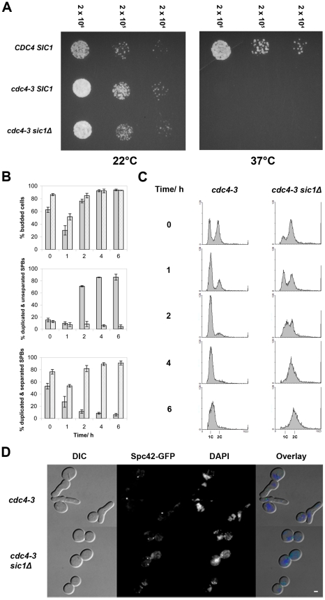
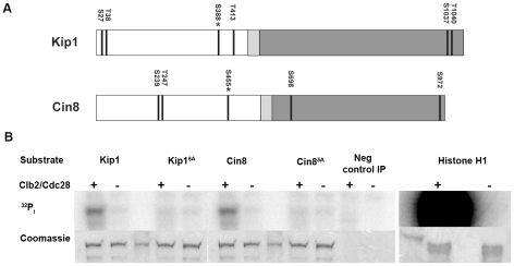
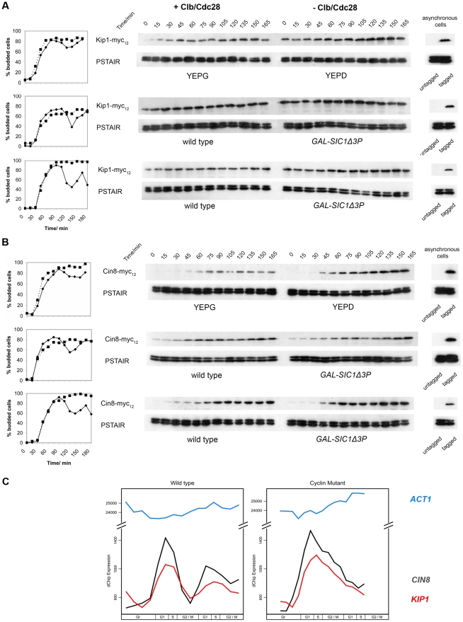
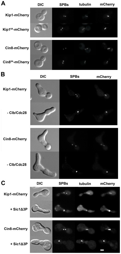
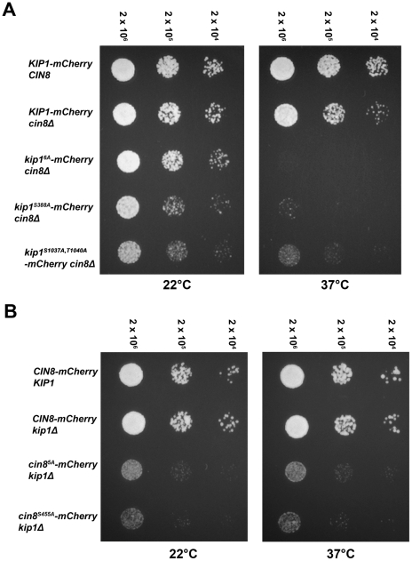
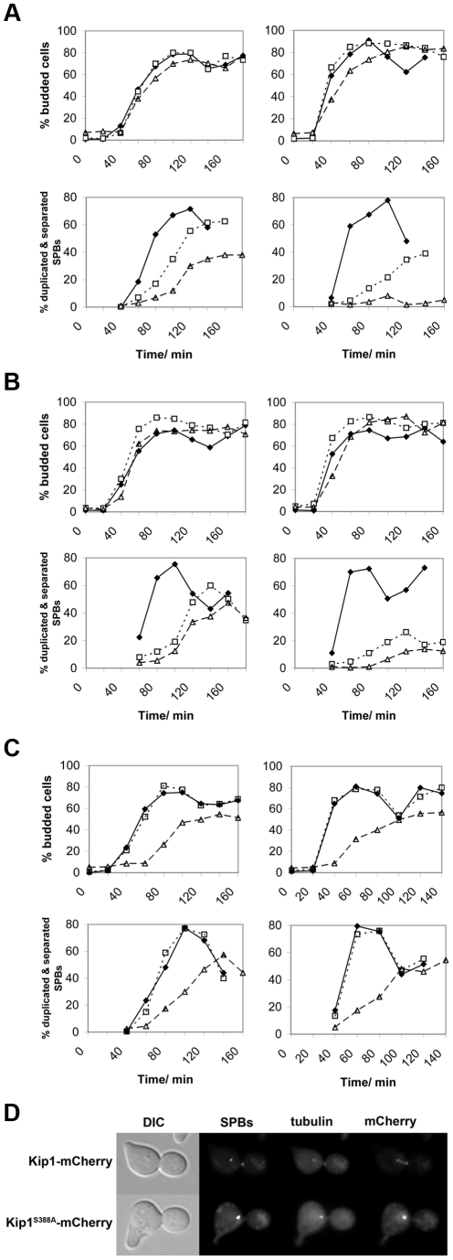
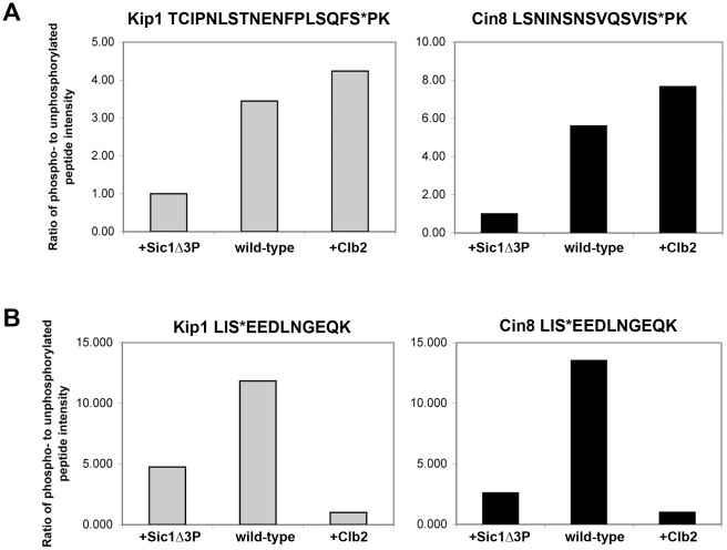
Although it has been known for many years that B-cyclin/CDK complexes regulate the assembly of the mitotic spindle and entry into mitosis, the full complement of relevant CDK targets has not been identified. It has previously been shown in a variety of model systems that B-type cyclin/CDK complexes, kinesin-5 motors, and the SCF(Cdc4) ubiquitin ligase are required for the separation of spindle poles and assembly of a bipolar spindle. It has been suggested that, in budding yeast, B-type cyclin/CDK (Clb/Cdc28) complexes promote spindle pole separation by inhibiting the degradation of the kinesins-5 Kip1 and Cin8 by the anaphase-promoting complex (APC(Cdh1)). We have determined, however, that the Kip1 and Cin8 proteins are present at wild-type levels in the absence of Clb/Cdc28 kinase activity. Here, we show that Kip1 and Cin8 are in vitro targets of Clb2/Cdc28 and that the mutation of conserved CDK phosphorylation sites on Kip1 inhibits spindle pole separation without affecting the protein's in vivo localization or abundance. Mass spectrometry analysis confirms that two CDK sites in the tail domain of Kip1 are phosphorylated in vivo. In addition, we have determined that Sic1, a Clb/Cdc28-specific inhibitor, is the SCF(Cdc4) target that inhibits spindle pole separation in cells lacking functional Cdc4. Based on these findings, we propose that Clb/Cdc28 drives spindle pole separation by direct phosphorylation of kinesin-5 motors.


Conflict of interest statement

The authors have declared that no competing interests exist.

Figures

Figure 1
Figure 1. SIC1 deletion allows SPB separation and DNA replication in cdc4-3(ts) cells at the restrictive temperature.
(A) Spot assay showing that cdc4-3 sic1Δ cells arrest at 37°C. Strains growing in log phase at permissive temperature were diluted to 2×10⁶ cells/ml, and further diluted serially to 2×10⁴ cells/ml. An equal volume of cells from each dilution was spotted on YEPD, and plates were incubated at either ambient temperature (∼22°C) or 37°C. (B) Percentages of budded cells, cells with duplicated and separated SPBs, and cells with duplicated but unseparated SPBs are shown for asynchronous log phase cultures shifted from permissive (24°C) to restrictive (37°C) temperature; each percentage shown is a percentage of that cell type over the total number of cells counted. The experiment was done in triplicate and the mean percentages are plotted; error bars indicate the standard deviation. Gray bars indicate cdc4-3 cells; spotted bars, cdc4-3 sic1Δ cells. Times after the shift to the restrictive temperature are shown. (C) Flow cytometric analysis of cells at the indicated times after the shift to the restrictive temperature; histograms show DNA content on the horizontal axis and the number of counts on the vertical axis. (D) Micrographs of arrested cdc4-3 and cdc4-3 sic1Δ cells showing bud morphology, DAPI-stained DNA, and Spc42-GFP-labeled SPBs. Scale bar: 2 µm.
Figure 2
Figure 2. Phosphorylation of Kip1 and Cin8 by Clb2/Cdc28 in vitro.
(A) Schematic showing the domain structure of Kip1 and Cin8 and the distribution of consensus CDK phosphorylation sites (S/T-P-X-X, indicated by dark lines) in each protein. The N-terminal motor domain is presented in white, the neck linker in light gray, and the C-terminal stalk and tail in dark gray; asterisk indicates the site that is conserved in almost all known kinesins-5 (Ser 388 in Kip1, Ser 455 in Cin8). (B) Wild-type Kip1 and Cin8, as well as their multiple consensus CDK site mutant forms (Kip16A, Cin85A) were immunoprecipitated from yeast lysates and mixed with soluble Clb2/Cdc28, also prepared from yeast, and 32P-γ-ATP. Soluble histone H1 (1.0 µg) was used as a control substrate. Proteins were subjected to SDS-PAGE after one hour at 30°C. PhosphorImages are shown on top and corresponding Coomassie-stained bands below. Unmarked lanes either contain molecular weight standards or had no protein loaded.
Figure 3
Figure 3. Kip1 and Cin8 protein and mRNA levels in the presence and the absence of active Clb/Cdc28 kinase.
Kip1 and Cin8 protein levels were determined by immunoblotting (see text) in three different sets of strains that were first synchronized with α-factor: BF264-15DU PGAL1-CLB1 Δclb1, 2, 3, 4, 5, 6 cells released into YEPG versus YEPD (top panels); wild-type BF264-15DU cells and mutants overexpressing Sic1Δ3P (GAL-SIC1Δ3P) to specifically inhibit Clb/Cdc28 (middle panels); wild-type W303a cells and mutants overexpressing Sic1Δ3P (bottom panels). Budding kinetics and corresponding immunoblots for strains with c-Myc-tagged Kip1 (A) and Cin8 (B) are shown; anti-PSTAIR was used as a loading control. Key: budding data for strains with active Clb/Cdc28 kinase (♦); lacking Clb/Cdc28 kinase activity (▪). (C) Transcript levels for KIP1 (red), CIN8 (black), and ACT1 (light blue) determined in wild-type and Δclb1-6 BF264-15DU cells synchronized in early G1 using centrifugal elutriation as reported in Orlando et al. (2008) .
Figure 4
Figure 4. Kip1 and Cin8 localization in the presence and the absence of phosphorylation by Clb/Cdc28.
Kip1 and Cin8 were visualized by fusion to a C-terminal mCherry tag and imaging with fluorescence microscopy. SPBs in the strains shown are marked with Spc42-GFP, and microtubules with CFP-Tub1. (A) Localization of wild-type Kip1 (top) and Cin8 (bottom), compared with that of Kip16A and Cin85A mutants, in live yeast cells. All consensus CDK sites in the mutant kinesins-5 have been mutated to non-phosphorylatable alanine. (B) Kip1 and Cin8 localization in PGAL1-CLB1 Δclb1, 2, 3, 4, 5, 6 cells 90 minutes after being released from α-factor arrest into medium containing either galactose (top panel; to induce Clb1 expression) or dextrose (bottom panel; to inhibit Clb1 expression). (C) Kip1 and Cin8 localization in wild-type cells versus cells with PGAL1-SIC1Δ3P integrated, 90 minutes after being released from α-factor arrest into galactose medium. Scale bar: 2 µm.
Figure 5
Figure 5. Impaired proliferation of strains with CDK site point mutants (Ser/Thr→Ala) of either (A) Kip1 or (B) Cin8 as their only kinesin-5.
Strains growing in log phase at permissive temperature were diluted to 2×10⁶ cells/ml, and then further diluted serially to 2×10⁴ cells/ml. An equal volume of cells from each dilution was spotted on YEPD, and plates were incubated at either ambient temperature (∼22°C) or 37°C. The number above each column of spots indicates the cell density (cells/ml). All alleles compared were tagged with mCherry at their C-terminus.
Figure 6
Figure 6. Impaired SPB separation in cells dependent on either Kip1 or Cin8 CDK site mutants as their only kinesin-5.
Cells were first synchronized in G1 with α-factor, and then released at either 25°C (permissive temperature, left column) or 37°C (restrictive temperature, right column). Time course experiments were carried out in triplicate, and both the percentage of budded cells (top panels for each group of strains) and the percentage of cells with duplicated and separated SPBs (bottom panels) from representative experiments are shown. The time elapsed following release from α-factor is indicated in minutes on the horizontal axis of each graph. (A) Cells expressing wild-type Kip1 and Cin8 (♦), cells expressing only Kip1 (□, dotted line) cells, and cells expressing only Kip1S388A (▵, dashed line); (B) cells expressing wild-type Kip1 and Cin8 (♦), cells expressing only Kip1 (□, dotted line) cells, and cells expressing only Kip1S1037A, T1040A (▵, dashed line); (C) cells expressing wild-type Cin8 and Kip1 (♦), cells expressing only Cin8 (□, dotted line), and cells expressing only Cin8S455A (▵, dashed line). (D) Fluorescence images of representative KIP1 cin8Δ and kip1S388A cin8Δ cells at 37°C 100 min after being released from α-factor arrest during the timecourse described in (A). Scale bar: 2 µm.
Figure 7
Figure 7. Relative abundance of specific phosphopeptides generated from Kip1-myc12 and Cin8-myc12 purified from yeast as measured by LC/MS/MS.
(A) Ratio of the intensity levels of the peaks corresponding to phosphopeptides with phosphates assigned to consensus CDK phosphorylation sites to that of their respective unphosphorylated forms. TCIPNLSTNENFPLSQFS*PK (asterisk denotes Ser 1037) was derived from Kip1 (gray bars), and LSNINSNSVQSVIS*PK (asterisk denotes Ser 972) from Cin8 (black bars). Kip1-myc12 and Cin8-myc12 were expressed from the GAL1 promoter in yeast (wild-type) for mass spectrometry analysis, and were also expressed in strains that simultaneously expressed either Sic1Δ3P (+Sic1Δ3P) or Clb2-HA3 (+Clb2) from the GAL1 promoter. (B) Relative intensity levels of peaks corresponding to phosphorylated and unphosphorylated forms of the peptide LIS*EEDLNGEQK, derived from the c-Myc epitope tags of Kip1-myc12 (gray bars) and Cin8-myc12 (black bars).
Figure 8
Figure 8. Homology models of Kip1 and Cin8 motor domains bound to MgADP.
(A) Ribbon structure overlay illustrating gross differences in the predicted structures of Kip1 (peach) and Cin8 (light blue), particularly in the length of the various loop regions. The human Eg5 (also known as Kif11) template structure (ExPDB 1ii6B) is shown in gray. The position of Ser 388 on Kip1 and Ser 455 on Cin8 is highlighted in red, while the location of the microtubule binding face and nucleotide binding pocket are indicated. (B) Close-up view of the surroundings of Ser 388 in Kip1 and Ser 455 in Cin8; both the serine residues are colored red. Residues with side chains lying within a 2.5 Å radius of the serine residue are colored light green for Kip1 and hot pink for Cin8; residues have also been labeled to highlight significant differences between the two model structures.

Similar articles

Cited by

References

    1. Morgan DO. Cyclin-dependent kinases: Engines, clocks, and microprocessors. Annual Review of Cell and Developmental Biology. 1997;13:261–291. - PubMed
    1. Murray AW. Recycling the cell cycle: Cyclins revisited. Cell. 2004;116:221–234. - PubMed
    1. Lacey KR, Jackson PK, Stearns T. Cyclin-dependent kinase control of centrosome duplication. Proceedings of the National Academy of Science USA. 1999;96:2817–2822. - PMC - PubMed
    1. Haase SB, Winey M, Reed SI. Multi-step control of spindle pole body duplication by cyclin-dependent kinase. Nature Cell Biology. 2001;3:38–42. - PubMed
    1. Adams IR, Kilmartin JV. Spindle pole body duplication: a model for centrosome duplication? Trends in Cell Biology. 2000;10:329–335. - PubMed

Publication types

MeSH terms

LinkOut - more resources