

Limitations of conventional approaches to identify myocyte nuclei in histologic sections of the heart
- PMID: 20457832
- PMCID: PMC2902296
- DOI: 10.1152/ajpcell.00435.2009
Limitations of conventional approaches to identify myocyte nuclei in histologic sections of the heart
Abstract
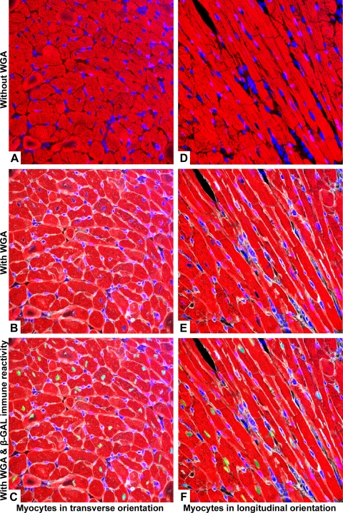
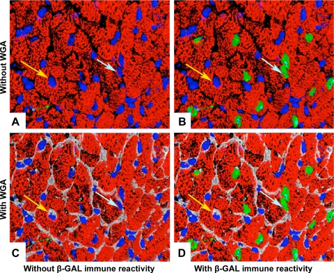
Accurate nuclear identification is crucial for distinguishing the role of cardiac myocytes in intrinsic and experimentally induced regenerative growth of the myocardium. Conventional histologic analysis of myocyte nuclei relies on the optical sectioning capabilities of confocal microscopy in conjunction with immunofluorescent labeling of cytoplasmic proteins such as troponin T, and dyes that bind to double-strand DNA to identify nuclei. Using heart sections from transgenic mice in which the cardiomyocyte-restricted alpha-cardiac myosin heavy chain promoter targeted the expression of nuclear localized beta-galactosidase reporter in >99% of myocytes, we systematically compared the fidelity of conventional myocyte nuclear identification using confocal microscopy, with and without the aid of a membrane marker. The values obtained with these assays were then compared with those obtained with anti-beta-galactosidase immune reactivity in the same samples. In addition, we also studied the accuracy of anti-GATA4 immunoreactivity for myocyte nuclear identification. Our results demonstrate that, although these strategies are capable of identifying myocyte nuclei, the level of interobserver agreement and margin of error can compromise accurate identification of rare events, such as cardiomyocyte apoptosis and proliferation. Thus these data indicate that morphometric approaches based on segmentation are justified only if the margin of error for measuring the event in question has been predetermined and deemed to be small and uniform. We also illustrate the value of a transgene-based approach to overcome these intrinsic limitations of identifying myocyte nuclei. This latter approach should prove quite useful when measuring rare events.
Figures

Similar articles
-
Deep Learning Identifies Cardiomyocyte Nuclei With High Precision.Circ Res. 2020 Aug 14;127(5):696-698. doi: 10.1161/CIRCRESAHA.120.316672. Epub 2020 Jun 3. Circ Res. 2020. PMID: 32486999 No abstract available.
-
Midbody Positioning and Distance Between Daughter Nuclei Enable Unequivocal Identification of Cardiomyocyte Cell Division in Mice.Circ Res. 2018 Oct 12;123(9):1039-1052. doi: 10.1161/CIRCRESAHA.118.312792. Circ Res. 2018. PMID: 30355161
-
Apela Promotes Cardiomyocyte Differentiation from Transgenic Human Embryonic Stem Cell Lines.Appl Biochem Biotechnol. 2019 Oct;189(2):396-410. doi: 10.1007/s12010-019-03012-2. Epub 2019 Apr 26. Appl Biochem Biotechnol. 2019. PMID: 31025171
-
Assessment of cardiomyocyte DNA synthesis in normal and injured adult mouse hearts.Am J Physiol. 1997 Jan;272(1 Pt 2):H220-6. doi: 10.1152/ajpheart.1997.272.1.H220. Am J Physiol. 1997. PMID: 9038941
-
Cardiac-specific deletion of Gata4 reveals its requirement for hypertrophy, compensation, and myocyte viability.Circ Res. 2006 Mar 31;98(6):837-45. doi: 10.1161/01.RES.0000215985.18538.c4. Epub 2006 Mar 2. Circ Res. 2006. PMID: 16514068
Cited by
-
A broadly applicable method for quantifying cardiomyocyte cell division identifies proliferative events following myocardial infarction.Cell Rep Methods. 2024 Sep 16;4(9):100860. doi: 10.1016/j.crmeth.2024.100860. Epub 2024 Sep 9. Cell Rep Methods. 2024. PMID: 39255794 Free PMC article.
-
Downregulation of doxorubicin-induced myocardial apoptosis accompanies postnatal heart maturation.Am J Physiol Heart Circ Physiol. 2012 Apr 15;302(8):H1603-13. doi: 10.1152/ajpheart.00844.2011. Epub 2012 Feb 10. Am J Physiol Heart Circ Physiol. 2012. PMID: 22328080 Free PMC article.
-
Isolation of Cardiomyocytes from Fixed Hearts for Immunocytochemistry and Ploidy Analysis.J Vis Exp. 2020 Oct 7;(164):10.3791/60938. doi: 10.3791/60938. J Vis Exp. 2020. PMID: 33104059 Free PMC article.
-
Musings on intrinsic cardiomyocyte cell cycle activity and myocardial regeneration.J Mol Cell Cardiol. 2023 Sep;182:86-91. doi: 10.1016/j.yjmcc.2023.07.007. Epub 2023 Jul 28. J Mol Cell Cardiol. 2023. PMID: 37517369 Free PMC article. Review.
-
Cardiomyocyte proliferation vs progenitor cells in myocardial regeneration: The debate continues.Glob Cardiol Sci Pract. 2013 Nov 1;2013(3):303-15. doi: 10.5339/gcsp.2013.37. eCollection 2013. Glob Cardiol Sci Pract. 2013. PMID: 24689031 Free PMC article. Review.
References
-
- Anversa P, Leri A, Kajstura J. Cardiac regeneration. J Am Coll Cardiol 47: 1769–1776, 2006 - PubMed
-
- Carter D. Practical considerations for collecting confocal images. Methods Mol Biol 122: 35–57, 1999 - PubMed
-
- Fleiss JL. Measuring nominal scale agreement among many raters. Psychol Bull 76: 378–382, 1971
Publication types
MeSH terms
Substances
Grants and funding
LinkOut - more resources
Full Text Sources
Other Literature Sources
Molecular Biology Databases