. 2010 May 17;189(4):755-67.
doi: 10.1083/jcb.200912011. Epub 2010 May 10.

Multiple Myo4 motors enhance ASH1 mRNA transport in Saccharomyces cerevisiae

Affiliations

Multiple Myo4 motors enhance ASH1 mRNA transport in Saccharomyces cerevisiae

Sunglan Chung et al. J Cell Biol. .

Abstract

In Saccharomyces cerevisiae, ASH1 mRNA is transported to the bud tip by the class V myosin Myo4. In vivo, Myo4 moves RNA in a rapid and continuous fashion, but in vitro Myo4 is a nonprocessive, monomeric motor that forms a complex with She3. To understand how nonprocessive motors generate continuous transport, we used a novel purification method to show that Myo4, She3, and the RNA-binding protein She2 are the sole major components of an active ribonucleoprotein transport unit. We demonstrate that a single localization element contains multiple copies of Myo4 and a tetramer of She2, which suggests that She2 may recruit multiple motors to an RNA. Furthermore, we show that increasing the number of Myo4-She3 molecules bound to ASH1 RNA in the absence of She2 increases the efficiency of RNA transport to the bud. Our data suggest that multiple, nonprocessive Myo4 motors can generate continuous transport of mRNA to the bud tip.


Figures

Figure 1.
Figure 1.
Purification of U3 RNP complex. (A) Schematic representation of constructs used for U3 RNP complex purification. The RNA-binding protein is expressed from the constitutive glycerol phosphate dehydrogenase (GPD) promoter, and contains U1Ap, GFP, TAP, and an NLS. The RNA partner is expressed from the inducible galactose (GAL) promoter, and consists of four repeats of U1A aptamer, the U3 localization element, and an ADH1 terminator. (B) GFP-bound U3 RNA localizes to the bud tip using the modified tagged RNA system. Expression of 4×U1A-tagged U3 RNA was induced in cells containing U1Ap-GFP-TAP, and GFP signals in live cells were observed by fluorescence microscopy. The outline was drawn based on the DIC image. The nuclear GFP signal is from U1Ap-GFP-TAP not bound to RNA. Bar, 10 µm. (C) Silver staining of TAP-purified complexes. Extracts prepared from cells expressing U1Ap-GFP-TAP only (lane 1), U1Ap-GFP-TAP and 4xU1A-tagged U3 RNA (lane2), or U1Ap-GFP-TAP and 4xU1A-tagged ADH2 (77 NT) RNA (lane 3) were used for TAP purification. The purified complexes were separated on a 4–15% gradient SDS-polyacrylamide gel, and silver stained. (D) Myo4, She3, and She2 specifically copurify with U3 RNA. Purified complexes from C were separated by 10% SDS-PAGE in the same order as in C and analyzed by Western blotting. U1Ap-GFP was detected by an anti-GFP antibody. (E) Mass spectrometry analysis of U3 RNA copurifying proteins. TAP-purified U3 RNP complexes were separated by SDS-PAGE and stained with Coomassie blue. Protein bands of interest were excised from gels and identified by mass spectrometry analysis (LC-MS/MS).
Figure 2.
Figure 2.
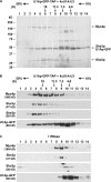
Myo4, She3, and She2 are the sole, major components of the U3 RNP complex. (A) Myo4, She3, and She2 co-migrate in velocity sedimentation analysis. TAP-purified U3 RNP complexes were resolved on a 10–50% sucrose gradient, and fractions were collected from the bottom (fraction 1). These fractions were analyzed by SDS-PAGE and silver stained. Positions of protein standards thyroglobulin (19S), catalase (11.3S), aldolase (7.3S), and albumin (4.6S) from parallel gradients are indicated. (B) Myo4, She3, and She2 migrate as a 20S RNP complex. TEV-eluted U3 RNP complexes were analyzed as in A, and Western blots were probed with antibodies as indicated. The sedimentation coefficient of U1Ap-GFP-TAP–bound U3 RNP complex was measured as 20.61 ± 0.56S (n = 3). U1Ap-GFP peaks twice, showing proteins bound to U3 RNP complex and unbound free U1Ap-GFP. (C) RNase dissociates the Myo4–She3–She2 complex. TEV-eluted U3 RNP complexes were treated with 0.3 mg/ml RNaseA and further analyzed along with intact U3 RNP complexes in B. The sedimentation coefficient value of Myo4–She3 is reduced to 7.76 ± 0.18S (n = 2) when not bound to RNA cargo.
Figure 3.
Figure 3.
Size estimation of the U3 RNP complex. (A) Comparison of U3 RNP protein profiles. U3 RNA was expressed with MYO4-TAP (lane 1) or U1Ap-GFP-TAP (lane 2). TAP-purified complexes from each cell extract were separated by 4–15% SDS-PAGE and silver stained. (B) Myo4–She3 shifts into a 15S RNP complex upon binding U3 RNA. U3 RNA expression was induced (top) or not induced (bottom) in cells expressing MYO4-1/2TAP, and the cell extracts were used for 1/2TAP-purification. The purified complexes were loaded on 10–50% sucrose gradients, and the collected fractions were analyzed by Western blotting. Myo4 bound to U3 RNA purifies as a complex with a size of 15.03 ± 0.90S (n = 6), whereas cargo-free Myo4 purifies at 7.81 ± 0.45S (n = 3). (C) Sedimentation coefficient values of Myo4 associated with U3 RNA and in soluble form. Size determination of U3 RNP complex and Myo4–She3 was based on TAP-purified complexes from cells expressing MYO4-1/2TAP with or without U3 RNA overexpression. Cells expressing MYO2-1/2TAP were used for Myo2p purification.
Figure 4.
Figure 4.
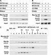
Multiple Myo4 motors are bound to a single localization element. (A) Myo4-HA coprecipitates with Myo4-myc when bound to U3 RNA. MYO4-HA was expressed in wild-type cells (lanes 1 and 2) or cells expressing MYO4-13xMYC (lanes 3–6), and U3 RNA expression was induced (lanes 1, 2, 5, and 6) or not induced (lanes 3 and 4). RNaseA was added to the indicated cell extracts (lanes 2, 4, and 6). Myo4-myc was immunoprecipitated from each cell extract, and the precipitants were analyzed by a Western blot with anti-HA or anti-She2 antibodies as indicated. Input shows equal amount of initial cell extracts probed with anti-HA antibody. (B) Myo4-myc and Myo4-HA coprecipitation requires She2. MYO4-HA was expressed with MYO4-13xMYC in the presence of SHE2 (lanes 1 and 2) or in she2Δ cells (lane 3), and U3 RNA expression was induced (lanes 2 and 3) or not induced (lane 1). Myo4-myc was immunoprecipitated and analyzed as in A. (C) The 15S U3 RNP complex consists of multiple Myo4 motors. MYO4-HA was expressed with MYO4-TAP, and U3 RNA expression was induced. TAP-purified complexes were loaded on 10–50% sucrose gradients and the collected fractions were analyzed by Western blotting. Note that the Myo4p blot detects both Myo4-TAP and Myo4-HA.
Figure 5.
Figure 5.
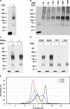
The oligomeric state of She2. (A) Cross-linking analysis of She2. SHE2-1/2TAP was expressed in wild-type cells, and purified She2 was treated with 40 mM EDC for 1 h at room temperature, separated by 4–15% SDS-PAGE, and probed with anti-She2 antibody. Note that She2 appears as a doublet in the absence of EDC due to oligomerization of wild-type and TAP-tagged She2. (B) She2 oligomerization is not concentration dependent. SHE2-1/2TAP was expressed from a high-copy plasmid in she2Δ cells, and cross-linking analysis was performed with purified She2 diluted to various concentrations. (C) She2 oligomerization does not require RNA binding. SHE2-1/2TAP was expressed in she2Δ, cells and U3 RNA expression was induced. Half of the cell extract was treated with 0.3 mg/ml RNaseA, and purified She2 from each cell extract was cross-linked. (D) Oligomerization is inhibited by mutations in the upper uncharged surface of She2. 1/2TAP-tagged SHE2 mutants (N36S, R63K, T47Y, and L130Y) were expressed in she2Δ cells, and the purified proteins were cross-linked and analyzed as described in A. (E) Analytical ultracentrifugation of She2. Purified wild-type She2 was diluted to 0.15 mg/ml (5.3 µM), 0.53 mg/ml (18.7 µM), 1.13 mg/ml (39.9 µM), and 1.90 mg/ml (67.1 µM), then subjected to analytical ultracentrifugation. A direct boundary modeling program from individual datasets using model-based numerical solutions to the Lamm equation was used to obtain data shown for the normalized continuous sedimentation coefficient, c(s), distribution plot. At 0.15 mg/ml, the weighted mean value of S obtained through integration of the c(s) curve was 5.95S.
Figure 6.
Figure 6.
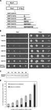
Increasing the number of Myo4 motors bound to RNA improves bud localization. (A) Model system for modifying Myo4 motor copy number on ASH1 RNA. She3 is expressed with the RNA-binding domain from U1Ap fused to its C terminus, and ASH1 is expressed with 0, 1, 2, 4, 6, or 8 U1Ap-binding sites positioned between the stop codon and 3′ UTR. Both constructs are expressed in ash1Δ she2Δ cells to ensure that Myo4 binds to ASH1 mRNA only through the interaction between She3-U1Ap and the U1Ap-binding sites tagged to ASH1 RNA. (B) Increasing the number of Myo4 bound to ASH1 RNA enhances bud localization of Ash1. She3-U1Ap and ASH1-HA tagged with the indicated number of U1A aptamers were expressed in ash1Δshe2Δ yeast cells where ADE was under the control of HO promoter. She2 was also coexpressed with She3-U1Ap and ASH1-HA as a control (bottom). Cells were spotted onto selective media plates with or without adenine. (C) U1A tags do not affect Ash1 expression. Protein extracts were prepared from cells expressing ASH1-HA tagged with the indicated number of U1A aptamers. Equal amounts of protein extracts were analyzed by Western blotting with anti-HA antibody. (D) Bud transport of ASH1 mRNA is improved by increasing Myo4 binding sites on ASH1 RNA. She3-U1Ap was expressed with U1A-tagged ASH1-HA in ash1Δshe2Δ cells. wt indicates cells coexpressing She2 with She3-U1Ap and ASH1 as a control. The cells were synchronized and fixed after release from synchronization. ASH1 mRNA was detected by FISH, and the percentage of cells showing bud-localized ASH1 mRNA among ∼100 cells was determined in each cell sample (n = 3). Bud localization was further subdivided to localization confined to the distal tip of buds and that dispersed throughout the bud.

Similar articles

Cited by

References

    1. Allain F.H., Gubser C.C., Howe P.W., Nagai K., Neuhaus D., Varani G. 1996. Specificity of ribonucleoprotein interaction determined by RNA folding during complex formulation. Nature. 380:646–650 10.1038/380646a0 - DOI - PubMed
    1. Beeg J., Klumpp S., Dimova R., Gracià R.S., Unger E., Lipowsky R. 2008. Transport of beads by several kinesin motors. Biophys. J. 94:532–541 10.1529/biophysj.106.097881 - DOI - PMC - PubMed
    1. Bertrand E., Chartrand P., Schaefer M., Shenoy S.M., Singer R.H., Long R.M. 1998. Localization of ASH1 mRNA particles in living yeast. Mol. Cell. 2:437–445 10.1016/S1097-2765(00)80143-4 - DOI - PubMed
    1. Block S.M., Goldstein L.S., Schnapp B.J. 1990. Bead movement by single kinesin molecules studied with optical tweezers. Nature. 348:348–352 10.1038/348348a0 - DOI - PubMed
    1. Böhl F., Kruse C., Frank A., Ferring D., Jansen R.P. 2000. She2p, a novel RNA-binding protein tethers ASH1 mRNA to the Myo4p myosin motor via She3p. EMBO J. 19:5514–5524 10.1093/emboj/19.20.5514 - DOI - PMC - PubMed

Publication types

MeSH terms