Mechanisms of the Hsp70 chaperone system
- PMID: 20453930
- PMCID: PMC5026485
- DOI: 10.1139/o09-175
Mechanisms of the Hsp70 chaperone system
Abstract
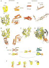
Molecular chaperones of the Hsp70 family have diverse functions in cells. They assist the folding of newly synthesized and stress-denatured proteins, as well as the import of proteins into organelles, and the dissociation of aggregated proteins. The well-conserved Hsp70 chaperones are ATP dependent: binding and hydrolysis of ATP regulates their interactions with unfolded polypeptide substrates, and ATPase cycling is necessary for their function. All cellular functions of Hsp70 chaperones use the same mechanism of ATP-driven polypeptide binding and release. The Hsp40 co-chaperones stimulate ATP hydrolysis by Hsp70 and the type 1 Hsp40 proteins are conserved from Escherichia coli to humans. Various nucleotide exchange factors also promote the Hsp70 ATPase cycle. Recent advances have added to our understanding of the Hsp70 mechanism at a molecular level.
Figures

Similar articles
-
The dissociation of ATP from hsp70 of Saccharomyces cerevisiae is stimulated by both Ydj1p and peptide substrates.J Biol Chem. 1995 May 5;270(18):10412-9. doi: 10.1074/jbc.270.18.10412. J Biol Chem. 1995. PMID: 7737974
-
Regulation of ATPase and chaperone cycle of DnaK from Thermus thermophilus by the nucleotide exchange factor GrpE.J Mol Biol. 2001 Feb 2;305(5):1173-83. doi: 10.1006/jmbi.2000.4373. J Mol Biol. 2001. PMID: 11162122
-
Human Hsp70 molecular chaperone binds two calcium ions within the ATPase domain.Structure. 1997 Mar 15;5(3):403-14. doi: 10.1016/s0969-2126(97)00197-4. Structure. 1997. PMID: 9083109
-
Nucleotide Exchange Factors for Hsp70 Molecular Chaperones: GrpE, Hsp110/Grp170, HspBP1/Sil1, and BAG Domain Proteins.Subcell Biochem. 2023;101:1-39. doi: 10.1007/978-3-031-14740-1_1. Subcell Biochem. 2023. PMID: 36520302 Review.
-
Hsp70 chaperones: cellular functions and molecular mechanism.Cell Mol Life Sci. 2005 Mar;62(6):670-84. doi: 10.1007/s00018-004-4464-6. Cell Mol Life Sci. 2005. PMID: 15770419 Free PMC article. Review.
Cited by
-
Plasmodium falciparum heat shock protein 110 stabilizes the asparagine repeat-rich parasite proteome during malarial fevers.Nat Commun. 2012;3:1310. doi: 10.1038/ncomms2306. Nat Commun. 2012. PMID: 23250440 Free PMC article.
-
Hsp110 is required for spindle length control.J Cell Biol. 2012 Aug 20;198(4):623-36. doi: 10.1083/jcb.201111105. J Cell Biol. 2012. PMID: 22908312 Free PMC article.
-
Involvement of molecular chaperones and the transcription factor Nrf2 in neuroprotection mediated by para-substituted-4,5-diaryl-3-thiomethyl-1,2,4-triazines.Cell Stress Chaperones. 2012 Jul;17(4):409-22. doi: 10.1007/s12192-011-0316-0. Epub 2012 Jan 4. Cell Stress Chaperones. 2012. PMID: 22212523 Free PMC article.
-
Proteasome-Rich PaCS as an Oncofetal UPS Structure Handling Cytosolic Polyubiquitinated Proteins. In Vivo Occurrence, in Vitro Induction, and Biological Role.Int J Mol Sci. 2018 Sep 14;19(9):2767. doi: 10.3390/ijms19092767. Int J Mol Sci. 2018. PMID: 30223470 Free PMC article. Review.
-
Chaperoning Endoplasmic Reticulum-Associated Degradation (ERAD) and Protein Conformational Diseases.Cold Spring Harb Perspect Biol. 2019 Aug 1;11(8):a033928. doi: 10.1101/cshperspect.a033928. Cold Spring Harb Perspect Biol. 2019. PMID: 30670468 Free PMC article. Review.
References
Publication types
MeSH terms
Substances
Grants and funding
LinkOut - more resources
Full Text Sources
Other Literature Sources

