. 2010 Apr 30;141(3):483-96.
doi: 10.1016/j.cell.2010.03.040.

NLRC5 negatively regulates the NF-kappaB and type I interferon signaling pathways

Affiliations

NLRC5 negatively regulates the NF-kappaB and type I interferon signaling pathways

Jun Cui et al. Cell. .

Abstract

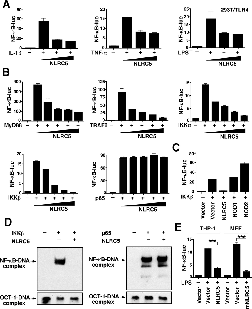
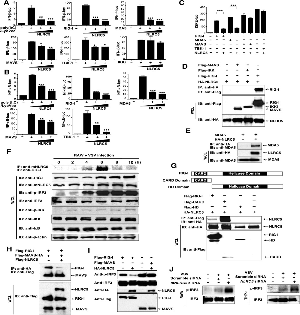
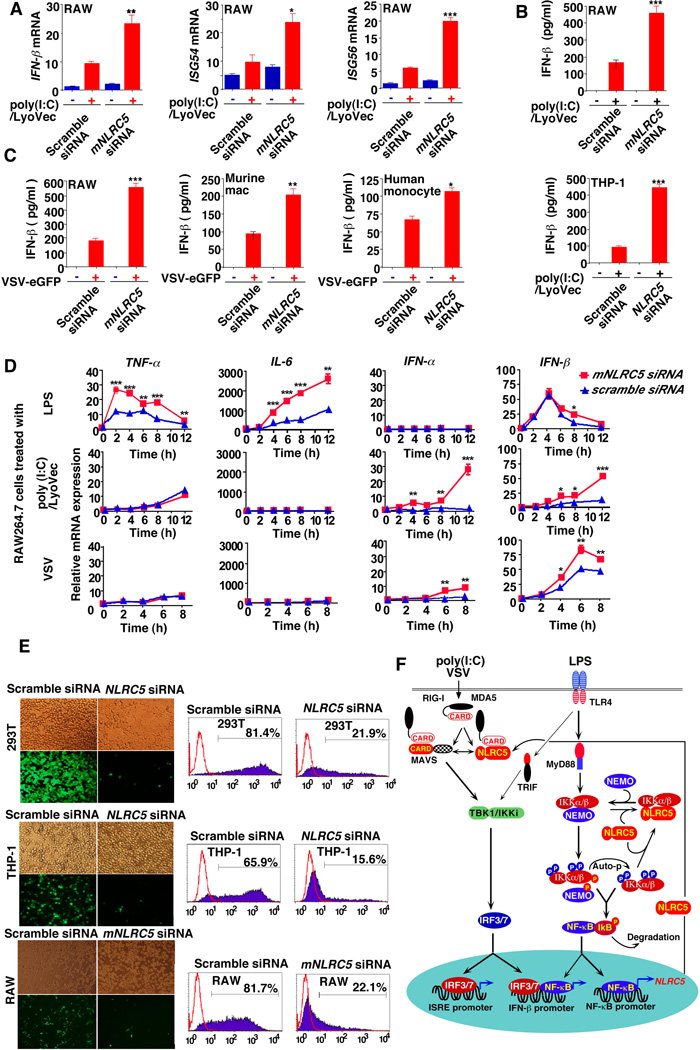
Stringent control of the NF-kappaB and type I interferon signaling pathways is critical to effective host immune responses, yet the molecular mechanisms that negatively regulate these pathways are poorly understood. Here, we show that NLRC5, a member of the highly conserved NOD-like protein family, can inhibit the IKK complex and RIG-I/MDA5 function. NLRC5 inhibited NF-kappaB-dependent responses by interacting with IKKalpha and IKKbeta and blocking their phosphorylation. It also interacted with RIG-I and MDA5, but not with MAVS, to inhibit RLR-mediated type I interferon responses. Consistent with these observations, NLRC5-specific siRNA knockdown not only enhanced the activation of NF-kappaB and its responsive genes, TNF-alpha and IL-6, but also promoted type I interferon signaling and antiviral immunity. Our findings identify NLRC5 as a negative regulator that blocks two central components of the NF-kappaB and type I interferon signaling pathways and suggest an important role for NLRC5 in homeostatic control of innate immunity.


Figures

Figure 1
Figure 1. Domain organization, expression and intracellular localization of human and mouse NLRC5
(A) Domain organization of human and mouse NLRC5 proteins. CARD, caspase recruitment domain; NOD, nucleotide binding domain; LRR, leucine rich repeats. (B) Western blot analysis of HA-tagged NLRC5 and mNLRC5 protein expression in 293T cells. (C) Human and mouse NLRC5 mRNAs were determined in different tissues determined by real-time PCR analysis. D) Expression of mNLRC5 mRNA in RAW264.7 cells was determined by real-time PCR analysis after LPS, intracellular poly (I:C) stimulation or VSV-eGFP infection. (E) Immunoblot analysis of mNLRC5 protein expression after LPS stimulation or VSV-eGFP infection. The relative protein levels of mNLRC5 were quantified by the band density scanning of Western blot images and plotted against time (h) after LPS treatment or VSV infection. (F) Both wild-type and MyD88-deficient macrophages were treated with LPS, and mNLRC mRNA expression was determined by real-time PCR analysis. See also Fig. S1.
Figure 2
Figure 2. NLRC5 inhibits NF-κB activation induced by IL-1β, LPS, TNF-α and their downstream signaling molecules
(A) 293T cells were transfected with an NF-κB-luc reporter plasmid and TLR4 plasmid (for LPS treatment), together with an empty vector or NLRC5 construct, and analyzed for NF-κB-dependent luciferase activity (fold induction) after treatment with IL-1β, LPS or TNF-α. (B) 293T cells were transfected with MyD88, TRAF6, IKKα, IKKβ, or p65, along with NF-κB-luc. (C) 293T cells were transfected with NOD1 or NOD2, along with NF-κB-luc. (D) Detection of endogenous NF-κB DNA binding activity in a gel-mobility shift assay. Oct-1/DNA-binding complexes served as a loading control for nuclear extracts. (E) Human THP-1 cells and murine embryonic fibroblasts (MEF) were transfected with the NF-κB-luc reporter plasmid, together with (or without) NLRC5 or mNLRC5 plasmid, and then analyzed for NF-κB-dependent luciferase activity after LPS treatment. See also Fig. S2.
Figure 3
Figure 3. NLRC5 interacts with IKKα and IKKβ to inhibit their phosphorylation
A) 293T cells transfected with Flag-IKKα, Flag-IKKβ, Flag-NEMO and HA-NLRC5. HA-tagged NLRC5 protein was immunoprecipitated with anti-HA beads, and blotted with anti-Flag. (B) 293T and RAW264.7 cell extracts were immunoprecipitated with IgG, anti-NLRC5 or anti-mNLRC5 antibody, respectively, and then analyzed together with whole cell extracts by Western blot with an anti-IKKα/β, anti-NEMO, anti-NLRC5 or anti-mNLRC5 antibody. (C) Cell extracts of RAW264.7 cells were fractionated on a size-exclusion column (HiPrep 16/60 Sephacryl S-300 HR). The collected factions with an equal volume were used for Western blot analysis with specific antibodies. The elution positions of calibration proteins with known molecular masses (kDa) were used to determine the size of complexes. (D) 293T cells were transfected with the indicated doses of Flag-NLRC5, HA-IKKβ and HA-NEMO. Whole cell extracts were immunoprecipitated with anti-Flag beads, and blotted with anti-HA. (E) 293T/TLR4 cells were transfected with empty vector or different doses of HA-NLRC5 (0, 50 or 200 ng) DNA and then treated with LPS. Cell extracts were collected at 30 min poststimulation and prepared for immunoprecipitation with anti-HA and anti-NEMO, followed by immunoblot (IB) with indicated antibodies or kinase assay (KA). (F) The domain structure of IKKβ. Numbers in parentheses indicate amino acid position in construct. LZ, leucine zipper; HLH, helix-loop-helix. (G) 293T cells were transfected with HA-NLRC5 and Flag-IKKβ or various Flag-IKKβ mutants. Whole cell extracts were immunoprecipitated with anti-Flag beads, and blotted with anti-HA. (H and I) 293T cells transfected with IKKα, IKKβ, JNK1, JNK2, and p38 with or without HA-NLRC5 were used to analyze the phosphorylation of IKKα/β, JNK and p38. (J) RAW264.7 cells were treated with LPS and collected at the indicated time points. Cell extracts were prepared for immunoprecipitation with anti-mNLRC5 or anti-NEMO, followed by immunoblot (IB) to determine the IKK phosphorylation with anti-p-IKK or anti-IKKα/βantibody and kinase activity of IKK. See also Fig. S3 and S4.
Figure 4
Figure 4. Interaction and functional analysis of NLRC5 deletions in the inhibition of NF-κB activation
(A) Four NLRC5 deletion constructs were generated. (B) and (C) 293T cells were transfected with NLRC5 and its mutations constructs, IKKβ or kinase domain of IKKβ and analyzed by coimmunoprecipitation and Western blot. (D) 293T cells were transfected with NF-κB-luc reporter, together with an empty vector, or with full-length NLRC5 and its deletion constructs, and analyzed for luciferase activity (fold induction). (E) 293T cells were transfected with various expression plasmids and the phosphorylation of IKKα or IKKβ was determined. See also Fig. S4G.
Figure 5
Figure 5. Knockdown of NLRC5 can significantly enhance NF-κB activation and inflammatory responses
(A) Specific knockdown of NLRC5 or mNLRC5 was evaluated in various types of cells transfected with NLRC5/mNLRC5- siRNA, or scrambled siRNA. (B) RAW264.7 cells were transfected with mNLRC5 siRNA or scrambled siRNA, and then treated with LPS. The cell extracts were harvested at different time points and used for Western blot of various kinases and signaling proteins. (C) The cell extracts of mNLRC5 knockdown and control RAW264.7 cells after LPS treatment were used for immunoprecipitation to obtain NEMO-associated IKK (IP-1) and NEMO-free IKK (free IKK and mNLRC5 associated IKK (IP-2). The phosphorylation of IKKα/β, the total amount of IKKα/β and NEMO proteins in different fractions were determined by anti-p-IKK, anti-IKKα/β or anti-NEMO antibody. (D) NLRC5 or mNLRC5 was knocked down in 293T/TLR4 and RAW264.7 cells. NF-κB-luc activity was determined after LPS treatment. (E) The mNLRC5 knockdown and control AW264.7 cells were treated with LPS for 1 h; the nuclear proteins were harvested for NF-κB binding activity determined by EMSA. Oct-1 DNA-binding complexes were served as a control. (F) Endogenous NLRC5 was knocked down in THP-1 and RAW264.7 cells and TNF-α and IL-6 production was measured after LPS treatment. Data in panels (D) and (F) are presented as means ± SEM. Asterisks indicate significant differences between groups (** P< 0.01, *** P<0.001 as determined by t-test analysis). See also Fig. S5.
Figure 6
Figure 6. NLRC5 negatively regulates IFN-β activation by inhibiting RIG-I and MDA5 function
(A–C): 293T cells were transfected with NF-κB-luc, INF-β-luc or ISRE-luc, NLRC5 plus poly(I:C)/Lyovec, RIG-I, MDA5, MAVS, TBK1 or IKKi plasmids and analyzed for INF-β or ISRE luciferase activity. Values are means ± SEM of three independent experiments. (D) 293T cells were transfected with HA-NLRC5 plus RIG-I, MAVS or IKKi. After immunoprecipitation with anti-HA beads, specific proteins were analyzed by Western blot with anti-Flag. (E) 293T cells were transfected with MDA5 with or without HA-NLRC5. After immunoprecipitation with anti-HA beads, specific proteins were analyzed by Western blot with anti-MDA5. (F) RAW264.7 cells were infected with VSV-eGFP, and cell extracts were harvested at different time points, immunoprecipitated with anti-mNLRC5 antibody and analyzed by Western blot with anti-RIG-I. (G) NLRC5 binds to the CARD domain of RIG-I. 293T cells were transfected with HA-NLRC5 plus Flag-RIG-I, Flag-RIG-I CARD domain and Helicase domain (HD). After immunoprecipitation with anti-Flag beads, specific proteins were analyzed by Western blot with anti-HA. (H) NLRC5 competitively binds to RIG-I with MAVS. HEK293T cells were transfected with Flag-MAVS-HA plus Flag-RIG-1, or Flag-NLRC5. After immunoprecipitation with anti-HA beads, specific proteins were analyzed by Western blot with anti-Flag. (I) 293T cells were transfected with Flag-RIG-I and Flag-MAVS, with or without HA-NLRC5, and used for Western blot analysis with anti-phospho-IRF3 and IRF3 antibodies. (J) RAW264.7 and THP-1 cells were transfected with NLRC5/mNLRC5-specific siRNA or scrambled siRNA, respectively, and then infected with VSV-eGFP. The cell extracts were harvested for Western blot with anti-phospho-IRF3 and IRF3 antibodies. See also Fig. S6.
Figure 7
Figure 7. Knockdown of NLRC5 enhances cytokine response and antiviral immunity
(A and B) RAW264.7 cells or THP-1 cells were transfected with mNLRC5/NLRC5-specific siRNA or scrambled siRNA, followed by poly(I:C)/Lyovec treatment. ISG-54, ISG-56, IFN-β mRNA and IFN-β protein were determined by real-time RT-PCR or ELISA. (C) NLRC5 or mNLRC5 knockdown and control cells were infected with VSV-eGFP. Cell supernatants were used to measure IFN-β protein secretion by ELISA. (D) RAW264.7 cells were transfected with mNLRC5 siRNA or scrambled siRNA for 36 h and then treated with LPS, poly (I:C)/LyoVec (1 µg/ml) or VSV-eGFP infection. Total RNAs from the treated cells were harvested at different time points and used for real-time PCR analysis to determine the expression of TNF-α, IL-6, IFN-α and IFN-β. The data in (A–D) are reported as means + SEM of three independent experiments. Asterisks indicate significant differences between groups (* P< 0.05, ** P<0.01, *** P<0.001 determined by t-test analysis). (E) 293T cells, THP-1 cells and RAW264.7 cells were transfected with NLRC5-, mNLRC5-specific siRNA or scrambled siRNA, and then infected with VSV-eGFP. Viral infections were analyzed by fluorescence microscopy (with phase contrast as a control) as well as FACS analysis. (F) Proposed model illustrating how NLRC5 negatively regulates both NF-κB and type I IFN signaling pathways. Auto-p, autophosphorylation. See also Fig. S7.

Similar articles

Cited by

References

    1. Akira S, Uematsu S, Takeuchi O. Pathogen recognition and innate immunity. Cell. 2006;124:783–801. - PubMed
    1. Arimoto K, Takahashi H, Hishiki T, Konishi H, Fujita T, Shimotohno K. Negative regulation of the RIG-I signaling by the ubiquitin ligase RNF125. Proc Natl Acad Sci U S A. 2007;104:7500–7505. - PMC - PubMed
    1. Chen G, Shaw MH, Kim YG, Nunez G. NOD-like receptors: role in innate immunity and inflammatory disease. Annu Rev Pathol. 2009;4:365–398. - PubMed
    1. Chen ZJ. Ubiquitin signalling in the NF-kappaB pathway. Nat Cell Biol. 2005;7:758–765. - PMC - PubMed
    1. Cui S, Eisenacher K, Kirchhofer A, Brzozka K, Lammens A, Lammens K, Fujita T, Conzelmann KK, Krug A, Hopfner KP. The C-terminal regulatory domain is the RNA 5'-triphosphate sensor of RIG-I. Mol Cell. 2008;29:169–179. - PubMed

Publication types