. 2010 Jun 15;21(12):2087-96.
doi: 10.1091/mbc.e09-12-1003. Epub 2010 Apr 28.

The NADPH oxidase subunit NOX4 is a new target gene of the hypoxia-inducible factor-1

Affiliations

The NADPH oxidase subunit NOX4 is a new target gene of the hypoxia-inducible factor-1

Isabel Diebold et al. Mol Biol Cell. .

Abstract

NADPH oxidases are important sources of reactive oxygen species (ROS), possibly contributing to various disorders associated with enhanced proliferation. NOX4 appears to be involved in vascular signaling and may contribute to the response to hypoxia. However, the exact mechanisms controlling NOX4 levels under hypoxia are not resolved. We found that hypoxia rapidly enhanced NOX4 mRNA and protein levels in pulmonary artery smooth-muscle cells (PASMCs) as well as in pulmonary vessels from mice exposed to hypoxia. This response was dependent on the hypoxia-inducible transcription factor HIF-1alpha because overexpression of HIF-1alpha increased NOX4 expression, whereas HIF-1alpha depletion prevented this response. Mutation of a putative hypoxia-responsive element in the NOX4 promoter abolished hypoxic and HIF-1alpha-induced activation of the NOX4 promoter. Chromatin immunoprecipitation confirmed HIF-1alpha binding to the NOX4 gene. Induction of NOX4 by HIF-1alpha contributed to maintain ROS levels after hypoxia and hypoxia-induced proliferation of PASMCs. These findings show that NOX4 is a new target gene of HIF-1alpha involved in the response to hypoxia. Together with our previous findings that NOX4 mediates HIF-1alpha induction under normoxia, these data suggest an important role of the signaling axis between NOX4 and HIF-1alpha in various cardiovascular disorders under hypoxic and also nonhypoxic conditions.


Figures
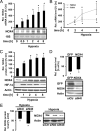
Figure 1.
Figure 1.
Hypoxia increases NOX4 expression in vitro. (A–C) Pulmonary artery smooth-muscle cells (PASMC) were stimulated for different time periods by hypoxia (1% O2). (A) Northern blot analyses were performed using a specific probe for NOX4; 18S staining served as loading control. Data represent % change of NOX4 mRNA levels versus normoxic control (100%; n = 3, *p < 0.05 vs. control). (B) Real-time PCR was performed with primers amplifying cDNA fragments specific for NOX4, PAI-1, or actin. Quantification was performed by ΔCT calculation. NOX4 and PAI-1 mRNA levels were normalized to actin levels. Normoxic control (0) was set to 100% (n = 3; *p < 0.05 vs. control). (C) NOX4 and HIF-1α protein levels were determined by Western blot analyses. Actin levels served as loading control. Data represent % change of NOX4 protein levels versus normoxic control (100%; n = 5, *p < 0.05 vs. control). (D) HEK293 cells were cotransfected with a plasmid encoding GFP-NOX4 and plasmids encoding shRNA against NOX4 (siN4I) or control shRNA (siCtr). Western blot analyses were performed with antibodies against NOX4 and GFP. Staining with an antibody against actin served as loading control. GFP or NOX4 protein levels in siCtr-expressing cells were set to 100%. Data represent % decrease of GFP or NOX4 protein levels compared with control (n = 3; *p < 0.05 vs. control). (E) PASMCs were transfected with vectors encoding two different shRNAs against NOX4 (siN4I, siN4II) or control shRNA (siCtr) and exposed to hypoxia for 4 h. Western blot analyses were performed with an antibody against NOX4. Actin staining served as loading control. NOX4 protein levels in hypoxic siCtr-expressing cells were set to 100% (n = 3; *p < 0.05 vs. control).
Figure 2.
Figure 2.
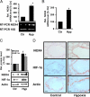
Hypoxia increases NOX4 expression in mouse lungs. Mice were exposed to normoxia (Ctr) or hypoxia (10% oxygen, Hyp) for 1 d, and lung tissue was obtained. (A and B) RNA was isolated from normoxic and hypoxic lung tissue. RT-PCR (A) or real-time PCR (B) were performed using specific primers for NOX4 or 18S. NOX4 mRNA levels under normoxia (Ctr) were set to 100%. Data represent % increase of NOX4 mRNA levels compared with control (n = 3; *p < 0.05 vs. Ctr). (C) Protein was isolated and Western blot analysis was performed with antibodies against NOX4 or HIF-1α. Actin levels served as loading control. Data represent % change of protein levels versus normoxic control (100%; n = 3, *p < 0.05 vs. control). (D) Immunohistochemistry was performed on lung tissue samples using antibodies against NOX4, HIF-1α, and α-actin (Actin). Images were taken with a 40x objective. Scale bars, 20 μm.
Figure 3.
Figure 3.
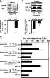
Hypoxia increases NOX4 transcription. (A and B) Pulmonary artery smooth-muscle cells (PASMCs) were treated with actinomycin D (Act, 5 μM) or DMSO (Ctr) for 1 h and exposed to hypoxia for 4 h. (A) Northern blot analyses were performed using a specific probe for NOX4, 18S staining served as loading control. NOX4 mRNA levels in DMSO-treated cells under hypoxia (Ctr) were set to 100%. Data represent % change of NOX4 mRNA levels versus hypoxic control (n = 3, *p < 0.05 vs. hypoxic control). (B) Western blot analyses were performed using antibodies against NOX4 or HIF-1α. Actin served as loading control. Protein levels in DMSO-treated cells under hypoxia (Ctr) were set to 100%. Data represent % change of protein levels versus hypoxic control (n = 3, *p < 0.05 vs. hypoxic control). (C) HEK293 cells were cotransfected with luciferase constructs containing either the wild-type NOX4 promoter (NOX4-730) or the NOX4 promoter mutated at the hypoxia-responsive element (HRE; NOX4-730m). Cells were exposed to hypoxia (Hyp) or were cotransfected with a plasmid coding for HIF-1α. Luciferase activities under the respective control conditions (Ctr) for each reporter plasmid were set equal to 100%. Data represent % induction of luciferase activity (n = 3; *p < 0.05 vs. control).
Figure 4.
Figure 4.
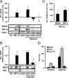
HIF-1α mediates NOX4 expression. (A–C) Pulmonary artery smooth-muscle cells (PASMCs) were transfected with vectors coding for HIF-1α or with vectors coding for shRNA against HIF-1α (siH1I, siH1II) or for control shRNA (siCtr) and exposed to hypoxia for 4 h. (A) Western blot analyses were performed using antibodies against NOX4 or HIF-1α. Actin was used as loading control. (B) Northern blots were performed using specific probes for NOX4 or 18S. Data represent % change of NOX4 protein (A) or NOX4 mRNA levels (B) versus the appropriate normoxic control set to 100% (n = 3, *p < 0.05 vs. control; #p < 0.05 vs. hypoxic control). (C) mRNA levels for NOX4 and PAI-1 were determined in HIF-1α–overexpressing cells by real-time PCR using primers specifically amplifying NOX4, PAI-1, or actin fragments. Quantification was performed by ΔCT calculation. NOX4 or PAI-1 mRNA levels were normalized to actin levels. Control cells (Ctr, siCtr) were set to 100%, and the relative change in HIF-1α–overexpressing cells is displayed (n = 3; *p < 0.05 vs. control). (D) HepG2 cells were exposed to hypoxia for 3 h. Chromatin immunoprecipitation (ChIP) was performed with an antibody against HIF-1α. Real-time PCR was performed on the precipitates using primers for the NOX4 promoter (black) or the PAI-1 promoter (gray) as positive control or for the third intron of β-actin lacking an HRE as negative control (dark gray, neg. Ctr). For background calculation, ChIP without antibody was performed. Quantification is shown in promille to chromatin input for all samples after background subtraction (n = 3, *p < 0.05 vs. control).
Figure 5.
Figure 5.
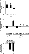
HIF-1α and NOX4 modulate ROS levels under normoxia and hypoxia. (A) Pulmonary artery smooth-muscle cells (PASMCs) were transfected with plasmids coding for NOX4 or for HIF-1α and were cotransfected with shRNA against NOX4 (siN4I) or with control shRNA (siCtr). ROS levels were evaluated by DHE fluorescence. ROS levels of cells transfected with control vectors were set to 100% (n = 3; *p < 0.05 vs. control, #p < 0.05 vs. HIF-1α). (B) PASMCs were transfected with a plasmid coding for NOX4 and were exposed to hypoxia for 30 min (0.5 h hypoxia), or with plasmids coding for shRNA against NOX4 (siN4I, siN4II), HIF-1α (siH1I, siH1II) or with control shRNA (siCtr) and exposed to hypoxia for 4 h. ROS levels were evaluated by DHE fluorescence thereafter. ROS levels of cells transfected with control vectors under normoxic conditions were set to 100% (n = 3; *p < 0.05 vs. normoxic controls, #p < 0.05 vs. hypoxic control). (C) PASMCs were transfected with plasmids coding for either shRNA against NOX4 (siN4I), HIF-1α (siH1I), or control shRNA (siCtr). Cells were exposed to hypoxia for 4 h, and ROS levels were measured by EPR using the spin-trap CMH. ROS levels of hypoxic control cells (siCtr) were set to 100% (n = 3; *p < 0.05 vs. hypoxic control).
Figure 6.
Figure 6.
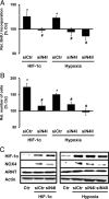
NOX4 mediates proliferative activity by HIF-1α and hypoxia. Pulmonary artery smooth-muscle cells (PASMCs) were transfected with vectors encoding different shRNAs against NOX4 (siN4I, siN4II) or control shRNA (siCtr) and were either cotransfected with a plasmid coding for HIF-1α or were exposed to hypoxia for 4 h. Proliferative activity was determined by (A) BrdU incorporation or (B) determination of cell numbers using a hemocytometer. Data are shown as relative change to normoxic control (100%; n = 3, *p < 0.05 vs. control; #p < 0.05 vs. HIF-1α–transfected or hypoxia-stimulated control). (C) Western blot analysis was performed with antibodies against HIF-1α, NOX4, or ARNT. Actin served as loading control.
Figure 7.
Figure 7.
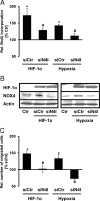
NOX4 mediates migration by HIF-1α and hypoxia. (A/B) HEK293 cells were transfected with vectors encoding shRNA against NOX4 (siN4I) or control shRNA (siCtr) and were cotransfected with an expression plasmid for HIF-1α or were exposed to hypoxia for 4 h. (A) Proliferative activity of HEK293 cells was determined by BrdU incorporation. Data are shown as relative change to normoxic control (100%; n = 3, *p < 0.05 vs. control; #p < 0.05 vs. HIF-1α–transfected or hypoxia-stimulated control). (B) Western blot analysis was performed with antibodies against HIF-1α or NOX4. Actin served as loading control. (C) Pulmonary artery smooth-muscle cells (PASMCs) were transfected with vectors encoding shRNA against NOX4 (siN4I) or control shRNA (siCtr) and were cotransfected with an expression plasmid for HIF-1α or were exposed to hypoxia for 4 h after wounding the cell layer with a 10-μl tip. The number of migrated cells into the wound was counted. Data are shown as relative change to control (n = 3; *p < 0.05 vs. control, #p < 0.05 vs. HIF-1α–transfected or hypoxia-stimulated control).
Figure 8.
Figure 8.
ROS mediate proliferation by hypoxia, NOX4, and HIF-1α. (A and B) Pulmonary artery smooth-muscle cells (PASMCs) were treated with vitamin C (VitC, 100 μM) for 60 min before exposure to hypoxia for 4 h. (A) ROS levels were evaluated by DHE fluorescence. (B) Proliferative activity was determined by BrdU incorporation. Untreated cells (Ctr) were set to 100% (n = 3; *p < 0.05 vs. control, #p <0.05 vs. hypoxic control). (C and D) PASMCs were transfected with plasmids encoding for HIF-1α or NOX4 or control plasmid (Ctr) and treated with VitC for 60 min. (C) ROS levels were evaluated by DHE fluorescence. (D) Proliferative activity was determined by BrdU incorporation. Untreated cells (Ctr) were set to 100% (n = 3; *p < 0.05 vs. control, #p < 0.05 vs. hypoxic control).

Similar articles

Cited by

References

    1. Aaronson P. I., Robertson T. P., Knock G. A., Becker S., Lewis T. H., Snetkov V., Ward J. P. Hypoxic pulmonary vasoconstriction: mechanisms and controversies. J. Physiol. 2006;570:53–58. - PMC - PubMed
    1. Archer S. L., Gomberg-Maitland M., Maitland M. L., Rich S., Garcia J. G., Weir E. K. Mitochondrial metabolism, redox signaling, and fusion: a mitochondria-ROS-HIF-1alpha-Kv1.5 O2-sensing pathway at the intersection of pulmonary hypertension and cancer. Am. J. Physiol. Heart Circ. Physiol. 2008;294:H570–H578. - PubMed
    1. Babior B. M. NADPH oxidase: an update. Blood. 1999;93:1464–1476. - PubMed
    1. Babior B. M., Lambeth J. D., Nauseef W. The neutrophil NADPH oxidase. Arch. Biochem. Biophys. 2002;397:342–344. - PubMed
    1. Bedard K., Krause K. H. The NOX family of ROS-generating NADPH oxidases: physiology and pathophysiology. Physiol. Rev. 2007;87:245–313. - PubMed

Publication types

MeSH terms

LinkOut - more resources