. 2010 Apr 27;3(119):ra33.
doi: 10.1126/scisignal.2000800.

Evolution of CASK into a Mg2+-sensitive kinase

Affiliations

Evolution of CASK into a Mg2+-sensitive kinase

Konark Mukherjee et al. Sci Signal. .

Abstract

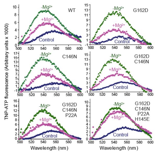
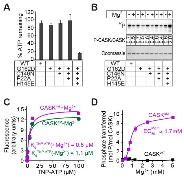
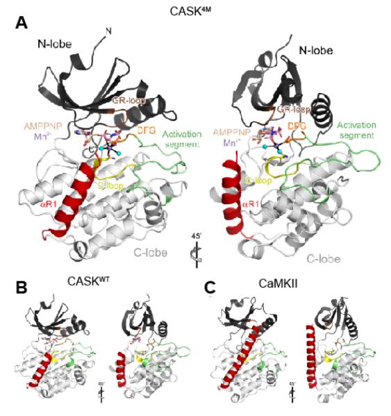
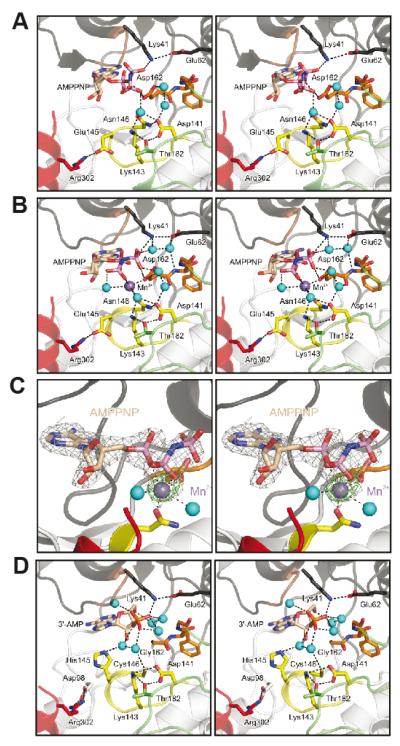
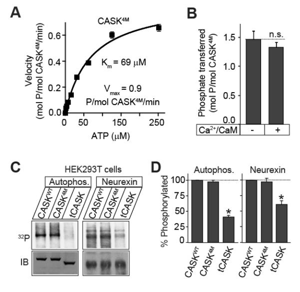
All known protein kinases, except CASK [calcium/calmodulin (CaM)-activated serine-threonine kinase], require magnesium ions (Mg(2+)) to stimulate the transfer of a phosphate from adenosine 5'-triphosphate (ATP) to a protein substrate. The CaMK (calcium/calmodulin-dependent kinase) domain of CASK shows activity in the absence of Mg(2+); indeed, it is inhibited by divalent ions including Mg(2+). Here, we converted the Mg(2+)-inhibited wild-type CASK kinase (CASK(WT)) into a Mg(2+)-stimulated kinase (CASK(4M)) by substituting four residues within the ATP-binding pocket. Crystal structures of CASK(4M) with and without bound nucleotide and Mn(2+), together with kinetic analyses, demonstrated that Mg(2+) accelerates catalysis of CASK(4M) by stabilizing the transition state, enhancing the leaving group properties of adenosine 5'-diphosphate, and indirectly shifting the position of the gamma-phosphate of ATP. Phylogenetic analysis revealed that the four residues conferring Mg(2+)-mediated stimulation were substituted from CASK during early animal evolution, converting a primordial, Mg(2+)-coordinating form of CASK into a Mg(2+)-inhibited kinase. This emergence of Mg(2+) sensitivity (inhibition by Mg(2+)) conferred regulation of CASK activity by divalent cations, in parallel with the evolution of the animal nervous systems.


Figures

Figure 1
Figure 1. Designing a Mg2+-coordinating version of CASK CaM-kinase domain
Fluorescence emission spectra of TNP-ATP in the presence of WT and mutant CASK. The protein analyzed (WT or mutant) is indicated in the upper right corner. Dark blue trace: Control spectrum of TNP-ATP (1 μM) in Tris-HCl buffer (pH 7.0) with EDTA (4 mM). Green trace: Spectra of samples containing 1 μM of the indicated recombinant CASK CaM-kinase domain, TNP-ATP (1 μM) and EDTA (4 mM) in Tris-HCl buffer (pH 7.0). Magenta trace: Spectra of samples containing 1 μM of the indicated recombinant CASK CaM-kinase domain, TNP-ATP (1 μM) and 100 μM MgCl2 in Tris-HCl buffer (pH 7.0). Samples were excited at 410 nm, and spectra were recorded between 500 and 600 nm. The spectra are representatives of experiments repeated three times with essentially identical results.
Figure 2
Figure 2. Effect of divalent ions on nucleotide-binding and hydrolysis
A. ATP consumption by WT or mutant CASK CaM-kinase domain. In Tris-HCl buffer (pH 7.0) supplemented with Ca2+ (1 mM), CaM (4 μM), and Mg2+ (2 mM), indicated variant of CASK CaM-kinase domain (1 μM), Autocamtide-2 (100 μM) and ATP (50 μM) were incubated for 60 min. The amount of ATP remaining was detected with KinaseGlotm. Data represents means ± standard deviation of three independent experiments. B. Autophosphorylation of WT and mutant CASK CaM-kinase domains. The indicated variants of CASK CaM-kinase domain (1 μM) were incubated in Tris-HCl buffer (pH 7.0) with Na+32P-ATP (50 cpm/pmol; -Mg2+) or 10 mM Mg2+-ATP (+Mg2+) at 30°C with shaking for 2 h. The proteins were separated by SDS-PAGE, transferred onto a nitrocellulose membrane, and visualized by phosphorimager scanning (upper panel). Ponceau staining was used for loading control (lower panel). Mean stoichiometry of phosphate incorporation (phosphates/CASK molecule) from three independent experiments are shown between the panels. C. TNP-ATP binding. Increasing amounts of TNP-ATP were added to cuvettes containing 10 mM Tris-HCl pH 7.0, 1 μM CASK4M, and either 4 mM EDTA (magenta symbols) or 200 μM Mg2+ (green symbols). The TNP-ATP fluorescence of the samples (excitation: 410 nm; emission: 541 nm) is plotted after subtracting background TNP-ATP fluorescence obtained with parallel samples, which contained the same TNP-ATP, Tris-HCl, EDTA, or Mg2+ concentrations but lysozyme instead of CASK. The plot is a representative of three independent experiments. D. Effect of Mg2+ on kinase activity of CASK4M. Indicated variants of CASK CaM-kinase domain (2 μM), autocamtide-2 (100 μM) and γ32P-ATP (250 μM; 250 cpm/pmol) in Tris-HCl buffer (pH 7.0) were incubated for 10 min with increasing amounts of Mg2+. Amount of phospho-autocamtide-2 generated was estimated by scintillation counting of dot-blots on nitrocellulose membrane. Data shown are means ± standard errors of the means (SEMs; n=3).
Figure 3
Figure 3. Overview of the CASK4M-Mn2+-AMPPNP crystal structure
A. Orthogonal ribbon plot of CASK4M CaM-kinase domain with landmark functional elements colored. Gly-rich loop (GR-loop) - brown, catalytic loop (C-loop) - yellow; DFG motif of the Mg2+-binding loop - orange; activation segment - green; C-terminal Ca2+/CaM-binding element - red. AMPPNP and residues Asn146 and Asp162, which coordinate the Mn2+ ion, are shown as sticks and colored by atom type. Carbon - beige; oxygen - red; nitrogen - blue; phosphorus - pink. B. Structure of CASK CaM-kinase domain in complex with AMPPNP (sticks) lacking a divalent metal ion (β and γ phosphates disordered; pdb ID 3C0H; (21)) in the same orientation as the CASK4M CaM-kinase domain in A. Cys146 is shown as sticks. Functional elements are colored as in A. C. Structure of CaMKII (pdb ID 2BDW; (28)) in the same orientation as the CASK4M CaM-kinase domain in A. Asn140 and Asp156, whose equivalents in CASK4M coordinate the Mn2+ ion, are shown as sticks. Functional elements are colored as in A.
Figure 4
Figure 4. Nucleotide-binding pocket of the CASK4M CaM-kinase domain
A. CASK4M CaM-kinase in complex with AMPPNP without a divalent metal ion. B. CASK4M CaM-kinase in complex with AMPPNP-Mn2+. Residues of the Mg2+-binding loop are shown in orange, residues of the catalytic loop are in yellow and residues of the C-terminal Ca2+/CaM-binding element are in red (as in Figure 3). Selected residues and the nucleotides are shown as sticks and colored by atom type; carbon - as the respective fragment; oxygen - red; nitrogen - blue; phosphorus - pink. Water molecules (cyan) and the Mn2+ ion (purple) are shown as spheres. The orientations are the same as in Figure 3A left panel. The orientations of the AMPPNP β and γ-phosphates differ in the complexes without and with Mn2+ (compare panels A and B). C. Stereo plot showing the final 2Fo-Fc electron density around the AMPPNP-Mn2+ complex contoured at the 1 σ level (gray mesh) and the anomalous difference Fourier map contoured at the 5 σ level (green mesh), indicating the position of the Mn2+ ion. AMPPNP is shown as sticks and colored by atom type as before. The Mn2+ ion (purple) and two coordinating water molecules (cyan) are shown as spheres. The orientation is the same as in Figure 3A left panel. D. CASKWT CaM-kinase domain in complex with co-purified 3′-AMP (21). Color-coding as above. The orientations are the same as in Figure 3B left panel.
Figure 5
Figure 5. Compensation for slow kinetics in CASK kinase activity
A. Catalytic kinetics of CASK4M. Purified CASK4M CaM-kinase domain (2 μM) was incubated with increasing amount of γ32P-ATP (400 cpm/pmol) in Tris-HCl buffer pH 7.0, containing Mg2+ (10 mM) and autocamtide (100 μM) as the substrate peptide for 10 min at 30°C. Amount of phospho-Autocamtide-2 generated was estimated by scintillation counting dot-blots on nitrocellulose membrane. Data shown are means ± SEMs (n=3). Michaelis constant (K ATP m ) and Vmax were calculated using Graph-Pad Prism software. Data shown are means ± SEMs (n=3). B. Effect of Ca2+/CaM on the catalytic rate. CASK4M CaM-kinase domain (2μM) was incubated with γ32P-ATP (200 μM, 800 cpm/pmol) in Tris-HCl buffer (pH 7.0) supplemented with Mg2+ (2 mM) and autocamtide-2 (100 μM), for 2 min in the presence or absence of Ca2+ (1 mM) and CaM (10 μM). Amount of phospho-autocamtide-2 generated was estimated with scintillation counting of dot blots on a nitrocellulose membrane. Data are represented as means ± SEMs, n = 3. n.s. (not significant) C, D. Neurexin phosphorylation. HEK293T cells were transfected with Flag epitope-tagged neurexin and either EGFP-CASK, EGFP-CASK4M or truncated EGFP-tCASK. 48 h later, transfected cells were labeled with 32Pi, followed by anti-Flag immunoprecipitation of neurexin. Immunoprecipitates were separated by SDS-PAGE and visualized by phosphorimager scanning. Autophosphorylation of the co-precipitated CASK (Autophos.) and phosphorylated neurexin (Neurexin) are shown. Immunoblotting (IB) for neurexin and CASK was performed to show expression. Bar-graph depicts the comparison of autophosphorylation or neurexin phosphorylation levels in cells co-expressing the indicated CASK variants. Data are represented as means ± SEMs, n = 3; asterisks indicate P < 0.05.
Figure 6
Figure 6. Evolution of CASK
A. Evolutionary changes in the nucleotide-binding pocket of CASK CaM-kinase domain. CASK CaM-kinase domain sequences from various animal species were aligned, and the residues corresponding to those mutated in CASK4M were identified and are shown. Corresponding human CaMKIIα residues are shown on the left for comparison. B. Sequence conservation (identities) of CASK domains between human and placozoan CASK (from T. adhaerens). See Supplemental Figure S7 for a full sequence alignment. C. Model comparing CASK and CaMKII catalytic cycles. Typically, CaM-kinases are held in an autoinhibited conformation by the autoregulatory domain (yellow) with an open, inactive nucleotide binding cleft. Upon binding of Ca2+ (purple)/CaM (green), this autoinhibition is released and the enzyme attains an active closed conformation amenable to Mg2+ (yellow)/ATP (blue) binding and substrate binding (lower panel). CASK CaM-kinase, on other hand, constitutively binds ATP, and is regulated by the recruitment of its substrates via the MAGUK scaffolding domains, especially the PDZ-domain.

Similar articles

Cited by

References

    1. Manning G, Whyte DB, Martinez R, Hunter T, Sudarsanam S. The protein kinase complement of the human genome. Science. 2002;298:1912–34. - PubMed
    1. Noble ME, Endicott JA, Johnson LN. Protein kinase inhibitors: insights into drug design from structure. Science. 2004;303:1800–5. - PubMed
    1. Huse M, Kuriyan J. The conformational plasticity of protein kinases. Cell. 2002;109:275–82. - PubMed
    1. Hanks SK, Quinn AM, Hunter T. The protein kinase family: conserved features and deduced phylogeny of the catalytic domains. Science. 1988;241:42–52. - PubMed
    1. Taylor SS, Radzio-Andzelm E. Three protein kinase structures define a common motif. Structure. 1994;2:345–55. - PubMed

Publication types

LinkOut - more resources