

Comparison of ATP-binding cassette transporter interactions with the tyrosine kinase inhibitors imatinib, nilotinib, and dasatinib
- PMID: 20423956
- PMCID: PMC2913625
- DOI: 10.1124/dmd.109.031302
Comparison of ATP-binding cassette transporter interactions with the tyrosine kinase inhibitors imatinib, nilotinib, and dasatinib
Abstract
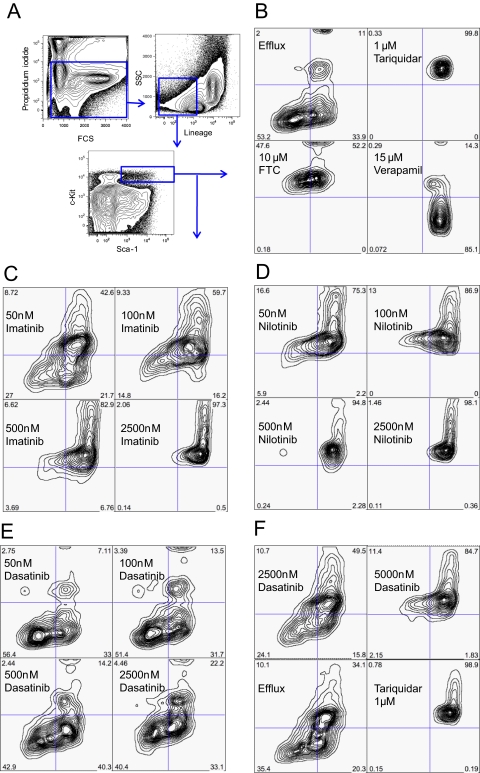
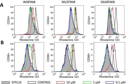
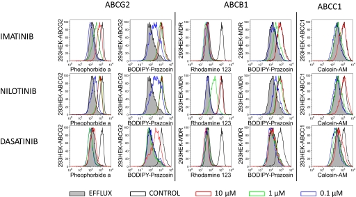
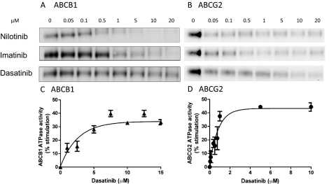
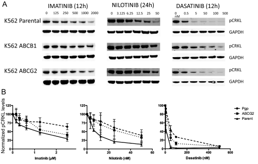
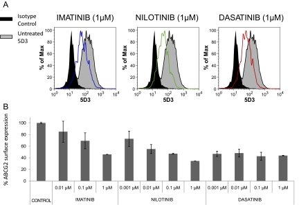
Although the development of tyrosine kinase inhibitors (TKIs) to control the unregulated activity of BCR-ABL revolutionized the therapy of chronic myeloid leukemia, resistance to TKIs is a clinical reality. Among the postulated mechanisms of resistance is the overexpression of ATP-binding cassette (ABC) transporters, such as P-glycoprotein (ABCB1) and breast cancer resistance protein (ABCG2), which mediate reduced intracellular drug accumulation. We compared the interactions of the TKIs imatinib, nilotinib, and dasatinib with ABCB1 and ABCG2 in ex vivo and in vitro systems. The TKIs inhibited rhodamine 123 and Hoechst 33342 efflux mediated by endogenous expression of the transporters in murine and human hematopoietic stem cells with potency order nilotinib >> imatinib >> dasatinib. Studies with ABCB1-, ABCG2-, and ABCC1-transfected human embryonic kidney 293 cells verified that nilotinib was the most potent inhibitor of ABCB1 and ABCG2. Cytotoxicity assays in stably transduced K562-ABCG2 and K562-ABCB1 cells confirmed that the TKIs were also substrates for the two transporters. Like imatinib, both nilotinib and dasatinib decreased ABCG2 surface expression in K562-ABCG2 cells. Finally, we found that all TKIs were able to compete labeling of ABCB1 and ABCG2 by the photo-cross-linkable prazosin analog [(125)I]iodoarylazidoprazosin, suggesting interaction at the prazosin-binding site of both proteins. Our experiments support the hypothesis that all three TKIs are substrates of ABC transporters and that, at higher concentrations, TKIs overcome transporter function. Taken together, the results suggest that therapeutic doses of imatinib and nilotinib may diminish the potential of ABCB1 and ABCG2 to limit oral absorption or confer resistance. Clinical data are required to definitively answer the latter question.
Figures
Similar articles
-
Interaction of nilotinib, dasatinib and bosutinib with ABCB1 and ABCG2: implications for altered anti-cancer effects and pharmacological properties.Br J Pharmacol. 2009 Oct;158(4):1153-64. doi: 10.1111/j.1476-5381.2009.00383.x. Epub 2009 Sep 28. Br J Pharmacol. 2009. PMID: 19785662 Free PMC article.
-
Interaction of the efflux transporters ABCB1 and ABCG2 with imatinib, nilotinib, and dasatinib.Clin Pharmacol Ther. 2014 Mar;95(3):294-306. doi: 10.1038/clpt.2013.208. Epub 2013 Oct 9. Clin Pharmacol Ther. 2014. PMID: 24107928 Review.
-
Tyrosine kinase inhibitor resistance in chronic myeloid leukemia cell lines: investigating resistance pathways.Leuk Lymphoma. 2011 Nov;52(11):2139-47. doi: 10.3109/10428194.2011.591013. Epub 2011 Jun 30. Leuk Lymphoma. 2011. PMID: 21718141
-
Degree of kinase inhibition achieved in vitro by imatinib and nilotinib is decreased by high levels of ABCB1 but not ABCG2.Leuk Lymphoma. 2013 Mar;54(3):569-78. doi: 10.3109/10428194.2012.715345. Epub 2012 Aug 21. Leuk Lymphoma. 2013. PMID: 22845311
-
Predicting the response of CML patients to tyrosine kinase inhibitor therapy.Curr Hematol Malig Rep. 2011 Jun;6(2):88-95. doi: 10.1007/s11899-011-0087-9. Curr Hematol Malig Rep. 2011. PMID: 21448598 Review.
Cited by
-
In vitro and ex vivo anti‑tumor effect and mechanism of Tucatinib in leukemia stem cells and ABCG2‑overexpressing leukemia cells.Oncol Rep. 2021 Mar;45(3):1142-1152. doi: 10.3892/or.2020.7915. Epub 2020 Dec 30. Oncol Rep. 2021. PMID: 33650639 Free PMC article.
-
Impact of ABCB1 1236C > T-2677G > T-3435C > T polymorphisms on the anti-proliferative activity of imatinib, nilotinib, dasatinib and ponatinib.Sci Rep. 2016 Jul 12;6:29559. doi: 10.1038/srep29559. Sci Rep. 2016. PMID: 27405085 Free PMC article.
-
Asciminib, a novel allosteric inhibitor of BCR-ABL1, shows synergistic effects when used in combination with imatinib with or without drug resistance.Pharmacol Res Perspect. 2024 Aug;12(4):e1214. doi: 10.1002/prp2.1214. Pharmacol Res Perspect. 2024. PMID: 39031848 Free PMC article.
-
ABC transporters in multi-drug resistance and ADME-Tox of small molecule tyrosine kinase inhibitors.Pharm Res. 2014 Sep;31(9):2237-55. doi: 10.1007/s11095-014-1389-0. Epub 2014 May 20. Pharm Res. 2014. PMID: 24842659 Review.
-
The effect of grape seed and green tea extracts on the pharmacokinetics of imatinib and its main metabolite, N-desmethyl imatinib, in rats.BMC Pharmacol Toxicol. 2020 Nov 16;21(1):77. doi: 10.1186/s40360-020-00456-9. BMC Pharmacol Toxicol. 2020. PMID: 33198812 Free PMC article.
References
-
- Ambudkar SV. (1998) Drug-stimulatable ATPase activity in crude membranes of human MDR1-transfected mammalian cells. Methods Enzymol 292:504–514 - PubMed
-
- Bhatia R, Holtz M, Niu N, Gray R, Snyder DS, Sawyers CL, Arber DA, Slovak ML, Forman SJ. (2003) Persistence of malignant hematopoietic progenitors in chronic myelogenous leukemia patients in complete cytogenetic remission following imatinib mesylate treatment. Blood 101:4701–4707 - PubMed
-
- Bradeen HA, Eide CA, O'Hare T, Johnson KJ, Willis SG, Lee FY, Druker BJ, Deininger MW. (2006) Comparison of imatinib mesylate, dasatinib (BMS-354825), and nilotinib (AMN107) in an N-ethyl-N-nitrosourea (ENU)-based mutagenesis screen: high efficacy of drug combinations. Blood 108:2332–2338 - PMC - PubMed
-
- Brendel C, Scharenberg C, Dohse M, Robey RW, Bates SE, Shukla S, Ambudkar SV, Wang Y, Wennemuth G, Burchert A, et al. (2007) Imatinib mesylate and nilotinib (AMN107) exhibit high-affinity interaction with ABCG2 on primitive hematopoietic stem cells. Leukemia 21:1267–1275 - PubMed
-
- Burger H, van Tol H, Boersma AW, Brok M, Wiemer EA, Stoter G, Nooter K. (2004) Imatinib mesylate (STI571) is a substrate for the breast cancer resistance protein (BCRP)/ABCG2 drug pump. Blood 104:2940–2942 - PubMed
Publication types
MeSH terms
Substances
Grants and funding
LinkOut - more resources
Full Text Sources
Other Literature Sources
Miscellaneous
