

SIRT1 decreases Lox-1-mediated foam cell formation in atherogenesis
- PMID: 20418343
- PMCID: PMC2938465
- DOI: 10.1093/eurheartj/ehq107
SIRT1 decreases Lox-1-mediated foam cell formation in atherogenesis
Abstract
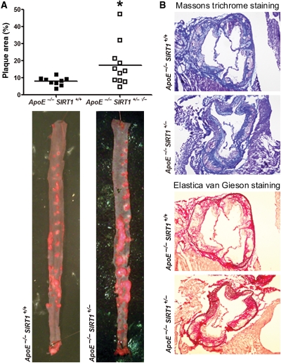
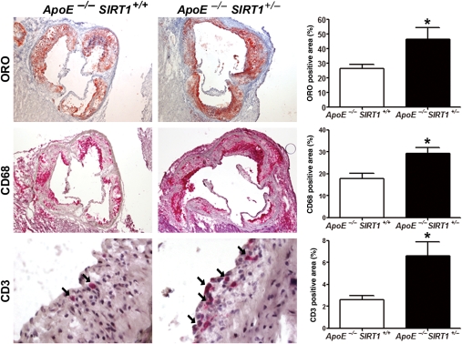
Aims: Endothelial activation, macrophage infiltration, and foam cell formation are pivotal steps in atherogenesis. Our aim in this study was to analyse the role of SIRT1, a class III deacetylase with important metabolic functions, in plaque macrophages and atherogenesis.
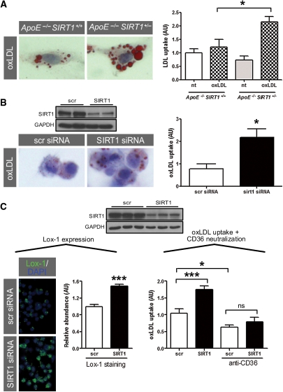
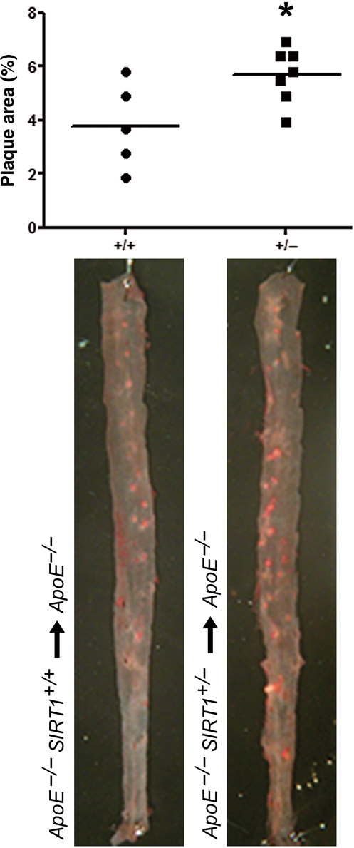
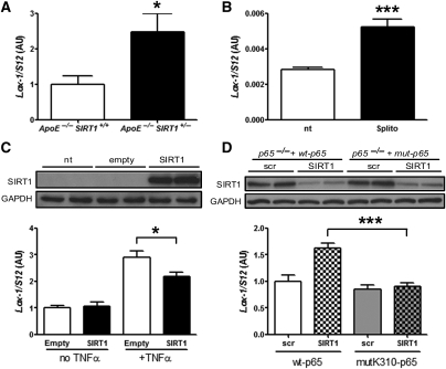
Methods and results: Using partial SIRT1 deletion in atherosclerotic mice, we demonstrate that SIRT1 protects against atherosclerosis by reducing macrophage foam cell formation. Peritoneal macrophages from heterozygous SIRT1 mice accumulate more oxidized low-density lipoprotein (oxLDL), thereby promoting foam cell formation. Bone marrow-restricted SIRT1 deletion confirmed that SIRT1 function in macrophages is sufficient to decrease atherogenesis. Moreover, we show that SIRT1 reduces the uptake of oxLDL by diminishing the expression of lectin-like oxLDL receptor-1 (Lox-1) via suppression of the NF-κB signalling pathway.
Conclusion: Our findings demonstrate protective effects of SIRT1 in atherogenesis and suggest pharmacological SIRT1 activation as a novel anti-atherosclerotic strategy by reducing macrophage foam cell formation.
Figures
Similar articles
-
Tanshinone II-A inhibits oxidized LDL-induced LOX-1 expression in macrophages by reducing intracellular superoxide radical generation and NF-κB activation.Transl Res. 2012 Aug;160(2):114-24. doi: 10.1016/j.trsl.2012.01.008. Epub 2012 Feb 2. Transl Res. 2012. PMID: 22677363
-
Inhibition of Orai1 Store-Operated Calcium Channel Prevents Foam Cell Formation and Atherosclerosis.Arterioscler Thromb Vasc Biol. 2016 Apr;36(4):618-28. doi: 10.1161/ATVBAHA.116.307344. Epub 2016 Feb 25. Arterioscler Thromb Vasc Biol. 2016. PMID: 26916730
-
Insulin-like growth factor I reduces lipid oxidation and foam cell formation via downregulation of 12/15-lipoxygenase.Atherosclerosis. 2015 Feb;238(2):313-20. doi: 10.1016/j.atherosclerosis.2014.12.024. Epub 2014 Dec 20. Atherosclerosis. 2015. PMID: 25549319 Free PMC article.
-
Protective roles of SIRT1 in atherosclerosis.Cell Cycle. 2011 Feb 15;10(4):640-7. doi: 10.4161/cc.10.4.14863. Epub 2011 Feb 15. Cell Cycle. 2011. PMID: 21293192 Review.
-
SIRT1 - an anti-inflammatory pathway at the crossroads between metabolic disease and atherosclerosis.Curr Vasc Pharmacol. 2012 Nov;10(6):693-6. doi: 10.2174/157016112803520756. Curr Vasc Pharmacol. 2012. PMID: 23259556 Review.
Cited by
-
Celastrol prevents atherosclerosis via inhibiting LOX-1 and oxidative stress.PLoS One. 2013 Jun 17;8(6):e65477. doi: 10.1371/journal.pone.0065477. Print 2013. PLoS One. 2013. PMID: 23799016 Free PMC article.
-
Peripheral blood monocyte Sirt1 expression is reduced in patients with coronary artery disease.PLoS One. 2013;8(1):e53106. doi: 10.1371/journal.pone.0053106. Epub 2013 Jan 29. PLoS One. 2013. PMID: 23382833 Free PMC article.
-
Novel Role of the SIRT1 in Endocrine and Metabolic Diseases.Int J Biol Sci. 2023 Jan 1;19(2):484-501. doi: 10.7150/ijbs.78654. eCollection 2023. Int J Biol Sci. 2023. PMID: 36632457 Free PMC article. Review.
-
Deletion of Sirt3 does not affect atherosclerosis but accelerates weight gain and impairs rapid metabolic adaptation in LDL receptor knockout mice: implications for cardiovascular risk factor development.Basic Res Cardiol. 2014 Jan;109(1):399. doi: 10.1007/s00395-013-0399-0. Epub 2013 Dec 27. Basic Res Cardiol. 2014. PMID: 24370889 Free PMC article.
-
Enhancement in efferocytosis of oxidized low-density lipoprotein-induced apoptotic RAW264.7 cells through Sirt1-mediated autophagy.Int J Mol Med. 2014 Mar;33(3):523-33. doi: 10.3892/ijmm.2013.1609. Epub 2013 Dec 27. Int J Mol Med. 2014. PMID: 24378473 Free PMC article.
References
-
- Libby P. Inflammation in atherosclerosis. Nature. 2002;420:868–874. doi:10.1038/nature01323. - DOI - PubMed
-
- Kaeberlein M, McVey M, Guarente L. The SIR2/3/4 complex and SIR2 alone promote longevity in Saccharomyces cerevisiae by two different mechanisms. Genes Dev. 1999;13:2570–2580. doi:10.1101/gad.13.19.2570. - DOI - PMC - PubMed
-
- Chen D, Steele AD, Lindquist S, Guarente L. Increase in activity during calorie restriction requires Sirt1. Science. 2005;310:1641. doi:10.1126/science.1118357. - DOI - PubMed
-
- Bordone L, Motta MC, Picard F, Robinson A, Jhala US, Apfeld J, McDonagh T, Lemieux M, McBurney M, Szilvasi A, Easlon EJ, Lin SJ, Guarente L. Sirt1 regulates insulin secretion by repressing UCP2 in pancreatic beta cells. PLoS Biol. 2006;4:e31. doi:10.1371/journal.pbio.0040031. - DOI - PMC - PubMed
-
- Picard F, Kurtev M, Chung N, Topark-Ngarm A, Senawong T, Machado De Oliveira R, Leid M, McBurney MW, Guarente L. Sirt1 promotes fat mobilization in white adipocytes by repressing PPAR-gamma. Nature. 2004;429:771–776. doi:10.1038/nature02583. - DOI - PMC - PubMed
Publication types
MeSH terms
Substances
LinkOut - more resources
Full Text Sources
Medical
