

The antipsychotics olanzapine, risperidone, clozapine, and haloperidol are D2-selective ex vivo but not in vitro
- PMID: 20410873
- PMCID: PMC3055486
- DOI: 10.1038/npp.2010.50
The antipsychotics olanzapine, risperidone, clozapine, and haloperidol are D2-selective ex vivo but not in vitro
Abstract
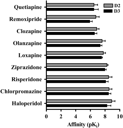
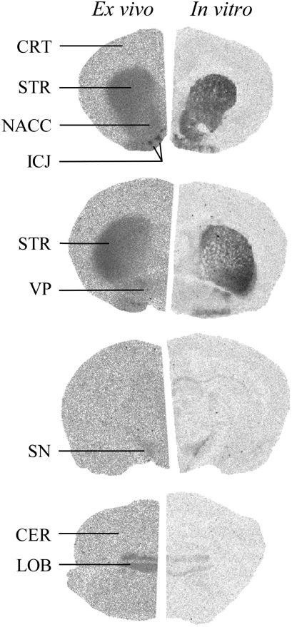
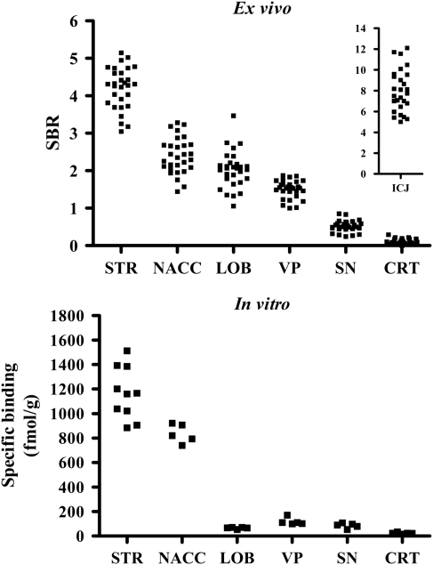
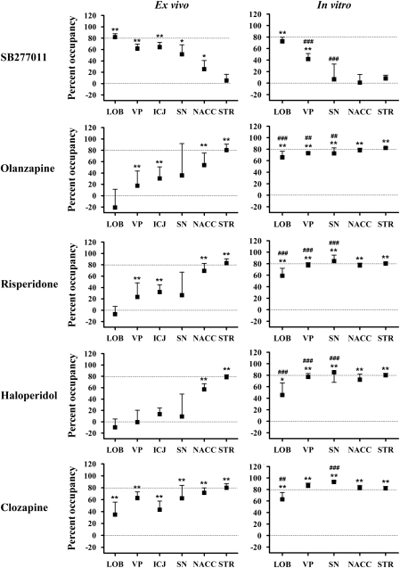
In a recent human [(11)C]-(+)-PHNO positron emission tomography study, olanzapine, clozapine, and risperidone occupied D2 receptors in striatum (STR), but, despite their similar in vitro D2 and D3 affinities, failed to occupy D3 receptors in globus pallidus. This study had two aims: (1) to characterize the regional D2/D3 pharmacology of in vitro and ex vivo [(3)H]-(+)-PHNO binding sites in rat brain and (2) to compare, using [(3)H]-(+)-PHNO autoradiography, the ex vivo and in vitro pharmacology of olanzapine, clozapine, risperidone, and haloperidol. Using the D3-selective drug SB277011, we found that ex vivo and in vitro [(3)H]-(+)-PHNO binding in STR is exclusively due to D2, whereas that in cerebellar lobes 9 and 10 is exclusively due to D3. Surprisingly, the D3 contribution to [(3)H]-(+)-PHNO binding in the islands of Calleja, ventral pallidum, substantia nigra, and nucleus accumbens was greater ex vivo than in vitro. Ex vivo, systemically administered olanzapine, risperidone, and haloperidol, at doses occupying approximately 80% D2, did not occupy D3 receptors. Clozapine, which also occupied approximately 80% of D2 receptors ex vivo, occupied a smaller percentage of D3 receptors than predicted by its in vitro pharmacology. Across brain regions, ex vivo occupancy by antipsychotics was inversely related to the D3 contribution to [(3)H]-(+)-PHNO binding. In contrast, in vitro occupancy was similar across brain regions, independent of the regional D3 contribution. These data indicate that at clinically relevant doses, olanzapine, clozapine, risperidone, and haloperidol are D2-selective ex vivo. This unforeseen finding suggests that their clinical effects cannot be attributed to D3 receptor blockade.
Figures
Similar articles
-
Acutely administered antipsychotic drugs are highly selective for dopamine D2 over D3 receptors.Pharmacol Res. 2013 Apr;70(1):66-71. doi: 10.1016/j.phrs.2013.01.002. Epub 2013 Jan 14. Pharmacol Res. 2013. PMID: 23327779
-
Risperidone compared with new and reference antipsychotic drugs: in vitro and in vivo receptor binding.Psychopharmacology (Berl). 1996 Mar;124(1-2):57-73. doi: 10.1007/BF02245606. Psychopharmacology (Berl). 1996. PMID: 8935801
-
Neuroendocrine responsivities of the pituitary dopamine system in male schizophrenic patients during treatment with clozapine, olanzapine, risperidone, sulpiride, or haloperidol.Eur Arch Psychiatry Clin Neurosci. 2001 Jun;251(3):141-6. doi: 10.1007/s004060170049. Eur Arch Psychiatry Clin Neurosci. 2001. PMID: 11697576 Clinical Trial.
-
[Cognition, schizophrenia and the effect of antipsychotics].Encephale. 2006 May-Jun;32(3 Pt 1):341-50. doi: 10.1016/s0013-7006(06)76162-0. Encephale. 2006. PMID: 16840928 Review. French.
-
Antipsychotic drugs which elicit little or no parkinsonism bind more loosely than dopamine to brain D2 receptors, yet occupy high levels of these receptors.Mol Psychiatry. 1998 Mar;3(2):123-34. doi: 10.1038/sj.mp.4000336. Mol Psychiatry. 1998. PMID: 9577836 Review.
Cited by
-
Prior haloperidol, but not olanzapine, exposure augments the pursuit of reward cues: implications for substance abuse in schizophrenia.Schizophr Bull. 2013 May;39(3):692-702. doi: 10.1093/schbul/sbs077. Epub 2012 Aug 27. Schizophr Bull. 2013. PMID: 22927669 Free PMC article.
-
Preferential binding to dopamine D3 over D2 receptors by cariprazine in patients with schizophrenia using PET with the D3/D2 receptor ligand [(11)C]-(+)-PHNO.Psychopharmacology (Berl). 2016 Oct;233(19-20):3503-12. doi: 10.1007/s00213-016-4382-y. Epub 2016 Aug 15. Psychopharmacology (Berl). 2016. PMID: 27525990 Free PMC article.
-
Effects of the T-type calcium channel antagonist Z944 on paired associates learning and locomotor activity in rats treated with the NMDA receptor antagonist MK-801.Psychopharmacology (Berl). 2018 Nov;235(11):3339-3350. doi: 10.1007/s00213-018-5040-3. Epub 2018 Sep 24. Psychopharmacology (Berl). 2018. PMID: 30251162
-
Comparative pharmacology of antipsychotics possessing combined dopamine D2 and serotonin 5-HT1A receptor properties.Psychopharmacology (Berl). 2011 Aug;216(4):451-73. doi: 10.1007/s00213-011-2247-y. Epub 2011 Mar 11. Psychopharmacology (Berl). 2011. PMID: 21394633 Review.
-
In vivo binding of antipsychotics to D3 and D2 receptors: a PET study in baboons with [11C]-(+)-PHNO.Neuropsychopharmacology. 2011 Mar;36(4):887-95. doi: 10.1038/npp.2010.228. Epub 2010 Dec 22. Neuropsychopharmacology. 2011. PMID: 21178982 Free PMC article.
References
-
- Bancroft GN, Morgan KA, Flietstra RJ, Levant B. Binding of [3H]PD 128907, a putatively selective ligand for the D3 dopamine receptor, in rat brain: a receptor binding and quantitative autoradiographic study. Neuropsychopharmacology. 1998;18:305–316. - PubMed
-
- Beckstead RM, Domesick VB, Nauta WJ. Efferent connections of the substantia nigra and ventral tegmental area in the rat. Brain Res. 1979;175:191–217. - PubMed
-
- Carson RE, Channing MA, Blasberg RG, Dunn BB, Cohen RM, Rice KC, et al. Comparison of bolus and infusion methods for receptor quantitation: application to [18F]cyclofoxy and positron emission tomography. J Cereb Blood Flow Metab. 1993;13:24–42. - PubMed
-
- Farde L, Nordstrom AL, Wiesel FA, Pauli S, Halldin C, Sedvall G. Positron emission tomographic analysis of central D1 and D2 dopamine receptor occupancy in patients treated with classical neuroleptics and clozapine. Relation to extrapyramidal side effects. Arch Gen Psychiatry. 1992;49:538–544. - PubMed
-
- Freedman SB, Patel S, Marwood R, Emms F, Seabrook GR, Knowles MR, et al. Expression and pharmacological characterization of the human D3 dopamine receptor. J Pharmacol Exp Ther. 1994;268:417–426. - PubMed
Publication types
MeSH terms
Substances
Grants and funding
LinkOut - more resources
Full Text Sources
