. 2010 Jun 18;285(25):19022-8.
doi: 10.1074/jbc.M110.113837. Epub 2010 Apr 19.

Proteins at the polypeptide tunnel exit of the yeast mitochondrial ribosome

Affiliations

Proteins at the polypeptide tunnel exit of the yeast mitochondrial ribosome

Steffi Gruschke et al. J Biol Chem. .

Abstract

Oxidative phosphorylation in mitochondria requires the synthesis of proteins encoded in the mitochondrial DNA. The mitochondrial translation machinery differs significantly from that of the bacterial ancestor of the organelle. This is especially evident from many mitochondria-specific ribosomal proteins. An important site of the ribosome is the polypeptide tunnel exit. Here, nascent chains are exposed to an aqueous environment for the first time. Many biogenesis factors interact with the tunnel exit of pro- and eukaryotic ribosomes to help the newly synthesized proteins to mature. To date, nothing is known about the organization of the tunnel exit of mitochondrial ribosomes. We therefore undertook a comprehensive approach to determine the composition of the yeast mitochondrial ribosomal tunnel exit. Mitochondria contain homologues of the ribosomal proteins located at this site in bacterial ribosomes. Here, we identified proteins located in their proximity by chemical cross-linking and mass spectrometry. Our analysis revealed a complex network of interacting proteins including proteins and protein domains specific to mitochondrial ribosomes. This network includes Mba1, the membrane-bound ribosome receptor of the inner membrane, as well as Mrpl3, Mrpl13, and Mrpl27, which constitute ribosomal proteins exclusively found in mitochondria. This unique architecture of the tunnel exit is presumably an adaptation of the translation system to the specific requirements of the organelle.


Figures

FIGURE 1.
FIGURE 1.
Proteins located around the mitochondrial ribosomal tunnel exit. A, bottom view of the large ribosomal subunit of bacteria. The localization of the conserved proteins located around the tunnel exit (TE) of the 50 S subunit is depicted. B, mitochondrial proteins that are homologous to the conserved tunnel exit proteins in bacteria. These mitochondrial proteins contain, in addition to a conserved region N-terminal mitochondrial targeting signals (MTS) as well as N- and C-terminal extension domains (NE-domain and CE-domain, respectively). Furthermore, Mrp20 and Mrpl40 have C-terminal domains that show a high propensity to form a coiled-coil structure. Numbers indicate amino acid positions. C, C-terminal His7 tags on Mrpl4, Mrp20, and Mrpl40 do not impair respiratory growth. Yeast cells of the indicated strains were plated in serial 10-fold dilutions onto media requiring (Glycerol) or not requiring respiration (Glucose) and incubated for 2 days. D, C-terminal His7 tags on Mrpl4, Mrp20, and Mrpl40 do not destabilize the proteins. Equal amounts of cell lysates of the indicated strains were separated on SDS-PAGE and analyzed by Western blotting.
FIGURE 2.
FIGURE 2.
Strategy to identify proteins located at the mitochondrial ribosomal tunnel exit. A, flow chart of the strategy to identify interaction partners of the tunnel exit proteins. B, purification of Mrpl40His7. Mitochondria from a strain expressing His7-tagged Mrpl40 were lysed in Triton X-100. Ribosomes of this lysate were centrifuged through a high density sucrose cushion. The resulting ribosomal pellet was resuspended in 1% SDS, and Mrpl40His7 was purified. Left panel, silver-stained gel; right panel, Western blotting using the indicated antibodies. T, total; S, supernatant after centrifugation; P, ribosomal pellet after centrifugation; NB, not bound material of the Ni-NTA purification; E, elution fraction. C, cross-linking of Mrpl40 in isolated mitochondria. Wild type mitochondria were incubated with a variety of cross-linking agents at different temperatures. DMSO was used as control. The mitochondria were reisolated, subjected to SDS-PAGE, and analyzed by Western blotting. A number of specific cross-linking products are indicated with their apparent molecular mass. * indicates nonspecific cross reactions. MBS, 3-maleimidobenzoyl-N-hydroxysuccinimide ester; BMH, bismaleimidohexane.
FIGURE 3.
FIGURE 3.
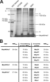
Identification of proteins located around the mitochondrial ribosomal tunnel exit. A, silver-stained gel of the elution fractions after a large scale experiment with cross-linking and purification using mitochondria that contain Mrpl40His7. Isolated mitochondria (40 mg each) were incubated with the indicated cross-linkers or a control (DMSO). Mrpl40His7 was purified from isolated ribosomes. In comparison with the control, the additional bands represent cross-linking products of Mrpl40His7 (indicated by the apparent molecular mass; X, not identified) and were excised from the gel and analyzed by LC-MS. B, identified cross-linking partners of Mrpl4, Mrp20, and Mrpl40. MWapp, apparent molecular weight.
FIGURE 4.
FIGURE 4.
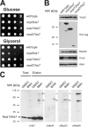
The identified components are located at the ribosomal tunnel exit of mitochondria. A, growth test of yeast strains harboring C-terminal His7-tagged variants of Mrpl3, Mrpl13, and Mrpl27. Yeast cells of the indicated strains were plated in serial 10-fold dilutions onto media requiring (Glycerol) or not requiring respiration (Glucose) and incubated for 2 days. B, C-terminal His7 tags on Mrpl13 and Mrpl27, but not on Mrpl3, allow the accumulation of mitochondrially encoded Cox2 to wild type level. Equal amounts of mitochondria isolated from the indicated strains were separated on SDS-PAGE and analyzed by Western blotting. C, Mrpl13 can be cross-linked to Mrpl4, Mrp20, and Mrpl40. Mitochondria containing Mrpl13His7 were incubated with the cross-linker bismaleimidohexane (BMH) or a control (DMSO). Next, Mrpl13His7 was purified following the scheme presented in Fig. 2A. The elution fraction was analyzed by Western blotting using antibodies against the His7 tag, Mrpl4, Mrp20, or Mrpl40, respectively.
FIGURE 5.
FIGURE 5.
Mba1 is located in proximity to the tunnel exit. A, a C-terminal His7 tag on Mba1 does not destabilize the protein. Lysates of the indicated strains were separated on SDS-PAGE and analyzed by Western blotting. B, large scale purification of cross-linking products to Mba1His7. The experiment was performed as depicted in Fig. 3A. MBS, 3-maleimidobenzoyl-N-hydroxysuccinimide ester. C, identified cross-linking partners of Mba1. MWapp, apparent molecular weight; DSG, disuccinimidyl glutarate.
FIGURE 6.
FIGURE 6.
Contacts of proteins located at the tunnel exit with nascent chains. A, strategy to cross-link nascent chains to proteins. B, Oxa1 and Mba1 can contact nascent chains independently of each other. Translation products of mitochondria isolated from wild type, Δmba1 or Δoxa1 cells were labeled with [35S]methionine in the presence of the cross-linking reagent DFDNB. Oxa1 and Mba1 with cross-linked nascent chains were precipitated from SDS-denatured lysates using specific antibodies or control IgGs. The samples were separated on SDS-PAGE, blotted to nitrocellulose, and analyzed by autoradiography. Nascent chains migrate as a diffuse smear above the position of Oxa1 (39 kDa) and Mba1 (28 kDa) in the gel. Total, 10% of total. C, contact of ribosomal proteins to nascent chains. The experiments were performed as in B with the exception that mitochondria containing His7-tagged proteins were used. The His7-tagged proteins were purified and eluted from the resin. Total, 1% of total. D, the experiment in C was repeated with mitochondria containing the indicated His7-tagged proteins. The radioactive signals of a region starting at the position of the His7-tagged protein plus an additional 10 kDa were densitometrically quantified in the elution fractions. The background signal of the control was subtracted from the signal of the DFDNB-cross-linked nascent chains. a.u., arbitrary units.
FIGURE 7.
FIGURE 7.
A comparison of interactions at the bacterial and mitochondrial ribosomal tunnel exit (TE). Left, the tunnel exit of bacterial ribosomes and the mapped binding sites of biogenesis factors are depicted. Right, a hypothetical model of the tunnel exit of mitochondrial ribosomes and the mitochondria-specific proteins as revealed in this study. Mba1 is positioned in proximity to Mrpl22 and Mrpl4. Oxa1 interacts with the mitochondrial ribosome close to Mrp20 and Mrpl40. The hypothetical distribution of the ribosomal proteins as suggested by the cross-linking analysis is delineated.

Similar articles

Cited by

References

    1. Borst P., Grivell L. A. (1978) Cell 15, 705–723 - PubMed
    1. Ott M., Herrmann J. M. (December2, 2009) Biochim. Biophys. Acta 10.1016/j.bbamcr.2009.11.010 - DOI
    1. Jia L., Dienhart M., Schramp M., McCauley M., Hell K., Stuart R. A. (2003) EMBO J. 22, 6438–6447 - PMC - PubMed
    1. Preuss M., Ott M., Funes S., Luirink J., Herrmann J. M. (2005) J. Biol. Chem. 280, 13004–13011 - PubMed
    1. Szyrach G., Ott M., Bonnefoy N., Neupert W., Herrmann J. M. (2003) EMBO J. 22, 6448–6457 - PMC - PubMed

Publication types

MeSH terms