. 2010 May 15;184(10):5485-92.
doi: 10.4049/jimmunol.0902219. Epub 2010 Apr 16.

TCR stimulation drives cleavage and shedding of the ITIM receptor CD31

Affiliations

TCR stimulation drives cleavage and shedding of the ITIM receptor CD31

Giulia Fornasa et al. J Immunol. .

Abstract

CD31 is a transmembrane molecule endowed with T cell regulatory functions owing to the presence of 2 immunotyrosine-based inhibitory motifs. For reasons not understood, CD31 is lost by a portion of circulating T lymphocytes, which appear prone to uncontrolled activation. In this study, we show that extracellular T cell CD31 comprising Ig-like domains 1 to 5 is cleaved and shed from the surface of human T cells upon activation via their TCR. The shed CD31 can be specifically detected as a soluble, truncated protein in human plasma. CD31 shedding results in the loss of its inhibitory function because the necessary cis-homo-oligomerization of the molecule, triggered by the trans-homophilic engagement of the distal Ig-like domain 1, cannot be established by CD31(shed) cells. However, we show that a juxta-membrane extracellular sequence, comprising part of the domain 6, remains expressed at the surface of CD31(shed) T cells. We also show that the immunosuppressive CD31 peptide aa 551-574 is highly homophilic and possibly acts by homo-oligomerizing with the truncated CD31 remaining after its cleavage and shedding. This peptide is able to sustain phosphorylation of the CD31 ITIM(686) and of SHP2 and to inhibit TCR-induced T cell activation. Finally, systemic administration of the peptide in BALB/c mice efficiently suppresses Ag-induced T cell-mediated immune responses in vivo. We conclude that the loss of T cell regulation caused by CD31 shedding driven by TCR stimulation can be rescued by molecular tools able to engage the truncated juxta-membrane extracellular molecule that remains exposed at the surface of CD31(shed) cells.


Figures

Figure 1
Figure 1. Flow cytometric analysis of CD31 expression on blood leukocytes
(A) Schematic representation of membrane-bound CD31 and of the monoclonal antibodies used to detect the CD31 domains 2 (WM59) and 6 (MBC 78.2) on peripheral blood leukocytes. (B) Representative example of flow cytometric analysis of human peripheral blood cells from a healthy donor. CD8+ and CD4+ subpopulations were gated within CD3+ cells and were further analyzed for the expression of CD45RA. The proportion of cells lacking domain 2 was dramatically increased in memory (CD45RA) cells compared to naïve (CD45RA+) cells. All leukocytes were positive for CD31 domain 6. Isotype controls of WM59 and MBC 78.2 antibodies are shown in the insets. (C) Fresh peripheral blood-derived resting CD4+ T-cells (top) lost their CD31 domain 2 upon TCR-activation (bottom).
Figure 2
Figure 2. Analysis of soluble and membrane-bound CD31 upon T-cell activation
Results from 6 independent experiments are shown in the mirrored bar histograms. Soluble CD31 (left panel) and membrane-bound CD31 (right panel) were measured respectively in the supernatant and in membrane lysates of Jurkat cells stimulated via their TCR by crosslinking CD3 molecules, as previously described (7). Supernatant and membrane lysates were collected at time 0, 5 and 20 minutes after TCR stimulation. For supernatant analysis, CD31 molecules were captured by cytometric beads coupled to JC70A antibodies (domain 1). For membrane lysates, the capture was performed with cytometric beads coupled to MBC 78.2-antibodies (domain 6). CD31 molecules captured by either type of bead were revealed using a three-color CD31 antibody cocktail (WM59-domain 2 coupled to PE; HC1/6-domain 5 coupled to FITC; and MBC 78.2 couped to Pacific Blue). The same sets of capture and detecting antibodies were used to obtain the standard curves using serial dilutions of recombinant human CD31 and data are expressed as ng/ml. Before TCR stimulation (Time=0), soluble CD31 is virtually absent in the supernatant, while all three detecting antibodies are able to detect membrane-bound CD31 molecules. Upon TCR stimulation (Time =5 and 20 minutes), virtually all CD31 molecules are cleaved upstream of the epitope recognized by MBC 78.2 (domain 6) antibodies since these are the only antibodies able to reveal the membrane bound CD31 molecules at these time points. The missing fragment comprising domain 1 (capture by JC70A), domain 2 (detection by WM59) through to domain 5 (detection by HC1/6) is shed from the membrane and detected in the supernatant. Bottom X-axis applies to time=0 while the top X-axis concerns times =5 and 20 minutes.
Figure 3
Figure 3. A homophilic peptide derived from the residual extracellular fragment on CD31shed inhibits T-cell activation and rescues CD31 signalling
(A) Proliferative response to TCR engagement of human peripheral blood mononuclear cells in the presence of increasing doses of human CD31 peptide. *p<0.05 vs dose “0”. CD31 peptide inhibits cell proliferation in a dose-dependent manner. (B) Real-time (BIAcore®) analysis of the human CD31 peptide homophilic interaction at two-fold stepwise dilutions of the peptide (0.63, 1.25, 2.5, 5, 10 and 20 μg/ml). Data are normalized with respect to the control channel (scrambled peptide) and expressed as ΔRU (Resonance Units). (C,D) Flow cytometric assessment of human CD31 pY686 (C) and of SHP2 pY542 (D) on cultured Jurkat cells. Unstim= membrane lysate from untreated Jurkat cells, pervanadate= incubation with sodium pervanadate (Na3VO8) at 100μM for 20 minutes, peptide= incubation with the peptide alone (100μg/ml) for 20 minutes, stim= crosslinking of CD3 molecules by a monoclonal mouse anti-human CD3ε + goat-anti-mouse IgG F(ab′)2 fragments, stim/cross= crosslinking of CD3 with CD31 molecules via their domain 6, stim/peptide= crosslinking of CD3 in the presence of 100μg/ml human CD31 peptide. Quantification of CD31 pY686 was performed on solubilized membrane-bound CD31; SHP2 pY542 was analyzed by intracellular staining. Data are expressed as Median Fluorescent Intensity (MFI). nd= not determined. The crosslinking of the TCR alone and with CD31 molecules induces a rapid and transient phosphorylation of the CD31 inhibitory motifs (ITIM’s) while the peptide treatment is able to sustain phosphorylation for at least 20 minutes after the stimulation (*p<0.001 between “stim peptide” and “stim” at 20 minutes). In parallel, the peptide increases the SHP2 Y542 phosphorylation to an extent comparable to that observed with CD31 antibody-mediated crosslinking. *p<0.001 between “stim peptide” and “stim”.
Figure 4
Figure 4. The mouse equivalent of the CD31 homotypic peptide inhibits T-cell responses in vitro and in vivo
(A) BIAcore® analysis of the mouse CD31 peptide homophilic interaction at different concentrations (0.63, 1.25, 2.5, 5, 10 and 20 μg/ml). Real time binding is evaluated as for the equivalent human CD31 peptide detailed in Figure 3. Data are normalized against the control channels and expressed as ΔRU (Resonance Units). (B) Intracellular calcium mobilization induced by crosslinking of mouse CD3ε and CD28, determined by flow cytometry in Fluo-3AM®-loaded spleen cells. Data are expressed as Median Fluorescence Intensity (MFI) detected in the FITC channel (BP 530/30 nm). Grey arrow = addition of anti-CD3/CD28 antibodies and crosslinker alone (■=control) or together with either mouse CD31 peptide at 100μg/ml (●) or CD31 antibody, clone Mab390 (○). The peptide treatment inhibits, upon TCR stimulation, the intracellular calcium influx to the same extent as CD31 crosslinking. (C) Inhibitory effect of the mouse peptide on the proliferative response of CD31+/+ and CD31−/− spleen cells. Crisscross column= 100μg/ml of the scrambled peptide. *p<0.05 vs previous peptide dose. (D) Immunosuppressive effect of the mouse peptide in vivo on a model of delayed type hypersensitivity. Mice were treated with different doses of the peptide (0, 25, 50, 100 μg) or with a scramble peptide (100μg). The graphic shows individual right ear thickness (average of 5 measurements±SEM) of each mouse, before and 24 hours after elicitation. The control group of mice did not receive the priming (no prime).
Figure 5
Figure 5. Peptide treatment affects surface CD31 redistribution and clustering upon TCR stimulation
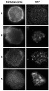
Epifluorescence and TIRF analysis of CD31 domain 6 membrane distribution on CD4+ Tcells isolated from peripheral blood. Cells were either were left in basal conditions (A) or stimulated by crosslinking the CD3 (B) in the presence of 10 μg/ml of anti-CD31 domain 6 antibody (clone MBC 78.2) directly coupled to AlexaFluor®546 (C) or 100μg/ml of human CD31 peptide (D). After 20 minutes all cells were fixed, rinsed and, for the conditions A, B and D, cells were stained with anti-CD31 domain 6 antibody (clone MBC 78.2) AlexaFluor®546-conjugated. Cells were then transferred to poly-D-lysine coated glass-bottom dishes and CD31 membrane clustering was visualized by TIRF. Antibody-mediated crosslinking of the CD31 domain 6 (C) did not change the appearance of the clustering spots as compared to T-cell stimulation alone (B). TIRF analysis of the cells stimulated in the presence of the peptide (D) showed larger clustering spots.
Figure 6
Figure 6. The CD31 peptide clusters on the plasma membrane and it accumulates between CD31 clusters upon TCR stimulation
Epifluorescence and TIRF analysis of CD31 domain 6 (A, B) and of CD31 fluorescent peptide (5,6 FAM) (C, D) distribution on CD4+ T-cell membrane. Cells were isolated from peripheral blood and stimulated by crosslinking the CD3 molecules in presence of 100 μg/ml 5,6 FAM human CD31 peptide. Twenty minutes after stimulation cells were fixed, rinsed and stained with anti-CD31 domain 6 antibody (clone MBC 78.2) AlexaFluor®546-conjugated. (F) Merge of B and D. (E) Exemple of the analysis of fluorescence intensity profiles obtained by line-scans across the cell in the red and green channels. The x-axis is the scanning coordinate in μm, and the y-axis plots the fluorescence intensities in arbitrary units. Upon TCR stimulation, the CD31 domain 6 and the CD31 human peptide form clusters on the plasma membrane (panels B and D) and, as shown in panel E, CD31 domain 6 clusters (arrows, red line) localize next to and in alternation with peptide clusters (arrow heads, green line).

Similar articles

Cited by

References

    1. Newman PJ. Switched at birth: a new family for PECAM-1. J Clin Invest. 1999;103:5–9. - PMC - PubMed
    1. Newman PJ, Berndt MC, Gorski J, White GC, 2nd, Lyman S, Paddock C, Muller WA. PECAM-1 (CD31) cloning and relation to adhesion molecules of the immunoglobulin gene superfamily. Science. 1990;247:1219–1222. - PubMed
    1. Muller WA, Weigl SA, Deng X, Phillips DM. PECAM-1 is required for transendothelial migration of leukocytes. J Exp Med. 1993;178:449–460. - PMC - PubMed
    1. Bird IN, Spragg JH, Ager A, Matthews N. Studies of lymphocyte transendothelial migration: analysis of migrated cell phenotypes with regard to CD31 (PECAM-1), CD45RA and CD45RO. Immunology. 1993;80:553–560. - PMC - PubMed
    1. Tada Y, Koarada S, Morito F, Ushiyama O, Haruta Y, Kanegae F, Ohta A, Ho A, Mak TW, Nagasawa K. Acceleration of the onset of collagen-induced arthritis by a deficiency of platelet endothelial cell adhesion molecule 1. Arthritis Rheum. 2003;48:3280–3290. - PubMed

Publication types

MeSH terms

Substances